Method and apparatus for sample preparation

a gene analysis and sample technology, applied in biochemistry apparatus and processes, specific use bioreactors/fermenters, after-treatment of biomass, etc., can solve the problems of reducing the reaction efficiency of a solid phase, reducing the reaction efficiency of a single phase, and not providing expected measurement samples, so as to save costs and labor costs. , the effect of preventing sample loss

Inactive Publication Date: 2008-10-02
HITACHI LTD
View PDF3 Cites 78 Cited by
  • Summary
  • Abstract
  • Description
  • Claims
  • Application Information

AI Technical Summary

Benefits of technology

[0030]According to the present invention, a large number of samples in small amounts such as all mRNAs contained in one cell can be amplified simultaneously and individually by PCR, and the obtained amplicons can be identified on the basis of fluorescence and collected as gelled droplets. This collection does not require providing a solid phase in a reaction solution. Therefore, cost and labors for this purpose are saved. Moreover, a sample loss and reduction in reaction efficiency attributed to a solid phase can be prevented.

Problems solved by technology

This method measures the amounts of a variety of mRNAs in an analog fashion and however, must take out mRNAs from many cells for measurement in terms of measurement sensitivity problems.
However, such methods have not been established so far.
The PCR amplification of these mRNAs or cDNA fragments by one operation merely produces a mixture of plural amplicons and does not provide expected measurement samples.
However, the amplification of 1,000,000 DNA fragments requires a larger number of reaction cells.
Moreover, it is impossible to exhaustively inject the whole target sample solution into small reaction cells.
However, this method uses spatially fixed reaction cells, some of which thus contain an expected amplicon but the others of which contain no amplicon.
Therefore, some target samples are unamplified.
Thus, the problem of this method is how to select the expected amplicon.
However, in the method using an emulsion, it is not easy to individually collect samples from individual droplets.
However, this technique, when applied to the amplification and sequencing of all mRNAs, presents a serious problem as expected.
Therefore, digital counting cannot be used in this technique.
Therefore, this approach is also inconvenient.
The collection of produced DNAs with solid beads is a good method, and this method is sufficiently available for genomic sequencing using overlapping DNA samples and however, is unsuitable for digital counting.
All the conventional methods had problems, as described above.
First, the technique using a microtiter plate does not give consideration to liquid handling during the isolation of amplicons derived from a large number of simultaneously treated samples.
Therefore, this technique had the problems of many sample vessels required according to the number of the samples and complicated handling procedures.
This method had the problem of reduction in amplification efficiency for obtaining amplicons on the solid surface using the primers immobilized on the solid phases as amplification primers.
This is because the degree of freedom of motion of DNA or RNA molecules as enzyme reaction substrates is lowered due to immobilization thereof, resulting in largely reduced reaction efficiency compared to solution systems.
Furthermore, this method had the problem of non-specific adsorption of DNAs or RNAs to solid phase surface.
Specifically, DNA fragments as initial amplification templates do not well work, when adsorbed to the solid phase.
However, no amplicon is obtained.
Particularly, when DNA or RNA samples with such an exceedingly low concentration as one molecule per reaction solution are used as starting materials for clone amplification, the influence of non-specific adsorption is relatively large and becomes a serious problem.
Furthermore, it is difficult to uniformly inject beads to individual reaction solutions in a droplet emulsion form, as described above.
Particularly, when an emulsion is prepared by stirring, it is impossible to inject the same numbers of solid phases such as beads to all droplets.
If the number of solid phases such as beads per droplet cannot be controlled, it is difficult to precisely perform single molecule measurement aimed at all molecules in a sample.
Thus, none of the conventional methods were suitable for the purpose of simultaneously amplifying and collecting all components constituting a DNA fragment pool (population of mRNAs or cDNA fragments obtained from one cell) as a sample.

Method used


Image

  • Method and apparatus for sample preparation
  • Method and apparatus for sample preparation
  • Method and apparatus for sample preparation

Examples


example 1

[0052]In this Example, the preparation of an emulsion containing agarose by stirring in oil is illustrated as an example.

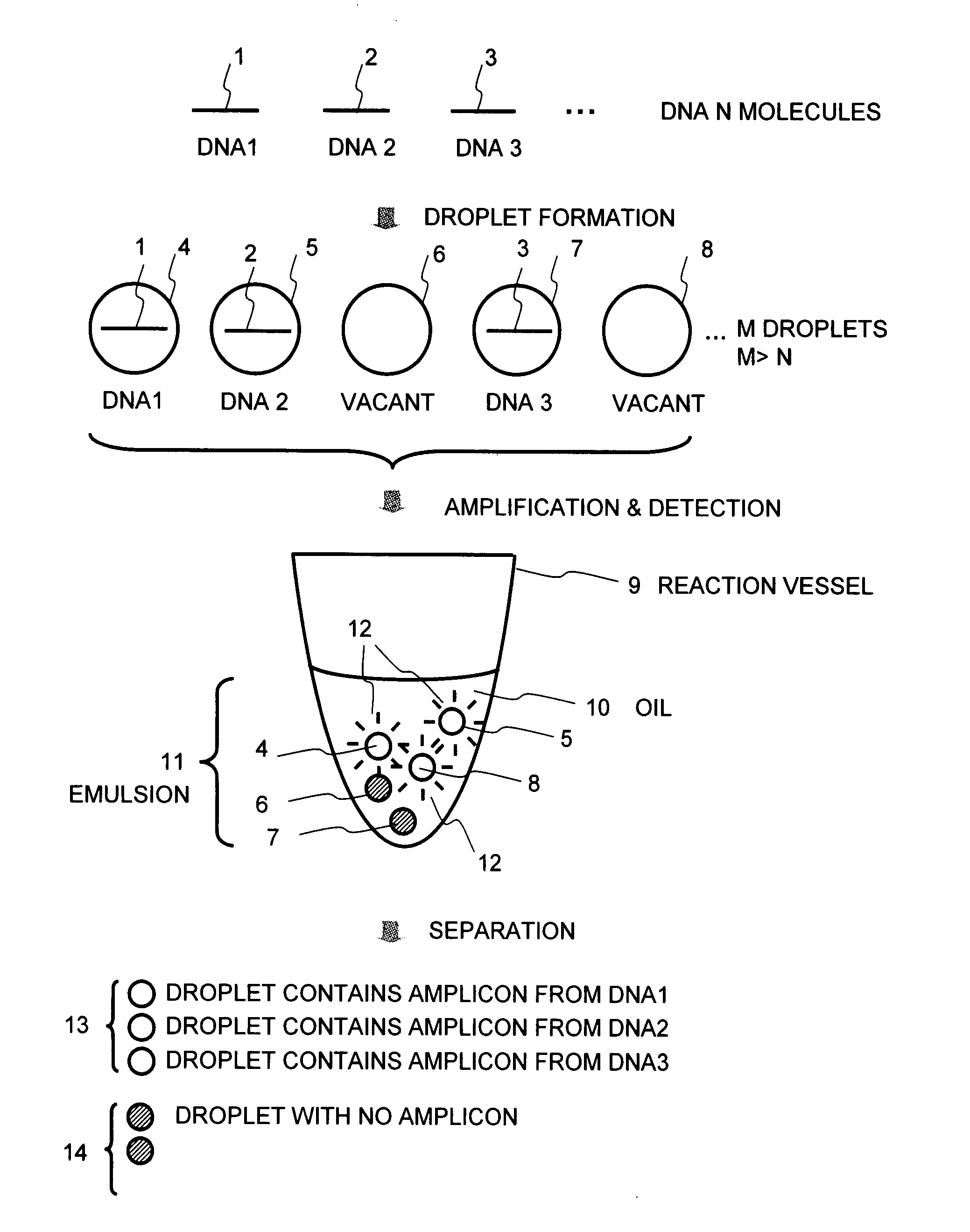
[0053]FIG. 1 shows the basic concept of the present method. A sample solution containing analyte DNA molecules 1 to 3 is fractionated into small droplets 4 to 8, wherein the number M of the droplets is greater than the total number N of the DNA molecules. As a result, the droplets 4, 5, and 7 containing the DNA and the droplets 6 and 8 containing no DNA are formed. The droplets 4 to 8 are dispersed into oil 10 in a reaction vessel 9 to form an emulsion 11. This emulsion containing the droplets is subjected to, for example, PCR amplification. Then, the presence or absence (amount) of an amplicon obtained in each droplet is detected by fluorescent detection using an intercalator or the like to make a separation between a droplet 13 that contains an amplicon from each of the DNAs 1 to 3, from which fluorescence 12 is detected, and a droplet 14 with no amplicon, from ...

example 2

Shape of Reaction Vessel

[0091]In this Example, the shape of a reaction vessel comprises a plate in which mutually separated small reaction cells are arranged.

[0092]This Example will be described with reference to FIGS. 8 to 10. As shown in FIG. 8, a plate 80 is provided with a large number of wells 83 for accommodating individual small droplets 81 and 82. The wells 83 are two-dimensionally arranged, as shown in FIG. 9, to constitute the plate 80. The droplet may be contained directly in the well 83 and covered with a hydrophobic solution 84 or may be contained in the hydrophobic solution 84 in the well 83.

[0093]In this case, the hydrophobic solution 84 is used for the purpose of forming an emulsion and further functions to prevent water evaporation from the reaction solution, to keep the shape of the droplets spherical, and to prevent the adhesion between the gel and the vessel surface during the isolation of the gel.

[0094]The droplets 81 and 82 must be separated mutually. However, ...

example 3

[0098]In this Example, another method for producing small droplets will be illustrated.

[0099]This Example will be described with reference to FIG. 11. In this Example, an ink jet unit 100 is used in droplet formation. The ink jet unit 100 comprises a tank 101 for storing a solution for preparation of droplets 103 and a nozzle 102 for spouting the formed droplets. The nozzle spouts a predetermined amount of a reaction solution by momentarily heating the reaction solution. The droplets 103 are placed in a vessel 105 so that the droplets 103 are directly spouted or allowed to fall into a hydrophobic solution 104. The droplets 103 are spouted or allowed to fall into the hydrophobic solution 104 to thereby prepare an emulsion 106.

[0100]This Example is suitable for controlling the size and quantity of the droplets and is particularly suitable for preparing approximately 0.5 pl to 10 pl droplets (approximately 10 μm to 30 μm in diameter). When the droplets are directly spouted into the hyd...


PUM

PropertyMeasurementUnit
diameteraaaaaaaaaa
diameteraaaaaaaaaa
temperaturesaaaaaaaaaa

Abstract

A method of the present invention comprises fractionating a sample solution containing analyte DNA molecules into small droplets, wherein the number M of the droplets is greater than the total number N of the DNA molecules, subjecting an emulsion containing the droplets to, for example, PCR amplification, and detecting the presence or absence (amount) of an amplicon obtained in each droplet by fluorescent detection using an intercalator or the like.

Description

CLAIM OF PRIORITY[0001]The present application claims priority from Japanese application JP 2007-093618 filed on Mar. 30, 2007, the content of which is hereby incorporated by reference into this application.BACKGROUND OF THE INVENTION[0002]1. Field of the Invention[0003]The present invention relates to a method for sample preparation for gene analysis techniques. More specifically, the present invention relates to a method for sample preparation for digital analysis of messenger RNAs (mRNAs) contained in one cell or for a method for analyzing a large number of target molecules simultaneously and individually.[0004]2. Background Art[0005]As the complete human genome sequence has been unveiled, the time has come when various genomic information has been examined energetically and exploited. Genomic information is transcribed to mRNAs and translated to proteins. Such gene expression profiling analysis is essential to examine details of life activity. Conventional mainstream analysis me...

Claims


Application Information

Patent Timeline
Patent Type & Authority Applications(United States)
IPC IPC(8): C12Q1/68C12P21/04C12M1/34
CPCC12Q1/6806C12Q1/6851C12Q2527/143C12Q2563/173
Inventor MURAKAWA, KATSUJITAKIGUCHI, SUMIYOKAMBARA, HIDEKI
Owner HITACHI LTD
Why Eureka
Try Eureka