Methods and devices for eye surgery

a technology for eye surgery and eye surgery, applied in the field of eye surgery, can solve the problems of reducing life quality, loss of vision ability, and none of them have been proved to be practical, effective, safe and routine clinical practice safe, and achieve the effect of successfully using the device and method and convenient integration in the lens extraction surgery

Inactive Publication Date: 2009-02-26
PHACO TREAT
View PDF8 Cites 71 Cited by
  • Summary
  • Abstract
  • Description
  • Claims
  • Application Information

AI Technical Summary

Benefits of technology

[0034]The method according to the invention comprises the insertion into the lens capsule of a filament that will form one or more loops inside the capsule. During insertion/removal of the filament and formation of loops, the filament is moved repeatedly back and forth, whereby material is shaved off from the capsule interior wall. The procedure is preferred to be performed after a capsulorhexis has been made, and before the lens has been separated from the capsule by other means, for a convenient integration in the lens extraction surgery. However, it is still possible to successfully use the device and method even when the lens has been separated from the capsule, and/or when the lens has been removed from the capsule. The method and device co...

Problems solved by technology

A cataract is defined as every form of opacity in the lens or its capsule; the lens becomes cloudy, resulting in a loss of visual ability.
A cataract is a painless phenomenon, but decreases the quality of life if the lens is not surgically extracted and replaced by an artificial lens.
However, none of them has been proved to be practical, effective, and safe enough for routine clinical practice.
Capsular polishing, aspiration of residual lens epithelial cells, ultrasound combined with aspiration, cryocoagulation, and osmolysis are examples of methods that have been developed and shown to remove or destroy remaining lens epithelial cells, but none of these methods have been proven ...

Method used


Image

  • Methods and devices for eye surgery
  • Methods and devices for eye surgery
  • Methods and devices for eye surgery

Examples


example 1

Experiments Using Different Filaments

[0132]Evaluation of lens-capsule dissection and shaving off lens epithelial cells from the inner capsular surface. Simulated lens extraction surgery in porcine cadaver eyes.

Young'sModulusDiameterNoFilament materialGPammOutcome1Polyethylene0.20.5Loop forms in the lens capsule, dissects0.1-1.2 GPathe lens and the capsule, simultaneouslyshaving off lens epithelial cells from theinner capsular surface.2Polyethylene0.50.4Outcome as in Experiment no 1, andeasier lens - capsule dissection, andmore efficient cell removal3Polypropylene1.00.25Outcome as in Experiment no 2, and1.0-2.0 GPaformation of a couple of loops, and evenmore efficient cell removal4Polyamid / nylon1.50.15Outcome as in Experiment no 3, and1.2-3.0 GPaformation of a several loops, and evenmore efficient cell removal5Polyvinylidenefluoride2.00.14Outcome as in Experiment no 4, and1.0-3.0 GPaeven more efficient cell removal6Reinforced2.50.12Outcome as in Experiment no 5, andPolyamid / nylonform...

example 2

Verification of the Invention by Showing the Different Parameters in Hand-Made Prototypes

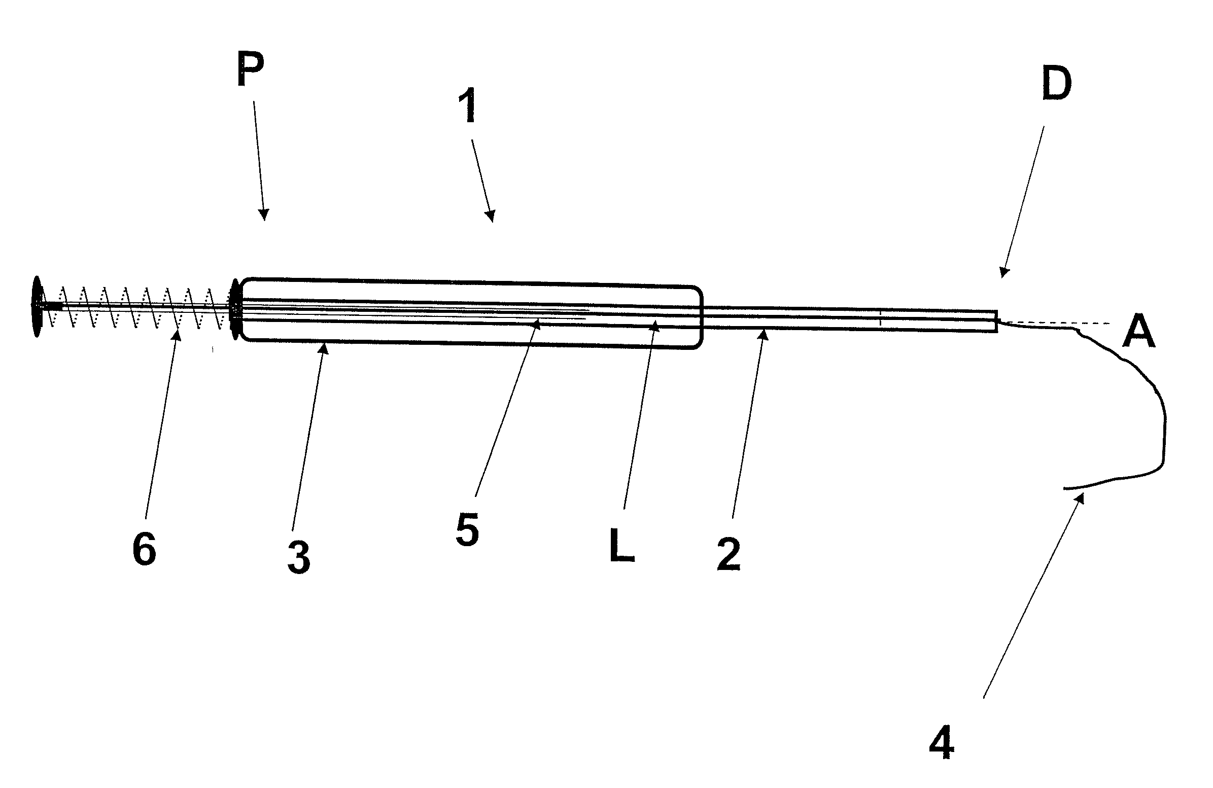
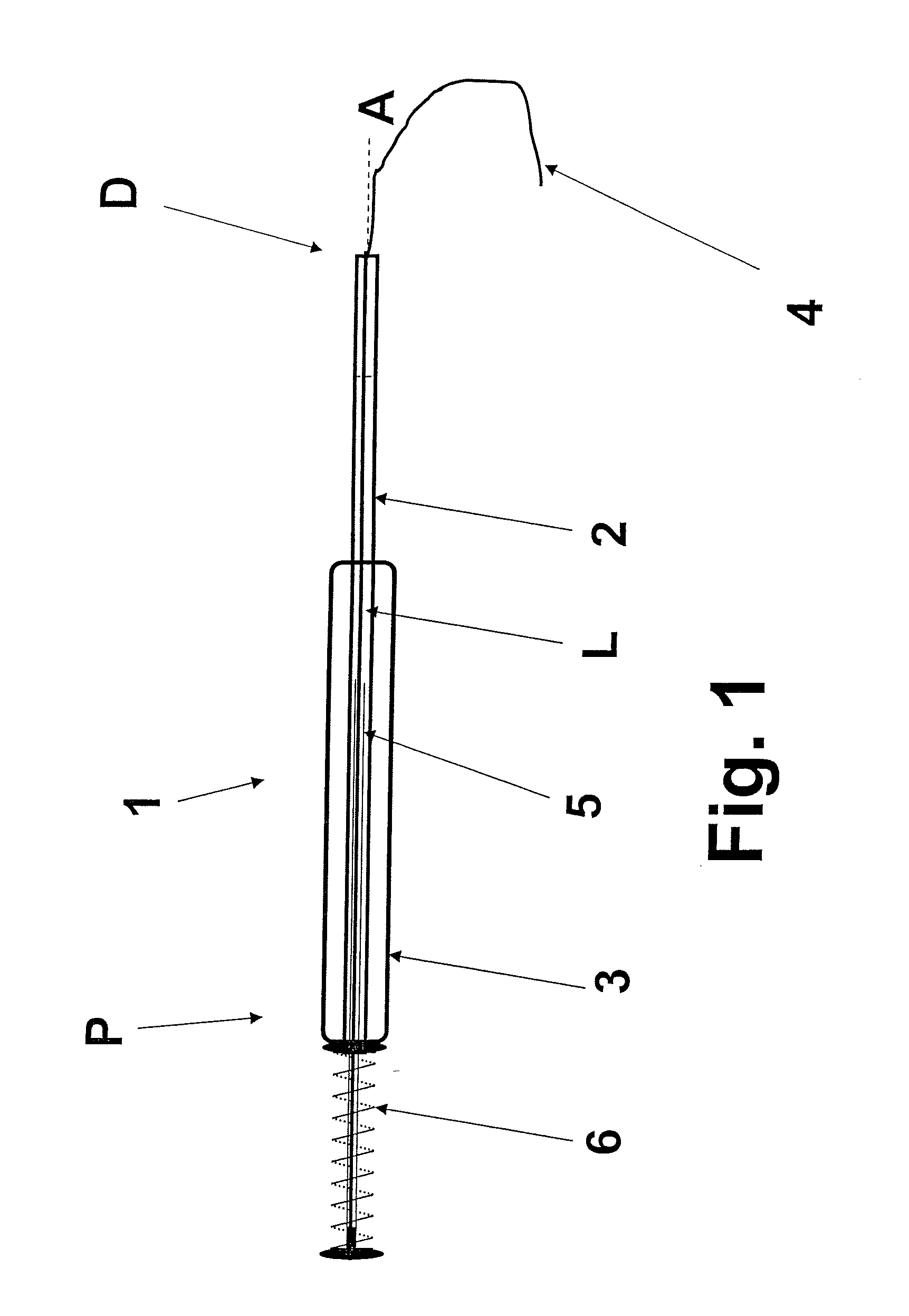
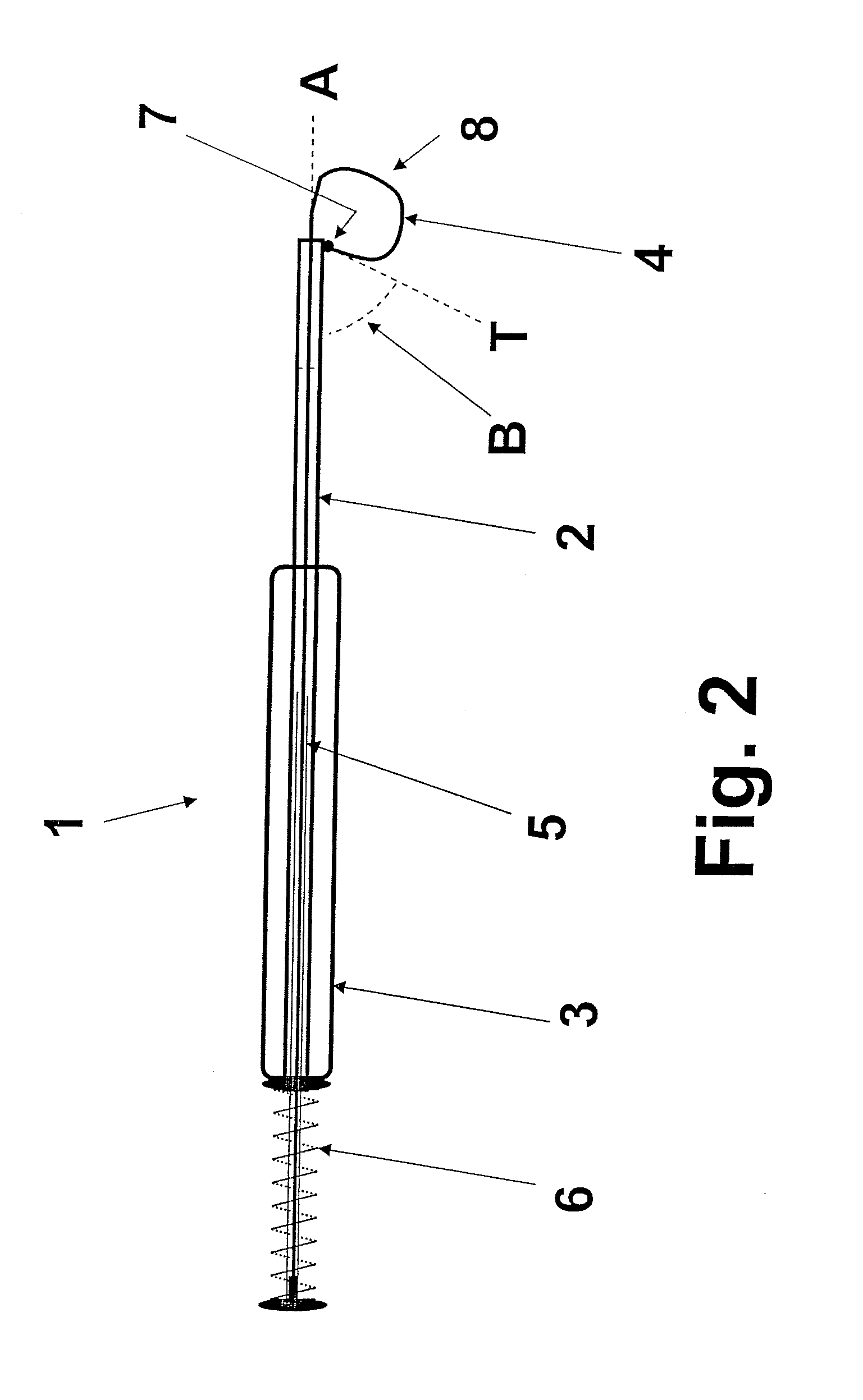
[0134]The total length (Lt), outer diameter (OD), inner diameter (ID) as well as the choice of materials are presented. The total length Lt means the distance between the proximal and distal ends of the component, if not others are mentioned. For the filament the effective length (Le) is also presented, meaning the length of the filament introduced in the lens capsule that forms the loops and the coil of loops. The use of prototypes was evaluated by simulated lens extraction surgery in fresh porcine cadaver eyes received from a nearby slaughter-house. After the lens was extracted from the eye globe, cornea and iris were removed and the amount of remaining lens epithelial cells in the lens capsule was assessed by observation, and compared to the amount of cells assessed in control eyes, i.e. conventional lens extraction.

Prototype I (Basic Concept)

[0135]Tube 2 stainless steel, L=100 mm, OD=0.81 mm...

example 3

Verification of the Idea, by Showing the Different Varying Options to Use the Device During the Surgery

[0158]The use of prototype VI, described in Experiment 2, after capsulorhexis, but before lens extraction.[0159]The use of prototype VI after lens extraction.[0160]The use of prototype VI both before and after lens extraction.

[0161]Results and conclusions: Confirmed the invention that the device can be used before and / or after the lens extraction. When using it before lens extraction, as an option to hydro-dissection, admit a second use of the device after completed lens extraction as well.


PUM


Abstract

A device for removing undesired tissue such as residual tissue, epithelial cells and/or other undesired material(s) from an inner surface of a lens capsule of an eye, includes an elongated body [2] having a proximal and a distal end and at least one central lumen extending between the ends. A flexible shaving filament (4) is movably provided in the lumen so as to be insertable into the lens capsule. The filament has an overall stiffness such that it will be able to conform to an inner surface of a lens capsule when inserted into the capsule, and also to enable shaving off of material from the inner surface. The invention also relates to a method for removing the undesired tissue is also disclosed.

Description

[0001]The present invention relates generally to eye surgery, in particular to prevention of secondary cataract (SC). More precisely the invention relates to a device for efficient removal of residual epithelial cells from the lens capsule in preparation for implanting an intraocular lens (IOL), and to a method for such removal.BACKGROUND OF THE INVENTION[0002]The crystalline lens of the human eye is located in the posterior chamber between the posterior iris surface and the vitreous body. It is a biconvex transparent tissue without nerves and blood vessels, weighing approximately 0.2 g. The lens is enveloped in a capsule, a structureless, transparent and elastic membrane bag. Approximately 80 zonular fibers, extending between the capsule and the ciliary body, suspend the lens. The inside of the lens capsule consists of lens epithelial cells and lens fibers. The lens epithelial cells form a monolayer underlying the capsule from the anterior pole to the equator of the lens. These cel...

Claims


Application Information

Patent Timeline
IPC IPC(8): A61F9/007
CPCA61B17/06166A61B17/221A61B17/320016A61F9/00736A61B2017/22034A61B2017/2212A61B2017/320733A61B17/32056
Inventor HOLMEN, JORGEN
Owner PHACO TREAT
Why Eureka
Try Eureka