Methods And Devices For Fractional Ablation Of Tissue For Substance Delivery

a fractional ablation and tissue technology, applied in the field of electromagnetic energy ablation of soft and hard tissues, can solve the problem of insufficient tissue damage, and achieve the effect of improving cosmetic and medical applications

Inactive Publication Date: 2009-03-12
PALOMAR MEDICAL TECH
View PDF6 Cites 135 Cited by
  • Summary
  • Abstract
  • Description
  • Claims
  • Application Information

AI Technical Summary

Benefits of technology

[0011]The present invention uses ablative fractional methods and devices to perform cosmetic and other treatments and functions on hard and soft tissue, including skin tissue. In various embodiments, examples of which are described in greater detail below, improved devices and systems for ablating tissue by producing lattices of EMR-treated islets in tissues are provided as well as improved cosmetic and medical applications of such devices and systems. For example, in one embodiment, methods and devices are described for creating lattices of ablation islets. In some embodiments, methods and devices are described for selectively damaging a portion of a tissue volume being treated by applying EMR radiation to produce a lattice of EMR-treated islets, which absorb an amount of EMR sufficient to damage the tissue by killing cells at the surface of the tissue or otherwise causing ablation of the tissue in the EMR-treated islets, but not sufficient to cause bulk tissue damage.

Problems solved by technology

In some embodiments, methods and devices are described for selectively damaging a portion of a tissue volume being treated by applying EMR radiation to produce a lattice of EMR-treated islets, which absorb an amount of EMR sufficient to damage the tissue by killing cells at the surface of the tissue or otherwise causing ablation of the tissue in the EMR-treated islets, but not sufficient to cause bulk tissue damage.

Method used


Image

  • Methods And Devices For Fractional Ablation Of Tissue For Substance Delivery
  • Methods And Devices For Fractional Ablation Of Tissue For Substance Delivery
  • Methods And Devices For Fractional Ablation Of Tissue For Substance Delivery

Examples


second embodiment

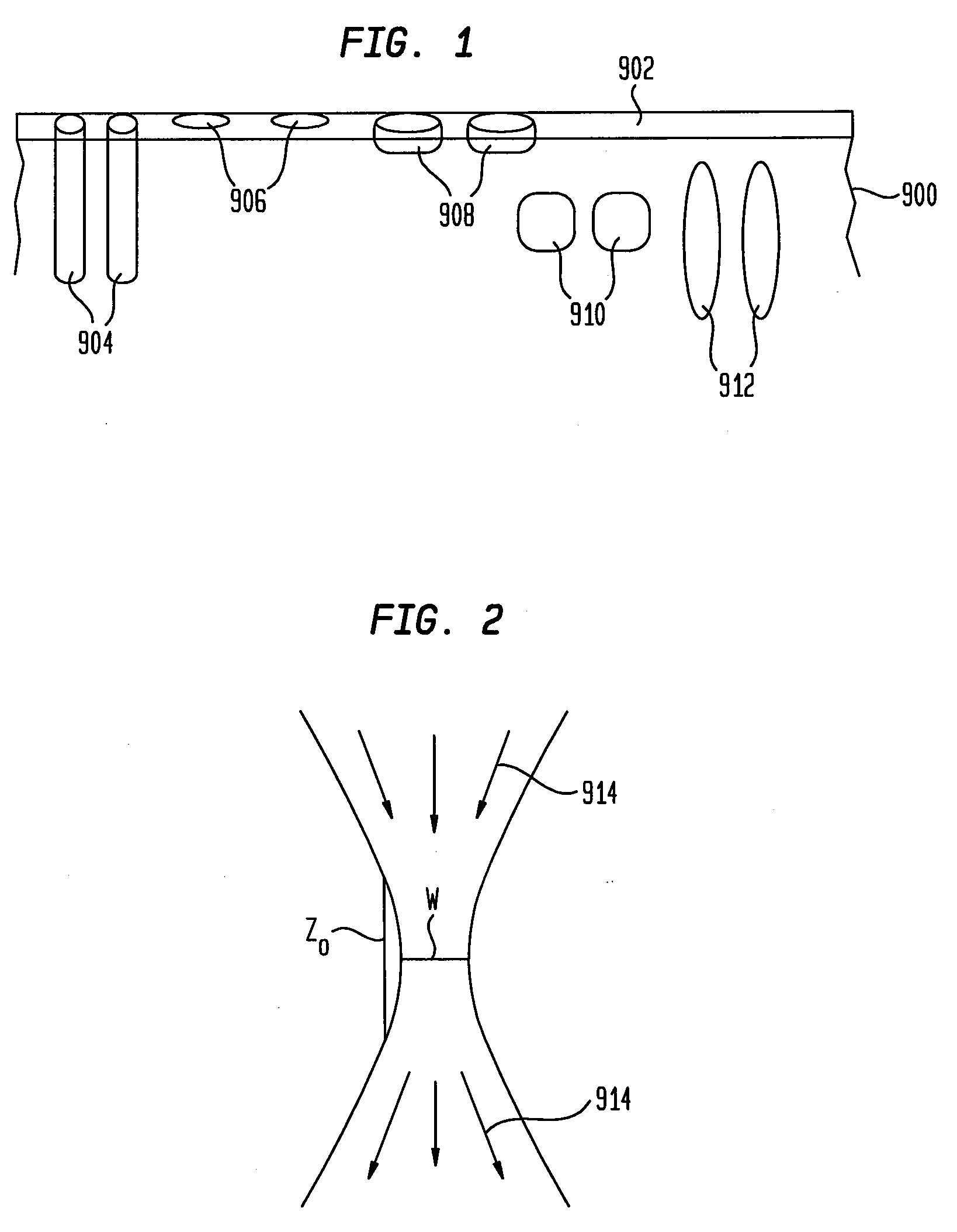
[0218]FIG. 21 depicts a hand piece 450 that uses a mirror in order to reflect portions of EMR, while allowing certain patterns of the EMR to pass through holes in order to create islets of treatment. The embodiment of FIG. 21 includes a light source 452 and, in some embodiments, beam-shaping optics 454 and a waveguide 456. These components can be in a hand piece 450, such as those hand pieces set forth above. In other embodiments, the light source 452 can be in a base unit outside of the hand piece 450. The light source 452 can be a laser, a flashlamp, a halogen lamp, an LED, or another coherent or thermal source. In short, the light source 452 can be any type of EMR source as set forth above. The beam-shaping optics 454 can be reflective or refractive and can serve to direct EMR downward toward the output of the hand piece. The beam-shaping optics 454 can generally be disposed above and to the sides of the light source 452. The waveguide 456 can be used, for example, for homogeniza...

experiment 2

[0428]2. Treatment of Tissue Using 2940 nm and a Pitch of 330 μm

[0429]In the following experiment, a sample of Yucatan black pig skin was treated in vitro using a device similar to device 500 of FIGS. 7-9. The device applied EMR at a wavelength of 2940 nm using a pitch of 30 micrometers to form the EMR islets. The skin was stored at −20C for approximately 3 months. The skin was defrosted prior to testing and warmed to room temperature. The skin was marked with a marking pen and treated with the EMR. The skin was then stretched and pinned down on a flat surface. A drop of black tattoo ink was placed on the treated area and massaged into the micro-holes. (In another test, red organic molecules in water (Eosin) were applied to the micro-holes in a method similar to the procedure described for tattoo ink.) The skin was released, and a 6 mm biopsy was obtained from the treated area. The biopsy was frozen and manually cut into 100-300 micron sections. The segments were examined with a BH2...

experiment 3

[0433]3. Treatment of Tissue Using 2940 nm and a Pitch of 220 μm

[0434]In this experiment, tissue from a Yucatan black pig in vitro was treated with a device similar to device 500 having beam spaced by 220 micrometers, and that irradiated the tissue at a wavelength of 2940 nm. The skin was defrosted prior to testing and warmed to room temperature. The skin was marked with a marking pen and treated with the EMR. The applied energy was verified after every shot of EMR. The glass window of the tip of the applicator was cleaned after each treatment. The energy readings varied by less than 5%.

[0435]The skin was then stretched and pinned down on a flat surface. A drop of black tattoo ink was placed on the treated area and massaged into the micro-holes. (In another test, red organic molecules in water (Eosin) were applied to the micro-holes in a method similar to the procedure described for tattoo ink.) The skin was released, and a 6 mm biopsy was obtained from the treated area. The biopsy ...


PUM


Abstract

Methods and devices for ablating portions of a tissue volume with electromagnetic radiation (EMR) to produce lattices of EMR-treated ablation islets in the tissue are disclosed, including lattices of micro-holes, micro-grooves, and other structures. Also, methods and devices for using the ablated islets are disclosed, including to deliver chromophores, filler, drugs and other substances to the tissue volume.

Description

RELATED APPLICATIONS[0001]This application claims the benefit of U.S. Provisional Application No. 60 / 970,420 filed Sep. 6, 2007. This application is a continuation-in-part of U.S. application Ser. Nos. 11 / 966,468, 11 / 966,538, 11 / 966,597, and 11 / 966,625 each of which was filed on Dec. 28, 2007 and which claim priority to U.S. Provisional Application No. 60 / 877,826, filed Dec. 29, 2006, and each of which is a continuation-in-part application of U.S. application Ser. Nos. 11 / 097,841, 11 / 098,000, 11 / 098,036, and 11 / 098,015, each of which was filed Apr. 1, 2005 and entitled “Methods and products for producing lattices of EMR-treated islets in tissues, and uses therefore” and each of which claims priority to U.S. Provisional Application No. 60 / 561,052, filed Apr. 9, 2004, U.S. Provisional Application No. 60 / 614,382, filed Sep. 29, 2004, U.S. Provisional Application No. 60 / 641,616, filed Jan. 5, 2005, and U.S. Provisional Application No. 60 / 620,734, filed Oct. 21, 2004.[0002]Each of the ap...

Claims


Application Information

Patent Timeline
Patent Type & Authority Applications(United States)
IPC IPC(8): A61M37/00A61B18/18
CPCA61B5/441A61N2007/0008A61B2017/00747A61B2017/00765A61B2018/00005A61B2018/00452A61B2018/00458A61B2018/0047A61B2018/207A61H23/0236A61H23/0245A61H2201/0207A61H2201/0214A61H2201/0242A61H2201/025A61H2201/0278A61H2201/0285A61H2201/10A61H2201/105A61H2201/5079A61H2230/50A61N1/30A61N1/306A61N1/327A61N2/00A61N7/00A61B18/203A61N5/062
Inventor ALTSHULER, GREGORY B.TUCHIN, VALERY V.YAROSLAVSKY, ILYACHILDS, JAMESTABATADZE, DAVIDEROFEEV, ANDREI V.
Owner PALOMAR MEDICAL TECH
Why Eureka
Try Eureka