
Immunochromatography method using fragmented antibody

a technology of immunochromatography and antibody, which is applied in the field of immunochromatography method using labeled antibodies, can solve the problems of false positive results, insufficient suppression of non-specific adsorption by the above-mentioned blocking method, etc., and achieves clear assay results, high sensitive results, and false positive results.

Active Publication Date: 2009-04-02
FUJIFILM CORP
View PDF2 Cites 15 Cited by
  • Summary
  • Abstract
  • Description
  • Claims
  • Application Information

AI Technical Summary

Benefits of technology

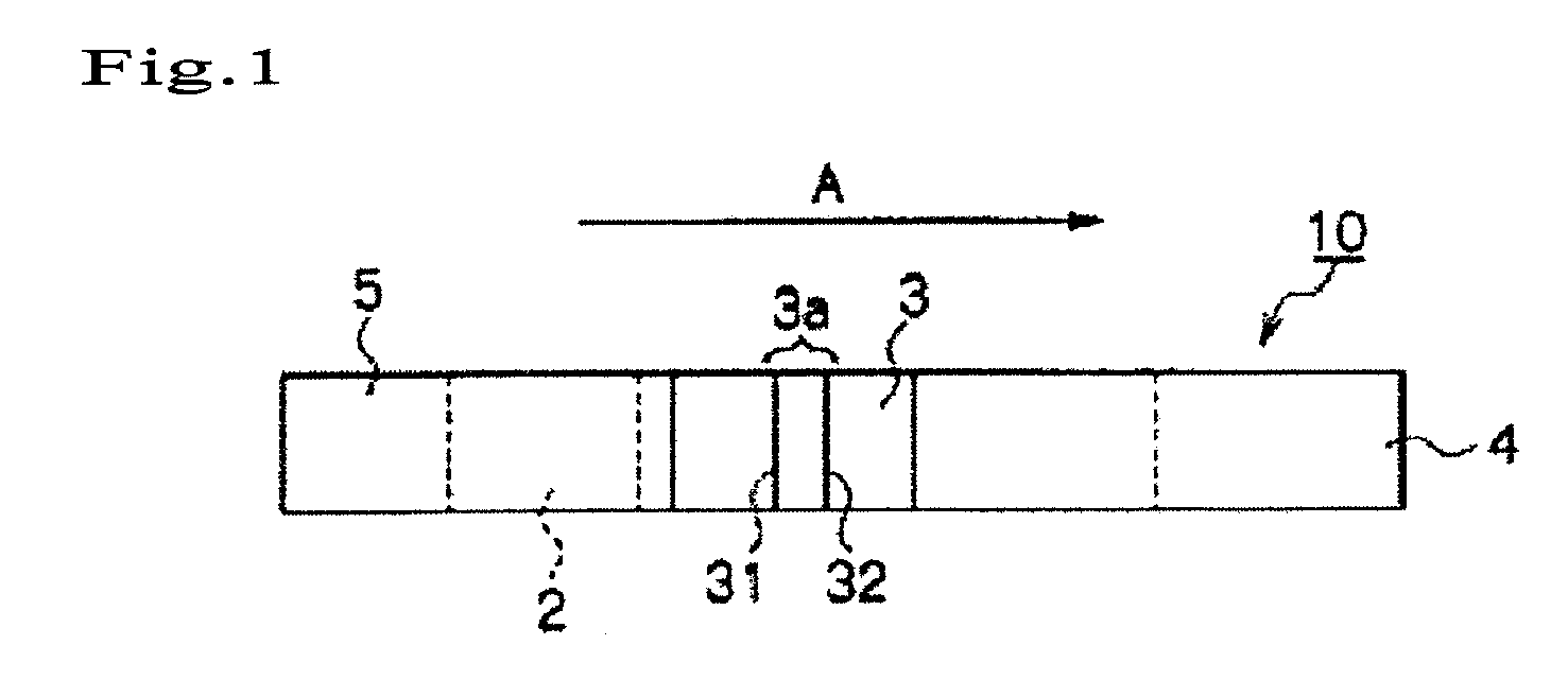
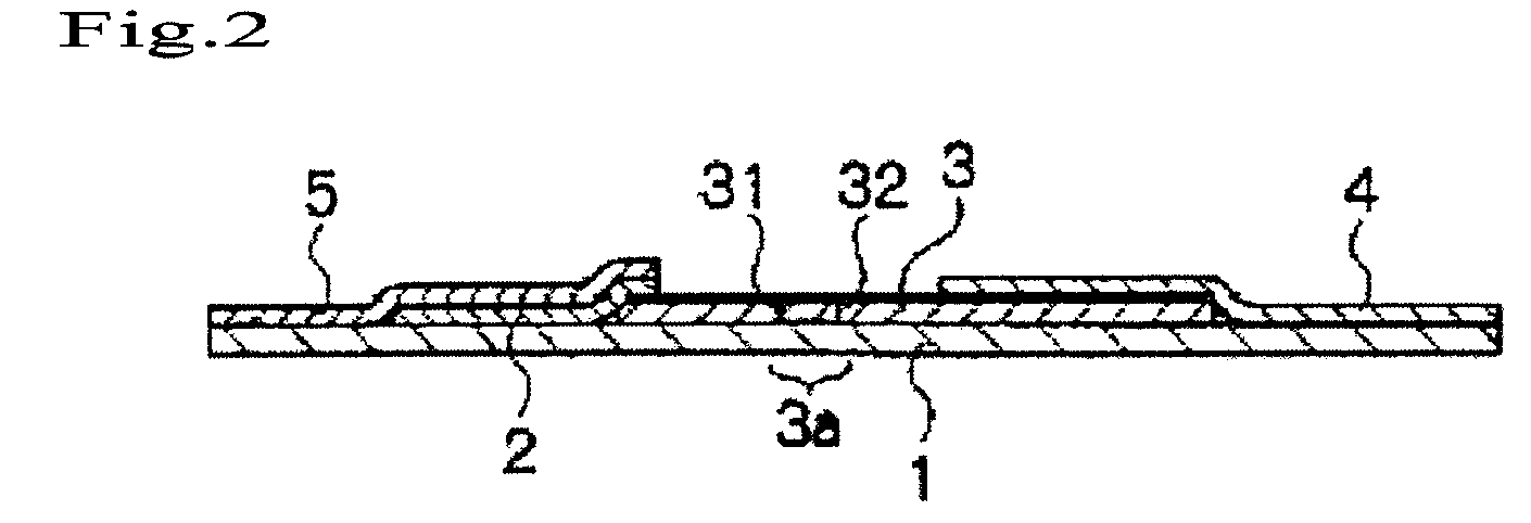
[0008]An object to be achieved by the present invention is to overcome the problem of nonspecific adsorption of a labeled antibody on the detection site, and to provide an immunochromatography method by which the false positive signal can be suppressed, and wherein the detection sensitivity is high and the highly reliable measurement can be performed.
[0016]According to the present invention, a false positive result can be suppressed, and high sensitive and clear assay result can be obtained in an immunochromatography method.

Problems solved by technology

However, the aforementioned conventional blocking method is not sufficient for suppressing non-specific adsorption.
As a result that a labeling substance is non-specifically adsorbed on the detection site, false positive result may be obtained.

Method used


Image

  • Immunochromatography method using fragmented antibody
  • Immunochromatography method using fragmented antibody
  • Immunochromatography method using fragmented antibody

Examples


example 1

Preparation and Evaluation of an Influenza Kit with Antibody Density of 0.11 μg / mm2, Gold Colloid Particle Diameter of 50 nm, and F(ab′)2 Fragment as an Antibody for Gold Colloid

1. Preparation of F(ab′)2 Fragment Anti-Influenza Type A Virus Antibody

[0137]An F(ab′)2 fragment anti-influenza type A virus antibody was prepared using an anti-influenza type A virus antibody (Product No. 7307, Medix Biochemica) and an ImmunoPure® IgG1 Fab and F(ab′)2 Preparation Kit (Product No. 44880, Pierce).

2. Preparation of Kit Using F(ab′)2 Fragment Anti-Influenza Virus Antibody-Modified Gold Colloid

[0138]An antibody-modified gold colloidal (50 nm) solution was similarly prepared using type A antibody prepared in 1, as follows.

[0139]1 mL of a 170 μg / mL F(ab′)2 fragment antibody solution was added to a gold colloidal solution having pH adjusted by addition of 1 mL of a 20 mM Borax buffer (pH 8.5) to 9 mL of a 50-nm diameter gold colloidal solution (EMGC50, BBI), followed by agitation. The mixture was a...

example 2

Preparation and Evaluation of an Influenza Kit with Antibody Density of 0.005 μg / mm2, Gold Colloid Particle Diameter of 100 nm, and F(ab′)2 Fragment as an Antibody for Antibody Immobilized Membrane

[0142]1. Preparation of F(ab′)2 Fragment Antibody Immobilized Membrane (Antibody Density of 0.005 μg / mm2)

[0143]F(ab′)2 fragment anti-influenza type A antibody was prepared in the same way as in Example 1, and the concentration was adjusted to 0.7 mg / mL. Using it, an antibody immobilized membrane was prepared as in Comparative example 1 (line width of 1 mm, and antibody density of 0.005 μg / mm2).

2. Preparation and Evaluation of a Kit with F(ab′)2 Fragment Anti-Influenza Virus Antibody-Modified Gold Colloid

[0144]An immunochromatography kit was prepared in the same way as in 1, 2 and 4 of Comparative example 1, provides that F(ab′)2 fragment antibody immobilized membrane prepared in 1 of Example 2 was used. This immunochromatography kit was evaluated in the same way as in 5 and 6 of Comparativ...

example 3

Preparation and Evaluation of an Influenza Kit with Antibody Density of 1.4 μg / mm2, Gold Colloid Particle Diameter of 15 nm, and F(ab′)2 Fragment as an Antibody for Gold Colloid

1. Preparation of Kit Using F(ab′)2 Fragment Anti-Influenza Virus Antibody-Modified Gold Colloid

[0145]F(ab′)2 fragment anti-influenza virus type A antibody was prepared in the same way as in 1 of Example 1, and an antibody-modified gold colloid was prepared as follows.

[0146]1 mL of a 500 μg / mL F(ab′)2 fragment antibody solution was added to a gold colloidal solution having pH adjusted by addition of 1 mL of a 50 mM KH2PO4 buffer (pH 8.0) to 9 mL of a 15-nm diameter gold colloidal solution (EMGC15, BBI), followed by agitation. The mixture was allowed to stand for 10 minutes and then 550 μL of 1% polyethylene glycol (PEG Mw. 20000, Product No. 168-11285, Wako Pure Chemical Industries, Ltd.) aqueous solution was added to the mixture, followed by agitation. Subsequently, 1.1 mL of a 10% bovine serum albumin (BSA ...


PUM


Abstract

An object of the present invention is to overcome the problem of nonspecific adsorption of a labeled antibody on the detection site and to provide an immunochromatography method by which the false positive signal can be suppressed, and wherein the detection sensitivity is high and the highly reliable measurement can be performed. The present invention provides an immunochromatography method, which comprises developing an analyte and a labeling substance modified with a first antibody against the analyte on a porous carrier in a state where the analyte and the labeling substance is mixed, and capturing the analyte and the labeling substance at the reaction site on the porous carrier having a second antibody against the analyte, so as to detect the analyte; wherein a fragmented antibody is used as the first antibody and / or the second antibody; an analyte is detected via amplification using an amplification solution which contains a compound containing silver and a reducing agent for silver ions; and the ratio of the non-specifically adsorbed labeling substance in the pre-amplification detection site and the non-detection site of the porous carrier is between 0.4 to 2.5 in a system where the density of the non-specifically adsorbed labeling substance is 106 / mm2 or less.

Description

TECHNICAL FIELD[0001]The present invention relates to an immunochromatography method using a labeled antibody.BACKGROUND ART[0002]Immunoassays are widely used as methods for qualitatively or quantitatively measuring the presence of an analyte existing in a biological sample such as urine or blood. Of these immunoassays, an immunochromatography method is generally used with high frequency since its implementation is simple and enables short-time measurement.[0003]The competitive reaction and the sandwich reaction are broadly used as immunoreactions to be employed in immunochromatography methods. In particular, the sandwich reaction is mainly employed for an immunochromatography method. In a typical example of the use of the sandwich reaction, the following procedures are performed to detect an analyte comprising an antigen in a sample. (1) A chromatographic medium having a reaction site is prepared by immobilizing a fine particle as a solid phase fine particle that has been sensitize...

Claims


Application Information

Patent Timeline
IPC IPC(8): G01N33/544
CPCG01N33/558Y10S435/962Y10T436/25375Y10T436/255Y10S436/826G01N33/54388
Inventor CHIKU, HIROYUKI
Owner FUJIFILM CORP
Learn More