Method for transcatheter intra-arterial perfusion magnetic resonance imaging

a magnetic resonance imaging and intra-arterial technology, applied in the field of biological tissues imaging, can solve the problems of increasing normal liver toxicity, inability to detect tumors, and inability to support optimal embolic endpoints of tae and tace procedures

Active Publication Date: 2009-05-07
NORTHWESTERN UNIV
View PDF2 Cites 27 Cited by
  • Summary
  • Abstract
  • Description
  • Claims
  • Application Information

AI Technical Summary

Benefits of technology

[0011]In one embodiment, a catheter is positioned in a hepatic artery under x-ray angiography. The subject is transferred to a magnetic resonance imaging device and a baseline transcatheter intra-arterial perfusion MRI is performed. A series of transcatheter arterial embolization (TAE) injections are performed in stages with a short time interval between each stage. A transcatheter intra-arterial perfusion (TRIP) MRI is performed after each embolic stage. The functional endpoint of the TAE embolic treatments are determined using TRIP MRI measurements. For each TRIP-MRI measurement, contrast agent is injected through the intra-arterial catheter. A time series of images is acquired to visualize contrast agent uptake within the tissue. A time series curve showing contrast agent uptake within each image voxel is evaluated to provide either semi-quantitative or fully quantitative perfusion measurements. Either the area under this curve (AUC) or maximum upslope (MUS) of the curve is calculated to provide a semi-quantitative perfusion measurement. Alternatively, the curve can be fit to a standard pharmacokinetic model to produce fully quantitative perfusion measurements or alternative measurements reflective if microvessel permeability (Ktrans and / or kep). The serial TAE and TRIP-MRI procedures are repeated until stasis.

Problems solved by technology

However, some tumors are unresectable and so other procedures must be used.
Currently, data supporting optimum embolic endpoints for the TAE and TACE procedures (sub-stasis endpoints or alternatively complete stasis of antegrade blood flow) remains conspicuously lacking.
Complete stasis of flow may increase normal liver toxicity and potentially induces release of growth factors promoting tumor growth.
Embolization to levels beyond therapeutic benefit may accelerate liver decompensation and subsequent failure.
However, accurate intra-procedural tumor perfusion measurements are currently not possible with conventional digital subtraction angiography (DSA) imaging techniques, thus representing a significant limitation for evaluating the efficacy of sub-stasis endpoints.
Rational arguments can be made in support of either of stasis or sub-stasis endpoints but there currently exist no quantitative data to support such hypotheses.
One reason for the lack of such data has been the inability to reproducibly assess sub-stasis endpoints of tumor perfusion using conventional DSA.
Although the spatial resolution of DSA permits superior visualization of vascular anatomy, small or hypo-vascular tumors can be poorly visualized.
While the recently introduced 3D x-ray angiography system has improved turn or visualization for optimal catheter placement, quantitative tumor perfusion measurements have yet to be demonstrated.
However, with IV contrast injection, the number of DCE-MRI measurements is limited by cumulative dose and the requisite wash-out times between injections.

Method used


Image

  • Method for transcatheter intra-arterial perfusion magnetic resonance imaging
  • Method for transcatheter intra-arterial perfusion magnetic resonance imaging
  • Method for transcatheter intra-arterial perfusion magnetic resonance imaging

Examples


Embodiment Construction

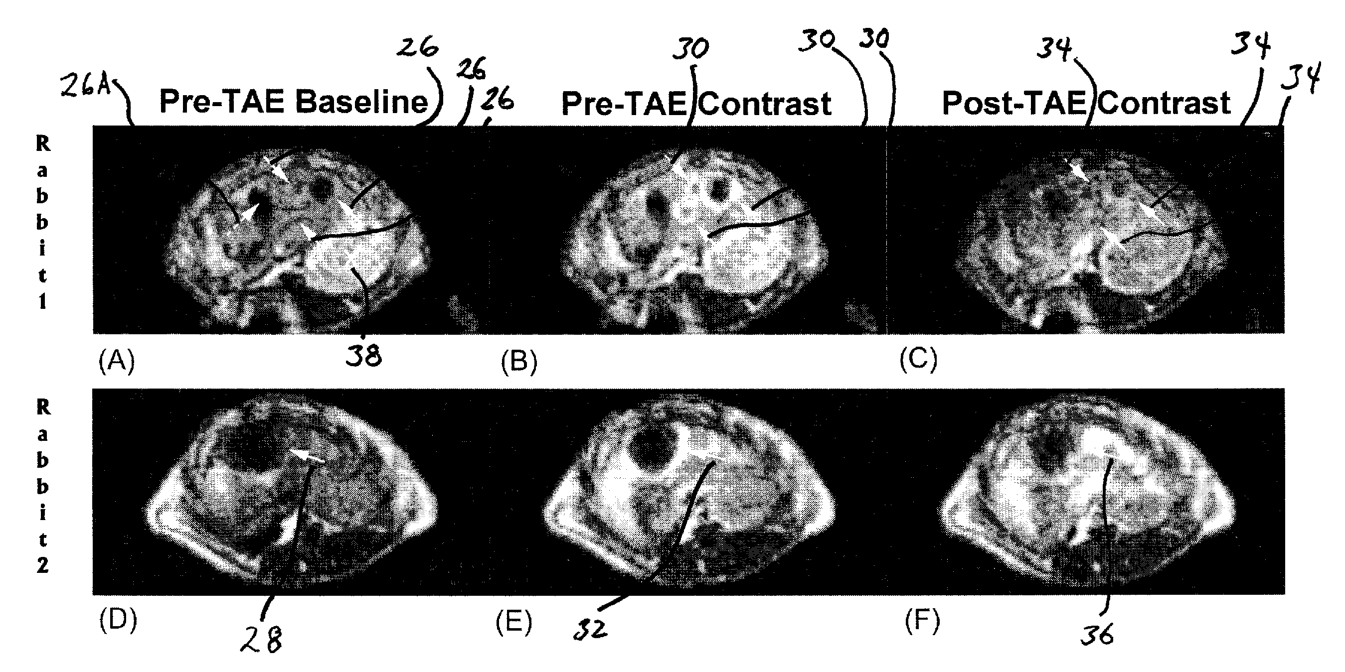
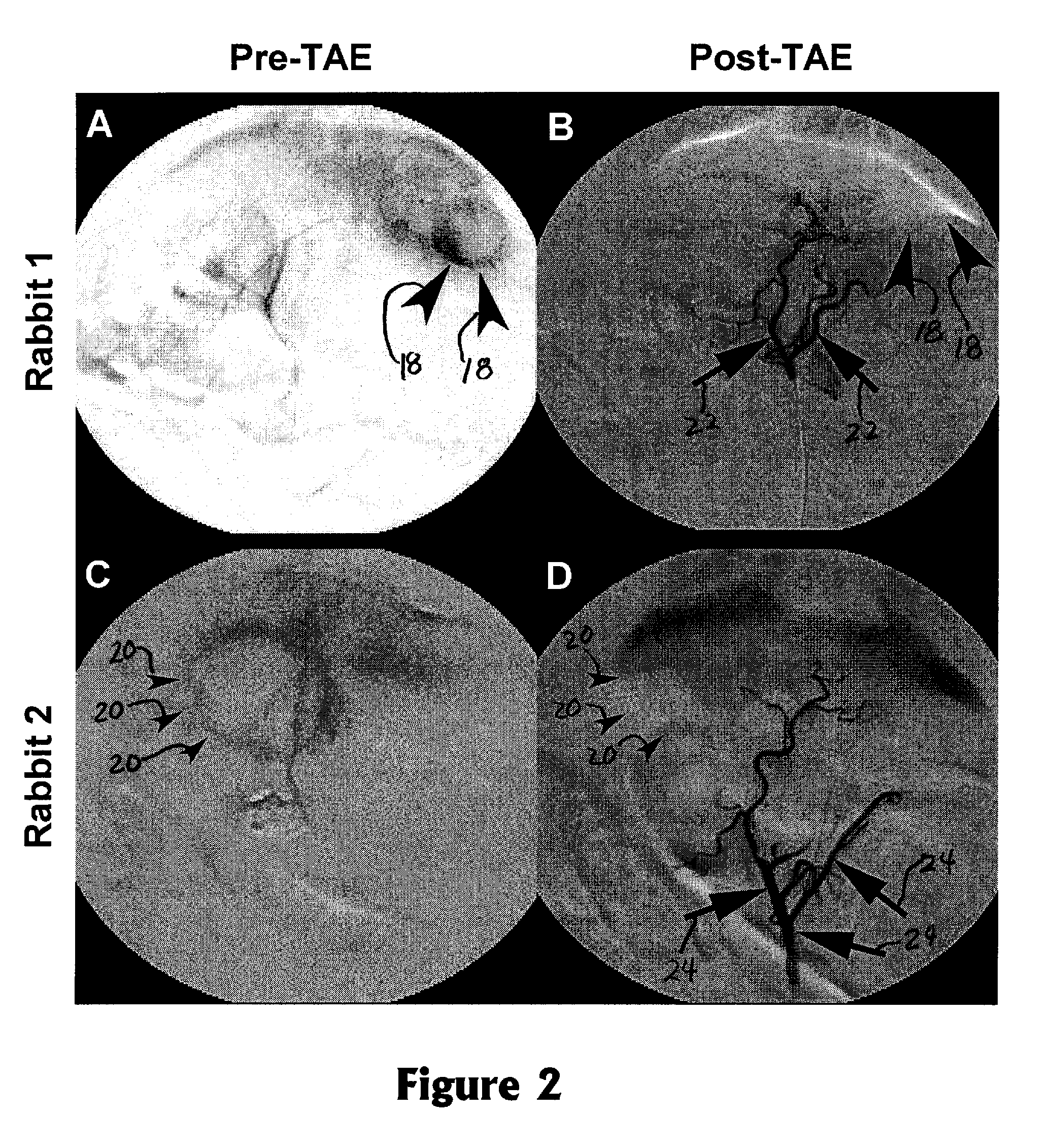
[0020]The invention provides a method for monitoring embolization procedures and in particular to determining embolic endpoints for treatment of tumors. An arterial catheter is used to inject embolizing particles into the blood vessels feeding the tumor in a series of injections, and transcatheter intra-arterial perfusion magnetic resonance imaging (TRIP-MRI) is performed following each injection of embolic particles until measurements indicate that stasis is reached (or alternatively that some selected sub-stasis endpoint is achieved).

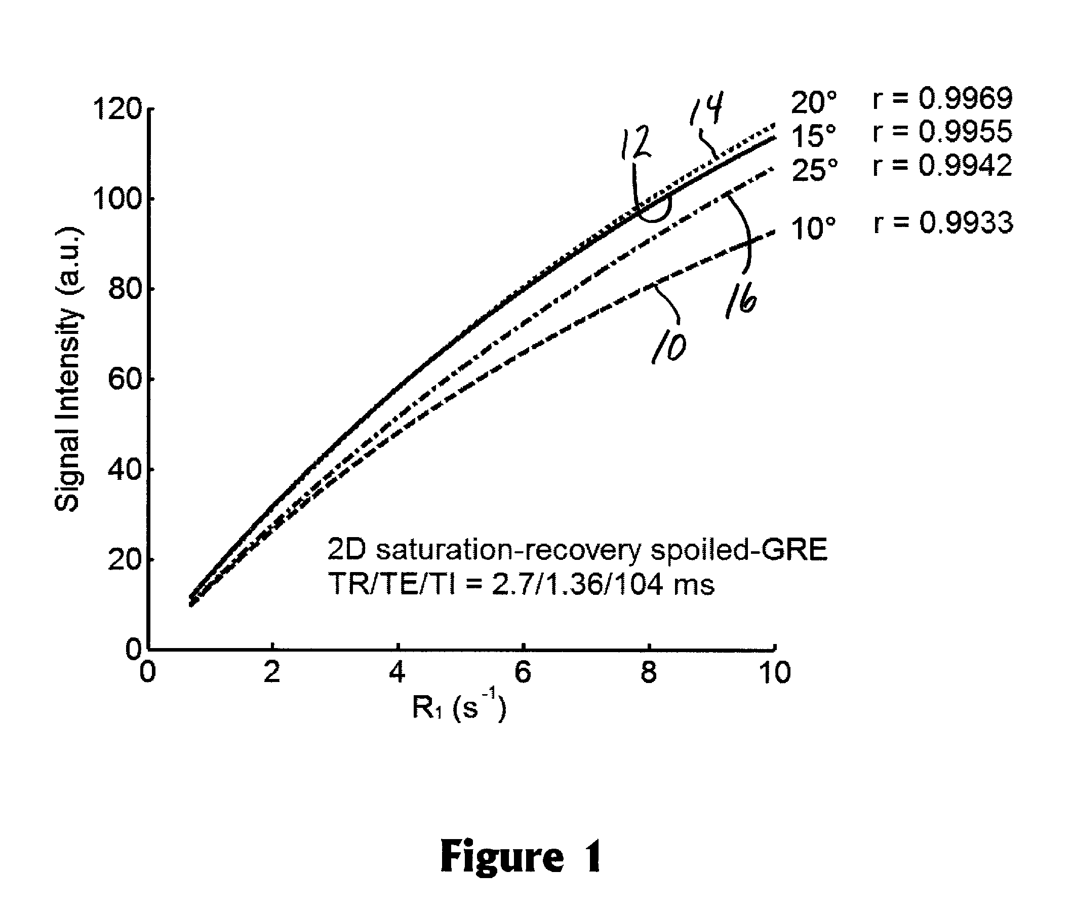
[0021]Perfusion refers to blood flow to tissues or an organ by way of the blood vessels. Perfusion MRI evaluates microscopic blood flow in the capillaries and venules using magnetic resonance image data (typically via IV injection of a contrast agent tracer). Transcatheter intra-arterial perfusion (TRIP) refers to the injection of the contrast agent being made through the lumen of a catheter inserted into the patient's artery.

[0022]The following tests...


PUM


Abstract

A method to serially determine changes in perfusion to tissues is provided. This method involves injecting contrast material into a catheter that is positioned in the blood supply proximal to the targeted tissue of interest, acquiring a time series of images that depicts the uptake of this contrast material within the tissue, deriving semi-quantitative or quantitative perfusion metrics based upon the time series of perfusion images, altering perfusion to the targeted tissue by means of injecting pharmacologic agents or embolic agents into the blood vessels supplying the targeted tissue, repeating the acquisition of perfusion images to serially monitor changes in tissue perfusion after each alteration, and calculating changes in perfusion metrics after each series of perfusion images. This method is used to monitor changes in perfusion to various tissues, including a diverse array of tumors. The perfusion imaging method can be acquired using magnetic resonance, x-ray computed tomography, or radionuclide imaging. The perfusion metric is serially measured during an embolization procedure as a means of measuring changes in tissue perfusion or to target an endpoint based upon a specific alteration in the calculated perfusion metric.

Description

CROSS-REFERENCE TO RELATED APPLICATION[0001]This application claims the benefit of U.S. Provisional Patent Application Ser. No. 60 / 984,081, filed Oct. 31, 2007, which is incorporated herein by reference.BACKGROUND OF THE INVENTION[0002]1. Field of the Invention[0003]The present invention relates generally to a method for imaging of biological tissues and in particular to a method for imaging tumors using a magnetic resonance imaging device to monitor an embolization procedure.[0004]2. Description of the Related Art[0005]Tumors may be surgically removed, or resected, if possible. However, some tumors are unresectable and so other procedures must be used. One such procedure is embolization, or the selective occlusion of blood vessels to slow or stop the blood flow to the tumor so as to reduce tumor size. One area for use of embolization is for the treatment of liver lesions, an example of which is hepatocellular carcinoma, one of the more common liver tumors. The procedure for emboliz...

Claims


Application Information

Patent Timeline
Patent Type & Authority Applications(United States)
IPC IPC(8): G06T7/00
CPCG06T7/0016Y10S128/922G06T2207/30104G06T2207/10088
Inventor LARSON, ANDREW CHRISTIANOMARY, REED ALIWANG, DINGXIN
Owner NORTHWESTERN UNIV
Why Eureka
Try Eureka