Method and Apparatus for Positioning a Medical Instrument

a medical instrument and positioning technology, applied in the direction of surgical instruments for heating, eye exercisers, chiropractic devices, etc., can solve the problems of less-invasive procedures, surgeons with limited ability to directly observe what they are doing, and the delivery of surgical tools to a treatment site not located within a body conduit, etc., to achieve the effect of improving the accuracy of surgical results

Inactive Publication Date: 2009-05-07
UC CARE
View PDF13 Cites 59 Cited by
  • Summary
  • Abstract
  • Description
  • Claims
  • Application Information

AI Technical Summary

Benefits of technology

[0016]According to further features in preferred embodiments of the invention described below, the positioning tool is a mechanical device operable to position the treatment tool at the second distance from the guiding element and in the second direction from the guiding element, or an electromechanical device operable to position the treatment tool at the second distance from the guiding element and in the second direction from the guiding element, or a position-reporting device operable to report distance and direction from the guiding element to the treatment tool, thereby providing information enabling a surgeon to position the treatment tool at a the second distance from the guiding element and in the direction from the guiding element.
[0036]According to further features in preferred embodiments of the invention described below, the positioning tool is a mechanical device operable to position the treatment tool at the second distance from the guiding element and in the second direction from the guiding element, or an electromechanical device operable to position the treatment tool at the second distance from the guiding element and in the second direction from the guiding element, or a position-reporting device operable to report distance and direction from the guiding element to the treatment tool, thereby providing information enabling a surgeon to position the treatment tool at a the second distance from the guiding element and in the direction from the guiding element.
[0055]The present invention successfully addresses the shortcomings of the presently known configurations by providing a method and apparatus enabling to accurately direct energies from a surgical and / or diagnostic tool to a treatment locus within a body of a patient, which treatment locus is distanced from that tool and not visible to a surgeon, without requiring use of real-time imaging modalities to guide placement of the tool and utilization thereof.
[0056]The present invention further successfully addresses the shortcomings of the presently known configurations by providing methods and apparatus for treating Benign Prostate Hyperplasia which are simpler and less costly to execute than are the methods of prior art.

Problems solved by technology

Minimally-invasive procedures are, however, by their nature, procedures wherein the surgeon has limited ability to directly observe what he is doing.
Delivering a surgical tool to a treatment site not located within a body conduit, however, is more complex.
Yet, use of such imaging modalities during a surgical procedure is often complex and in some cases quite difficult.
Each known imaging modality presents certain disadvantages: extended periods of fluoroscopy, for example, require extended exposure to pathogenic x-rays.
Squeezing of the prostatic urethra causes patients to experience a frequent urge to urinate because of incomplete emptying of the bladder, and a burning sensation or similar discomfort during urination.
The obstruction of urinary flow can also lead to a general lack of control over urination, including difficulty initiating urination when desired, as well as difficulty in preventing urinary flow because of the residual volume of urine in the bladder, a condition known as urinary incontinence.
Left untreated, the obstruction caused by BPH can lead to acute urinary retention (complete inability to urinate), serious urinary tract infections and permanent bladder and kidney damage.
In particular, patients with two or three or more negative transrectal biopsies who have one or more cancer risk factors such as a rising PSA, falling free PSA, strong family history, atypia, or high-grade prostatic intraepithelial neoplasia (PIN) on previous transrectal biopsies pose a diagnostic dilemma that repeated transrectal biopsy—even using an extended 10- to 12-core multisite technique (J Urol 2000; 163:152-7)—has left unresolved.
This approach, however, suffers from inaccuracy of mapping and unrepeatability, since the needle penetrations are not mechanically guided.
The setup is analogous or equivalent to the brachytherapy model.” A disadvantage of this approach is the requirement for complex ultrasound equipment and the presence of an operator skilled in its use.

Method used


Image

  • Method and Apparatus for Positioning a Medical Instrument
  • Method and Apparatus for Positioning a Medical Instrument
  • Method and Apparatus for Positioning a Medical Instrument

Examples


Embodiment Construction

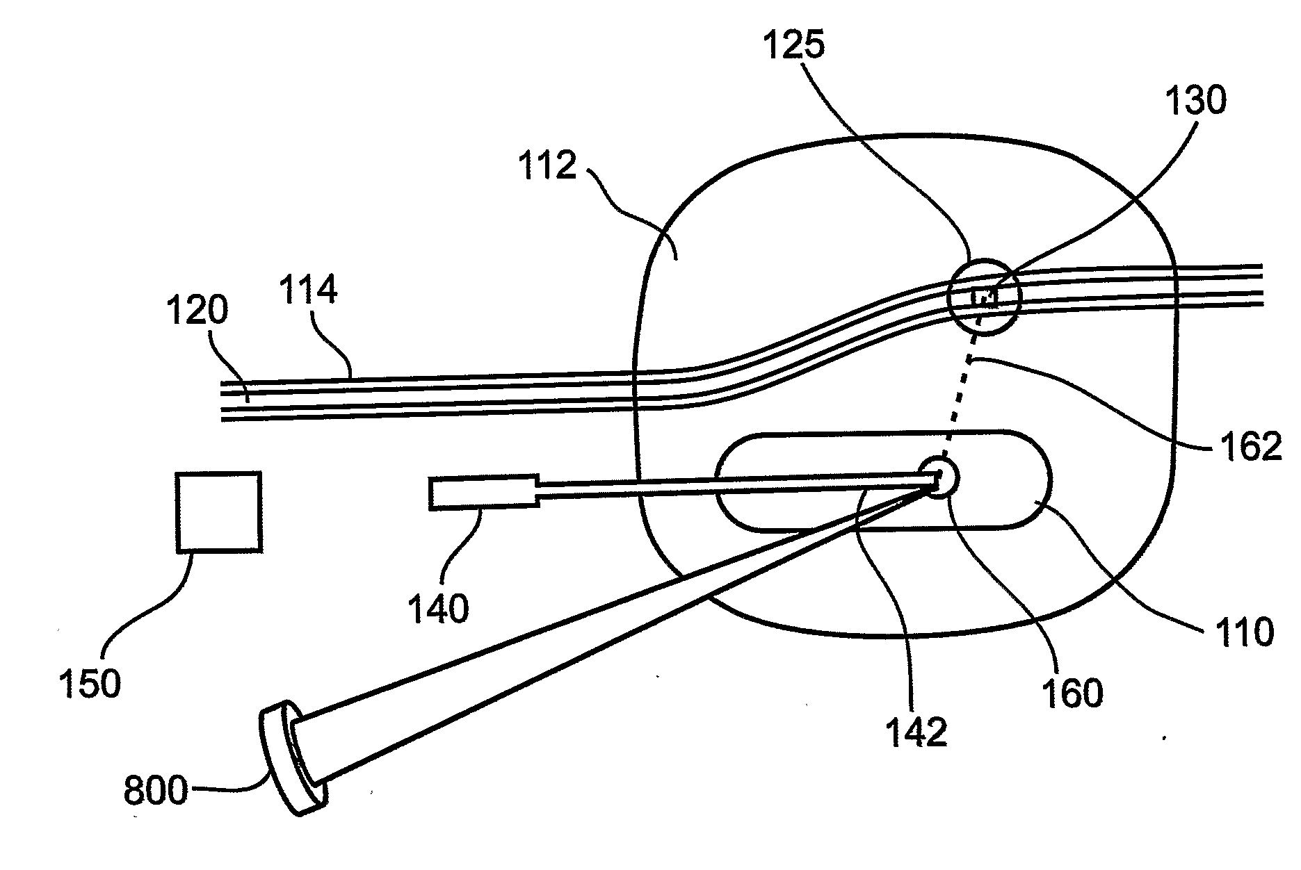
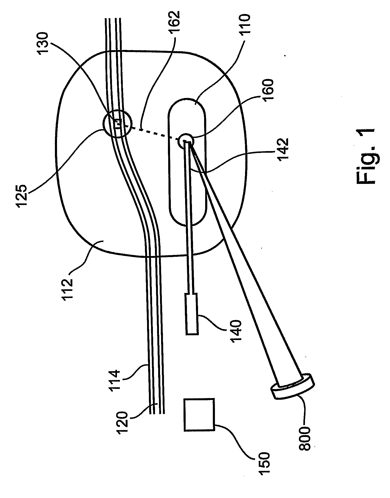
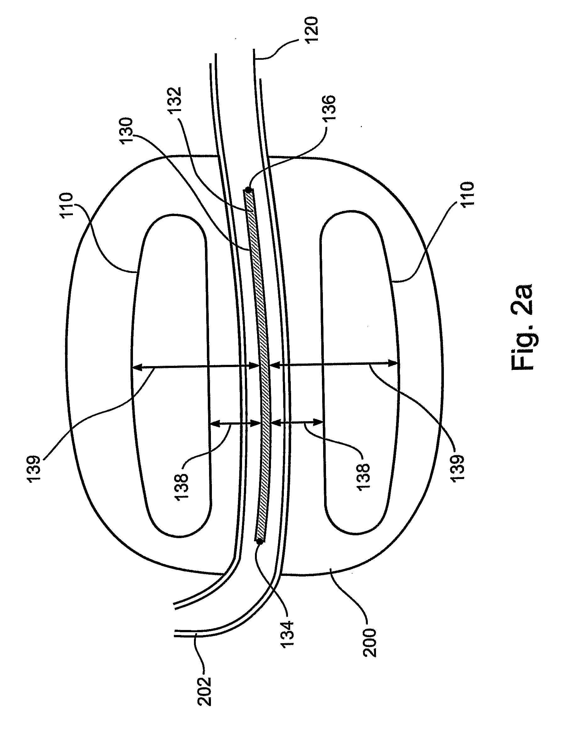
[0094]The present invention is of a method and apparatus for positioning a surgical tool at a treatment site within the body of a patient. Specifically, the present invention can be used during a minimally-invasive surgical procedure to direct a surgical treatment tool to a desired treatment site, for diagnosis or for surgical treatment at that site, while reducing dependence on real-time use of imaging modalities during positioning of the tool.

[0095]The principles and operation of a surgical treatment tool placement system according to the present invention may be better understood with reference to the drawings and accompanying descriptions.

[0096]Before explaining at least one embodiment of the invention in detail, it is to be understood that the invention is not limited in its application to the details of construction and the arrangement of the components set forth in the following description or illustrated in the drawings. The invention is capable of other embodiments or of be...


PUM


Abstract

Presented are methods and apparatus for delivering a surgical instrument to a treatment site within the body of a subject, enabling accurate placement of surgical tools in areas not directly visible to a surgeon during a surgical procedure, while reducing or eliminating need for real-time imaging modalities to guide placement of those surgical tools. A treatment tool is guided to a treatment site by placing a guiding element at a reference site within a body of a subject, the reference site having a known spatial relationship to the treatment site, and utilizing a positioning tool to guide a treatment tool to a locus so positioned with respect to that guiding element that the spatial relationship between that guiding element and that locus is substantially similar to the spatial relationship known to exist between the reference site and the treatment site, thereby positioning the treatment tool substantially at the treatment site. Methods and apparatus for focusing energy at a treatment site, methods and apparatus for treatment of Benign Prostate Hyperplasia, and methods and apparatus for performing a saturation biopsy of an organ are also presented.

Description

FIELD AND BACKGROUND OF THE INVENTION[0001]The present invention relates to methods and apparatus for diagnosing and treating a site within the body of a patient. More particularly, the present invention serves to simplify surgical procedures for treating a variety of ailments, by enabling accurate placement of surgical and diagnostic tools in areas not directly visible to a surgeon during a surgical procedure, and by accurately directing energies of surgical and diagnostic tools to a treatment locus distanced from those tools and not visible to a surgeon, while reducing or eliminating need for real-time imaging modalities to guide placement of those surgical and diagnostic tools and utilization thereof. Hence, the present invention finds uses in, for example, interventional cardiology, interventional gastrology, interventional urology, interventional gynecology, endoscopy and laparoscopy, as well as other medical disciplines.[0002]The apparatus and methods described hereinbelow are...

Claims


Application Information

Patent Timeline
Patent Type & Authority Applications(United States)
IPC IPC(8): A61B18/04A61B10/02A61B19/00A61N7/00
CPCA61B19/201A61N7/02A61B2018/00547A61B2017/00274A61B90/11
Inventor ZVULONI, RONISCHATZBERGER, SHAIKECYTRON, SHMUEL
Owner UC CARE
Why Eureka
Try Eureka