
System And Method For Non-Contrast Agent MR Angiography

a magnetic resonance angiography and contrast agent technology, applied in the field of magnetic resonance angiography, can solve the problems of limiting the diagnostic information in the image, unable to determine if a stenosis is hemodynamically, and the contrast agent that must be administered to enhance the blood vessel carries a significant financial cost, and achieves a high, often maximal signal-to-noise ratio (snr)

Inactive Publication Date: 2009-06-04
NORTHSHORE UNIV HEALTHSYST RES INST
View PDF26 Cites 43 Cited by
  • Summary
  • Abstract
  • Description
  • Claims
  • Application Information

AI Technical Summary

Benefits of technology

[0020]It is widely recognized that an SSFP pulse sequence offers the benefit of high, often maximal signal-to-noise ratio (SNR) for the blood vessels. However, it is highly sensitive to the presence of static magnetic field inhomogeneities, as typically occur near bone or air-containing structures (e.g. the base of the skull or paranasal sinuses). Intravascular spins passing through such an inhomogeneous region experience a loss of signal intensity and flow artifacts are observed in the SSFP images. The spoiled gradient-echo pulse sequence is relatively insensitive to these static magnetic field inhomogeneities. However, the SNR is substantially lower than with SSFP. Moreover, images acquired with a spoiled gradient-echo pulse sequence suffer from substantial saturation effects which result in a loss of vessel signal intensity as the blood passes inside of the 3D imaging volume and is exposed to multiple repetitions of the excitatory RF pulse.

Problems solved by technology

If the central lines of k-space are acquired prior to the arrival of contrast, severe image artifacts can limit the diagnostic information in the image.
While CE MRA is a highly effective means for noninvasively evaluating suspected vascular disease, the technique suffers from several additional drawbacks.
First, the contrast agent that must be administered to enhance the blood vessel carries a significant financial cost.
Third, CE MRA does not provide hemodynamic information, so that it is not always feasible to determine if a stenosis is hemodynamically significant.
Fourth, the signal-to-noise ratio (SNR) and, therefore, spatial resolution is limited by the need to acquire data quickly during the first pass of contrast agent through a target vessel.
An alternative technique known as pulsed arterial spin labeling (PASL) was first applied to image intracranial circulation years ago; however, image quality never approached that of 3D TOF and the method has had little clinical utility.
The use of TOF MRA is generally limited to imaging of intracranial circulation, however, because of sensitivity to patient motion and flow artifacts.
The STAR technique is ideally suited for imaging blood vessels containing fast blood flow, such as arteries, and is not well suited for imaging of veins containing slow blood flow.
It relies on inflow of spins into the selected region and is not suitable for imaging of veins.
Moreover, it is highly sensitive to magnetization transfer effects that can result in imperfect image subtraction.

Method used


Image

  • System And Method For Non-Contrast Agent MR Angiography
  • System And Method For Non-Contrast Agent MR Angiography
  • System And Method For Non-Contrast Agent MR Angiography

Examples


Embodiment Construction

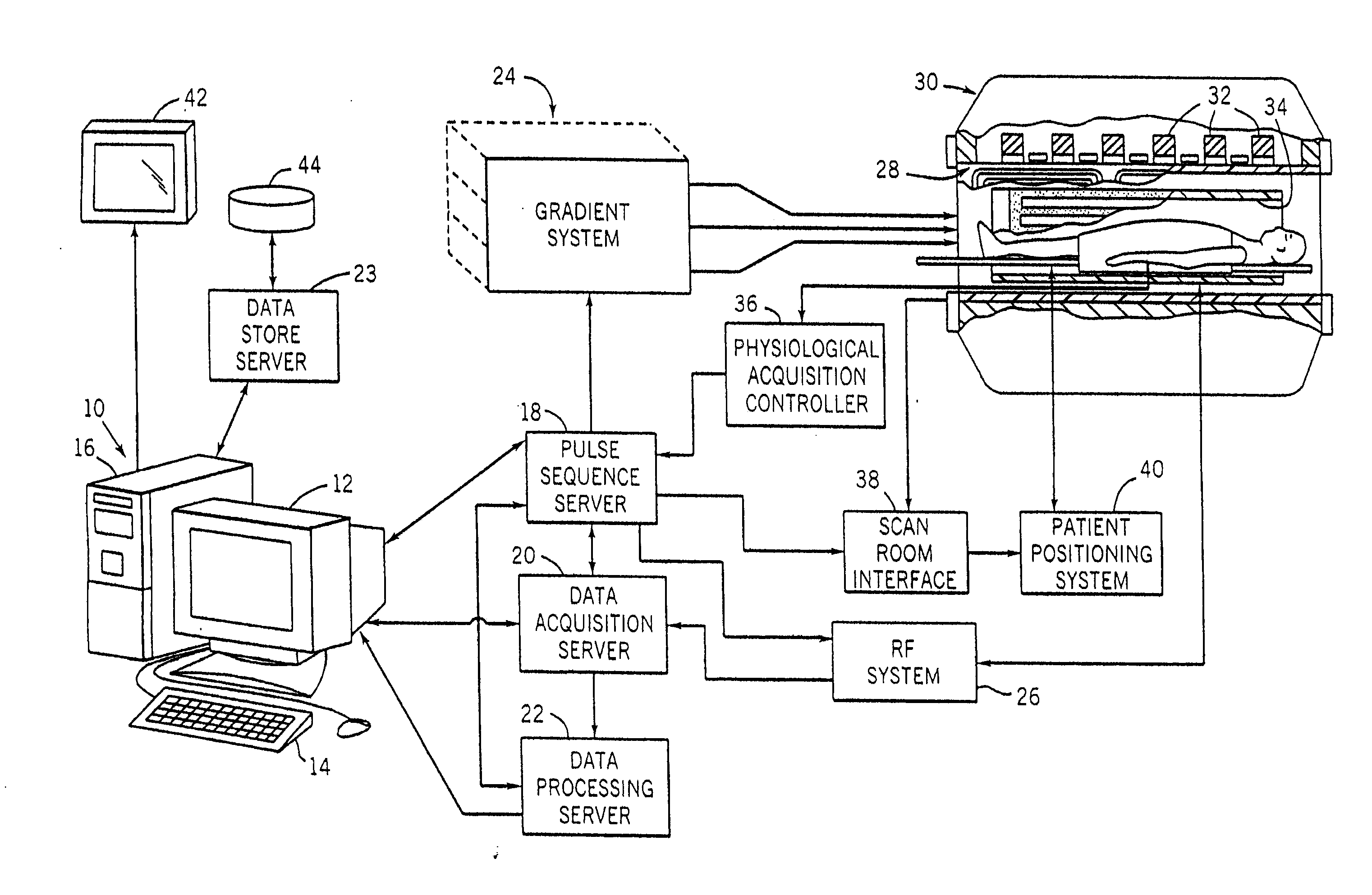
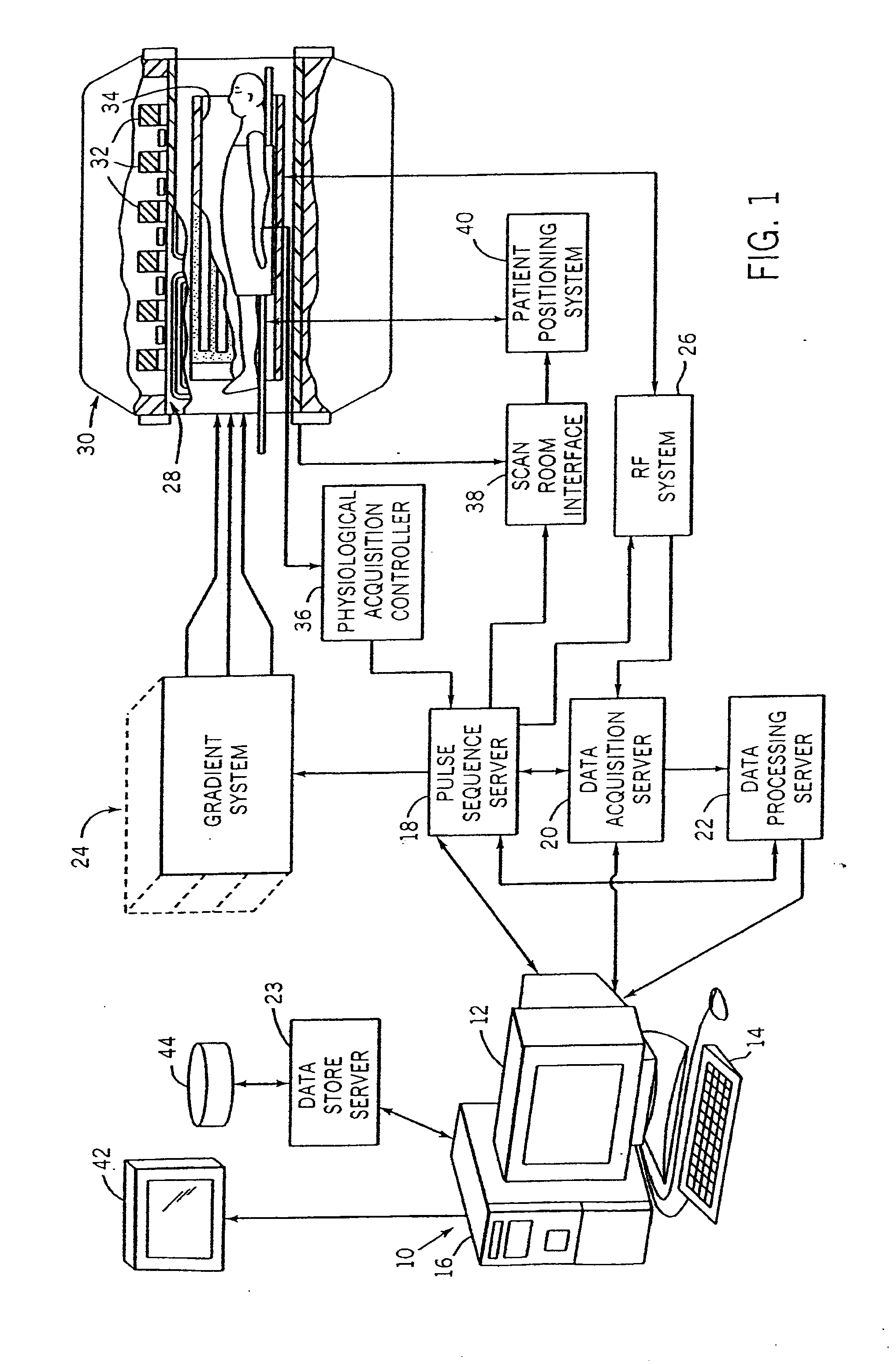
[0031]Referring particularly to FIG. 1, the preferred embodiment of the invention is employed in a MRI system. The MRI system includes a workstation 10 having a display 12 and a keyboard 14. The workstation 10 includes a processor 16 that is a commercially available programmable machine running a commercially available operating system. The workstation 10 provides the operator interface which enables scan prescriptions to be entered into the MRI system.

[0032]The workstation 10 is coupled to at least four servers, including a pulse sequence server 18, a data acquisition server 20, a data processing server 22, and a data store server 23. In one embodiment, the data store server 23 is performed by the workstation processor 16 and associated disc drive interface circuitry and the remaining three servers 18, 20, 22 are performed by separate processors mounted in a single enclosure and interconnected using a backplane bus. The pulse sequence server 18 employs a commercially available micr...


PUM


Abstract

A system and method for imaging a desired region of the circulatory system uses the subtraction of data from two acquisitions using substantially different RF pulses and / or pulse sequence timing parameters. In one or both data sets, the longitudinal magnetization of spins within a selected imaging volume has been altered by the application of one or more RF preparatory (prep) pulses. The prep is applied in such a way that subtraction eliminates signals from static background spins, such as fat, while maintaining the signal intensity of intravascular spins.

Description

CROSS-REFERENCE TO RELATED APPLICATIONS[0001]This application is based on, incorporates herein by reference, and claims the benefit of provisional application Ser. No. 60 / 991,002, filed Nov. 29, 2007, and entitled “SYSTEM AND METHOD FOR NON-CONTRAST AGENT MR ANGIOGRAPHY.”STATEMENT REGARDING FEDERALLY SPONSORED RESEARCH[0002]Not applicable.FIELD OF THE INVENTION[0003]The invention relates to a system and method for performing magnetic resonance angiography (MRA) and, more particularly, to a system and method for performing MRA without the need of a contrast agent.BACKGROUND OF THE INVENTION[0004]When a substance such as human tissue is subjected to a uniform magnetic field (polarizing field B0), the individual magnetic moments of the nuclear spins in the tissue attempt to align with this polarizing field, but precess about it in random order at their characteristic Larmor frequency. Usually the nuclear spins are comprised of hydrogen atoms, but other NMR active nuclei are occasionall...

Claims


Application Information

Patent Timeline
Patent Type & Authority Applications(United States)
IPC IPC(8): A61B5/055
CPCA61B5/055G01R33/4828G01R33/5602G01R33/5635G01R33/5611G01R33/5613G01R33/56325G01R33/5607
Inventor EDELMAN, ROBERT R.KOKTZOGLOU, IOANNIS
Owner NORTHSHORE UNIV HEALTHSYST RES INST
Learn More