Systems and methods for treating glaucoma and systems and methods for imaging a portion of an eye

a technology of glaucoma and system and method, applied in the field of system and method for treating glaucoma in the eye, can solve the problems of increased intraocular pressure, intraocular pressure, vision loss, etc., and achieve the effect of accurately monitoring the course of surgery, avoiding damage to these portions, and reducing the acquisition time for oct-image acquisition

Active Publication Date: 2009-06-18
CARL ZEISS MEDITEC AG
View PDF13 Cites 106 Cited by
  • Summary
  • Abstract
  • Description
  • Claims
  • Application Information

AI Technical Summary

Benefits of technology

[0012]According to an exemplary embodiment, the treating the volume portion by directing the laser beam to the volume portion comprises adjusting at least one of a) a laser output energy, b) a positioning of the laser beam and c) a spot size of the laser beam. This adjusting is based on at least one of the first OCT-image, the second OCT-image, and a difference between the first and the second OCT-images. Thus, the effect of a treating the volume portion by the laser beam in a previous step is taken into account to control the laser beam with respect to plural parameters to optimally adapt the laser beam for a treating in a next step of the surgery.
[0013]According to an exemplary embodiment, the acquiring at least one of the first and the second OCT-images comprises lateral scanning a measurement beam across the volume portion according to a scanning path and detecting light intensities emanating from scanned parts of the volume portion, wherein a geometry of the scanning path is based on the acquired optical microscopic image. Thus, before the first OCT-image is acquired, an optimal scan path for acquiring it may be designed based on the acquired optical microscopic image, and in particular based on an analysis of the acquired optical microscopic image. Thus, the scanning path for acquiring the OCT-image can be optimally adapted to the volume portion to be imaged three-dimensionally and to the desired accuracy.
[0020]According to an exemplary embodiment, a method for treating glaucoma in an eye of a patient is provided, comprising locating a volume portion of the eye, acquiring a first OCT-image of the located volume portion of the eye by performing a first interferometric measurement and then treating the volume portion by inserting an implant at the volume portion based on the first OCT-image. Thus, a method for treating glaucoma is provided, wherein a manual surgery is performed utilizing three-dimensional data of the volume portion which data are acquired by performing optical coherence tomography. Thus, the surgeon has three-dimensional data of the volume portion at hand, to navigate to a particular structure to be manipulated. In particular, an implant is inserted by the surgeon at a particular location of the volume portion of the eye. As in a method according to other embodiments, the locating the volume portion of the eye comprises acquiring an optical microscopic image of at least a part of the eye, identifying a distinguishable anatomical structure in the acquired optical microscopic image, and predicting a location of the volume portion based on the identified anatomical structure. Thus, it is avoided to acquire OCT-data of regions of the eye that are not required to be imaged to perform the current surgery. Thus, damage to these portions is avoided and an acquisition time to acquire the OCT-image may be shortened. Also, an accuracy of the OCT-image can be adapted, to optimally image in three dimensions that volume portion of the eye most important for performing the surgery.
[0021]According to an exemplary embodiment, the method further comprises acquiring a second OCT-image of the volume portion of the eye by performing a second interferometric measurement, and further treating the volume portion based on at least one of the first and the second OCT-images. Thus, a surgeon can accurately monitor a course of the surgery and based on the current stage of the surgery he or she can continue treating the volume portion.
[0027]According to an exemplary embodiment, a method for imaging a volume portion of an eye of a patient is provided, wherein the method comprises acquiring an optical microscopic image of a part of the eye, identifying a distinguishable anatomical structure in the acquired optical microscopic image, predicting a location of the volume portion based on the identified anatomical structure, and acquiring an OCT-image of merely the volume portion of the eye by performing an interferometric measurement based on the predicted location. The optical microscopic image of the part of the eye serves as an auxiliary image to visualize and distinguish anatomical structures in the eye. By using anatomical knowledge, in particular by analyzing the acquired optical microscopic image by performing image processing using a library of images of anatomical structures, a location of the volume portion comprising structures to be imaged three dimensionally can be predicted. The predicted location can then be used to design an optimally designed scanning path for acquiring an OCT-image.

Problems solved by technology

Glaucoma may lead to vision loss.
One of the major risk factors for developing glaucoma is the intraocular pressure.
In many cases an increased intraocular pressure is due to an impaired balance between the production of aqueous humor in the ciliary body of the posterior chamber of the eye and the drainage of the aqueous humor within the anterior chamber of the eye through the trabecular meshwork and Schlemm's canal.
Although considerably successes have been achieved during recent years, the accuracy with which surgical interventions can be performed is often not high enough.

Method used


Image

  • Systems and methods for treating glaucoma and systems and methods for imaging a portion of an eye
  • Systems and methods for treating glaucoma and systems and methods for imaging a portion of an eye
  • Systems and methods for treating glaucoma and systems and methods for imaging a portion of an eye

Examples


Embodiment Construction

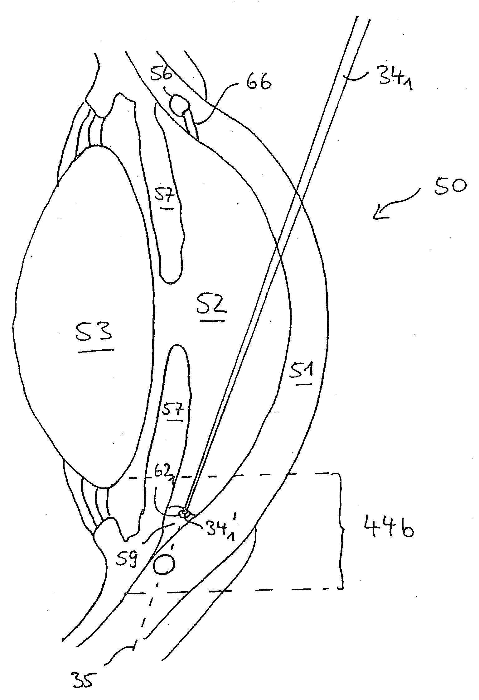
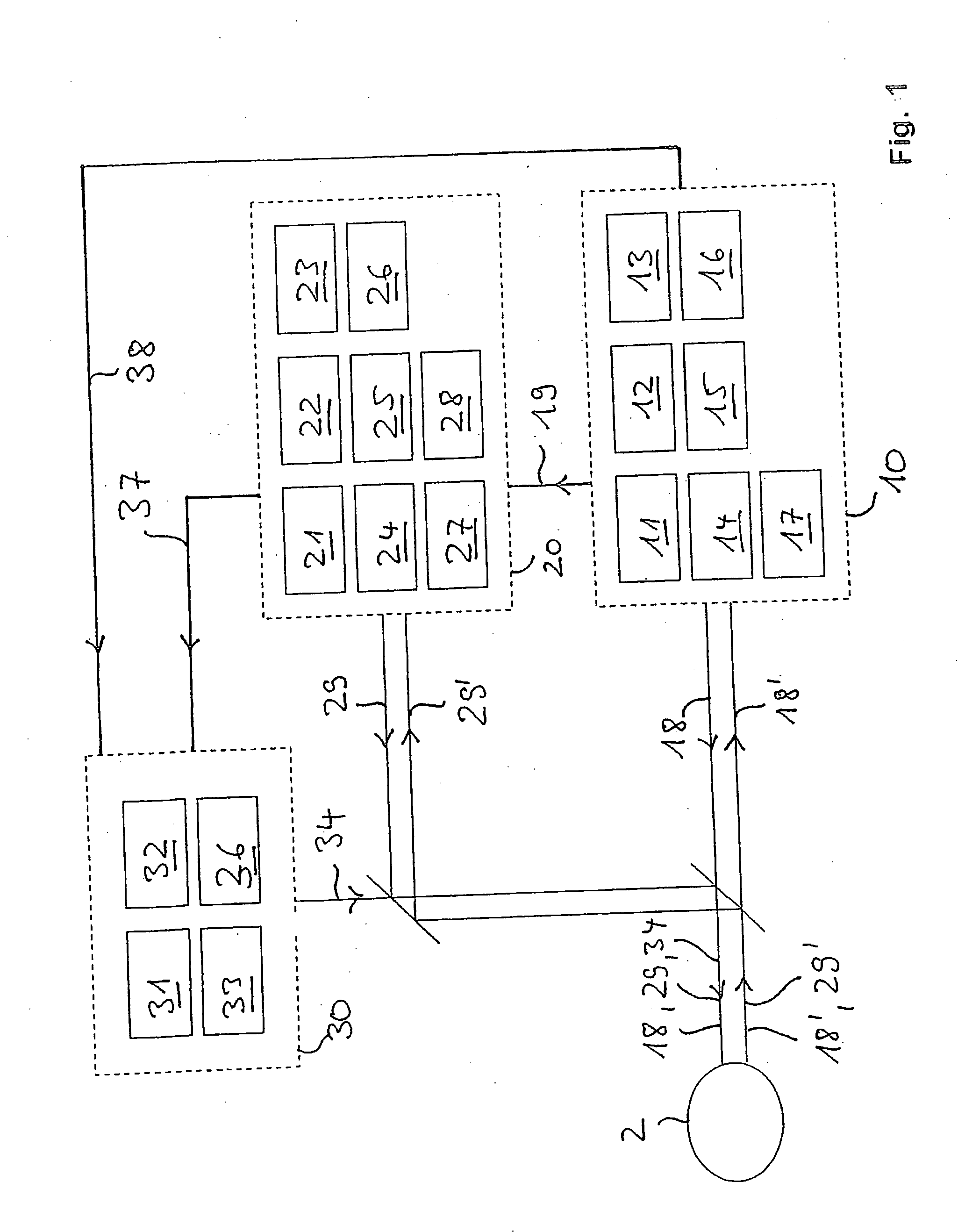
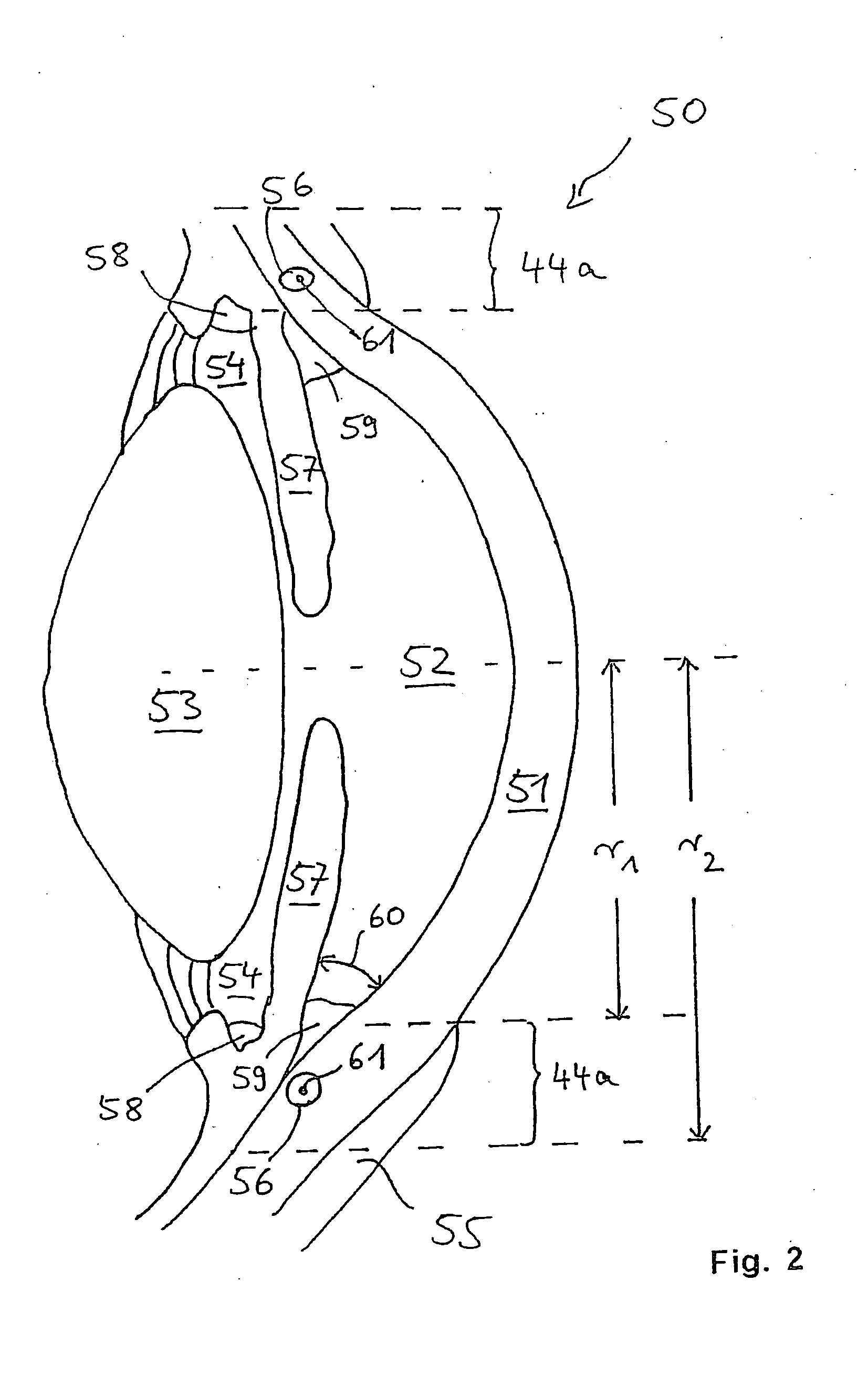
[0046]FIG. 1 schematically illustrates a system 1 of an embodiment according to the present invention for performing embodiments of methods according to the present invention. The system 1 is adapted to treat glaucoma in an eye 2 of a patient. The system 1 comprises an optical microscopic apparatus 10, an optical coherence tomography (OCT) apparatus 20, and a laser treatment apparatus 30.

[0047]Information regarding the general construction and details about optical elements of such a system are described in the European patent application EP 0 697 611 A2 the full content of which is incorporated by reference into this application. However, the inventive system 1 additionally comprises elements and devices for controlling the system 1 as set forth below.

[0048]The optical microscopic apparatus 10 comprises a light source 11 for producing a light beam 18 for illumination of at least a part of the eye 2 for which the light source also comprises illumination optics. A light beam 18′ eman...


PUM


Abstract

Systems and methods are described for imaging an eye portion or for treating glaucoma in an eye of a patient. In a first step an optical microscopic image of a portion of the eye is acquired. In the optical microscopic image a distinguishable anatomical structure is identified to predict a location of a volume portion to be imaged three-dimensionally. Three-dimensional imaging of the located volume portion is performed by acquiring an optical coherence tomography image of the located volume portion. The volume portion is treated by either directing a laser beam to the volume portion or inserting an implant based on the OCT-image.

Description

BACKGROUND[0001]1. Field of the Disclosure[0002]The present invention relates to systems and methods for treating glaucoma in an eye of a patient. In particular, the present invention relates to systems and methods for treating glaucoma in an eye of a patient using a laser beam, systems and methods for treating glaucoma by inserting implants to an eye of a patient, and methods and systems for three dimensionally imaging a portion of an eye.[0003]2. Background Information[0004]Glaucoma is a severe disease of the eye, where the optic nerve is impaired. Glaucoma may lead to vision loss. One of the major risk factors for developing glaucoma is the intraocular pressure. Increased intraocular pressure can be treated by applying drugs. In cases where applying medication does not result in the desired decrease of intraocular pressure, surgery is another therapy. In such cases both laser surgery and conventional surgery are performed.[0005]In many cases an increased intraocular pressure is d...

Claims


Application Information

Patent Timeline
Patent Type & Authority Applications(United States)
IPC IPC(8): A61F9/008A61B18/20A61M27/00A61B3/13A61B3/14
CPCA61B3/102A61B3/13A61F2009/00868A61F9/008A61B3/14
Inventor HAUGER, CHRISTOPHANDREWS, PETERNAHM, WERNERKUSCHNEREIT, RALFHACKER, MARTINSCHARIOTH, GABOR
Owner CARL ZEISS MEDITEC AG
Why Eureka
Try Eureka