System and method for image based multiple-modality cardiac image alignment

a multi-modality, cardiac image technology, applied in image enhancement, image analysis, instruments, etc., can solve the problems of archiving, storage and data transfer, unable to fuse information from multiple dynamic clinical images, and unable to meet the needs of patients,

Active Publication Date: 2009-07-16
GENERAL ELECTRIC CO
View PDF29 Cites 9 Cited by
  • Summary
  • Abstract
  • Description
  • Claims
  • Application Information

AI Technical Summary

Benefits of technology

[0009]Certain embodiments of the present invention provide a method for temporally aligning a plurality of cardiac image sequences. The method comprises performing a locally linear embedding algorithm on a first set of cardiac image sequences and on a second set of cardiac image sequences. The locally linear embedding algorithm yields a first set of motion patterns corresponding to the first set of cardiac image sequences and a second set of motion patterns corresponding to the second set of cardiac image sequences. The method also comprises mapping the first set of motion patterns to a first graphical representation and mapping the second set of motion patterns to a second graphical representation. The method also comprises determining whether the first set of cardiac image sequences and the second set of cardiac image sequences were generated from a similar point of view. If the first set of cardiac image sequences and the second set of cardiac image sequences were generated from a similar point of view, the first graphical representation is aligned with the second graphical representation by minimizing the error between at least one point on the first graphical representation and at least one point on the second graphical representation. The graphical representations are further aligned by rotating the end-diastolic point for the first graphical representation until the first graphical representation and the second graphical representation are aligned according to a threshold value

Problems solved by technology

It is difficult, however, to fuse information from multiple dynamic clinical images, especially when the clinical images are captured at different time points, with different modalities and with different setups.
For example, because of the rapid progresses in imaging techniques, the amount of image data involved in a comprehensive patient study is massive.
Gated cardiac MRI or CT sequences recorded from a complete cardiac cycle, for example, comprise 1500-5000 two dimensional images and pose challenges to archiving, storage and data transfer.
Alignment of inter-modality and / or intra-modality images is also challenging because images are captured in various orientations and positions and at different scales.
Typically, it is a time consuming process to manually align the data.
Generally, gating techniques used in the clinical environment, coupled with noise and error, are not consistent with each other.
Moreover, gating information is usually not available when these images are inspected.

Method used


Image

  • System and method for image based multiple-modality cardiac image alignment
  • System and method for image based multiple-modality cardiac image alignment
  • System and method for image based multiple-modality cardiac image alignment

Examples


Embodiment Construction

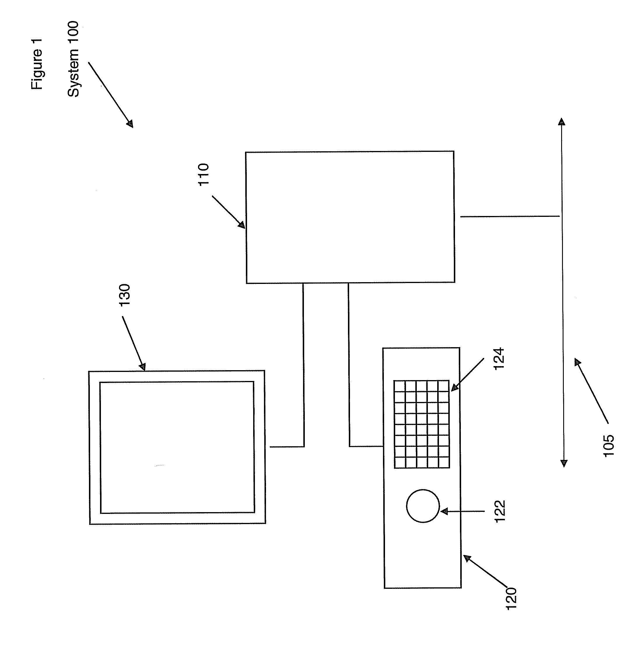
[0022]FIG. 1 illustrates a system 100 for manipulating and viewing medical images. The system 100 includes a computer unit 110. The computer unit 110 may be any equipment or software that permits electronic medical images, such as ultrasound, CT, EBT, MR, PET, or SPECT for example, to be electronically acquired, stored, or transmitted for viewing and operation. The computer unit 110 may have at least one processor and memory. The computer unit may receive input from a user. The computer unit 110 may be connected to other devices as part of an electronic network. In FIG. 1, the connection to the network is represented by line 105. The computer unit 110 may be connected to network 105 physically, by a wire, or through a wireless medium. In an embodiment, the computer unit 110 may be, or may be part of, a picture archival communication system (PACS).

[0023]The system 100 also includes an input unit 120. The input unit 120 may be a console having a track ball 122 and keyboard 124. Other ...


PUM


Abstract

Certain embodiments of the present invention provide a system and method for temporally aligning a plurality of cardiac image sequences. The method includes performing a locally linear embedding algorithm on a first set of cardiac image sequences and on a second set of cardiac image sequences. A graphical representation is created for the first set of cardiac image sequences and the second set of cardiac image sequences. A determination is made whether the first set of cardiac image sequences and the second set of cardiac image sequences were generated from a similar point of view. If a similar point of view is found, the first graphical representation and the second graphical representation are aligned using a minimization function. If a similar point of view is not found, the graphs are aligned with a template and then aligned with each other using the minimization function.

Description

RELATED APPLICATIONS[0001][Not Applicable]FEDERALLY SPONSORED RESEARCH OR DEVELOPMENT[0002][Not Applicable]MICROFICHE / COPYRIGHT REFERENCE[0003][Not Applicable]BACKGROUND OF THE INVENTION[0004]The present invention generally relates to a system and method for improved cardiac image alignment. Particularly, the present invention relates to a system and method for temporally aligning a plurality of cardiac image sequences.[0005]Medical diagnostic imaging systems encompass a variety of imaging modalities, such as x-ray systems, computerized tomography (CT) systems, ultrasound systems, electron beam tomography (EBT) systems, magnetic resonance (MR) systems, positron emission tomography (PET), single photon emission computed tomography (SPECT), and the like. Medical diagnostic imaging systems generate images of an object, such as a patient, for example, through exposure to an energy source, such as x-rays passing through a patient, for example. The generated images may be used for many pu...

Claims


Application Information

Patent Timeline
Patent Type & Authority Applications(United States)
IPC IPC(8): A61B5/02G06V10/24
CPCG06K9/32G06T7/0028G06T7/0038G06T2207/30048G06T2207/10076G06T2207/10081G06T2207/10088G06T7/20G06T7/33G06T7/38G06V10/24
Inventor LI, SHUOMAHESH, PRAKASHROEDER, DAVERANKIN, RICHARD
Owner GENERAL ELECTRIC CO
Why Eureka
Try Eureka