Kits for detecting cervical dysplasia

a cervical dysplasia and kit technology, applied in the field of kits for detecting cervical dysplasia, can solve the problems of high inter- and intra-observer variance, high rate of false positive and false negative results in screening tests, and laborious and time-consuming morphologic examination, and thus expensiv

Inactive Publication Date: 2009-07-16
VENTANA MEDICAL SYST INC
View PDF9 Cites 18 Cited by
  • Summary
  • Abstract
  • Description
  • Claims
  • Application Information

AI Technical Summary

Problems solved by technology

Thus, for examination of swabs, cytologists and pathologists have to be especially trained and even experienced examiners have a high inter- and intra-observer variance in the assessment of a diagnosis based on cytological specimens.
As a result the rate of false positive and false negative results in the screening tests remains unsatisfying high.
The morphologic examination remains laborious and time consuming and thus expensive, even when supported by the molecular methods, that make the results more reliable.
So the methods for diagnosis of conditions on a molecular level only, without the support of cell based information, are restricted to cases, where there are suitable markers, that are non-ambiguously specific for the condition to be characterized.
However, the problem with the preservation and preparation of the samples may not be overcome by just additionally using molecular markers.
This is in part due to the instability of the cell based morphological information and in part to the instability of the molecular markers to be detected during the tests.
If the samples are not prepared, transported or stored in the appropriate manner, the cell based information, or even the molecular information may get lost, or may be altered.
So the diagnosis may be impossible, or may be prone to artefacts.
For example, the interpretation of biopsies or cytological preparations is frequently made difficult or impossible because of damaged (physically or biochemically) cells.
First, the methods are highly dependent on individual perception of the examiners.
Secondly the morphological information is quite sensitive to decay processes and thus may cause artefacts after preparation of the samples.
Both aspects contribute to improper reproducibility of the results.

Method used


Image

  • Kits for detecting cervical dysplasia
  • Kits for detecting cervical dysplasia
  • Kits for detecting cervical dysplasia

Examples


example 1

Specific Immunohistochemical Detection of Endocervical and Ectocervical Epithelial Cells in Cervical Sections

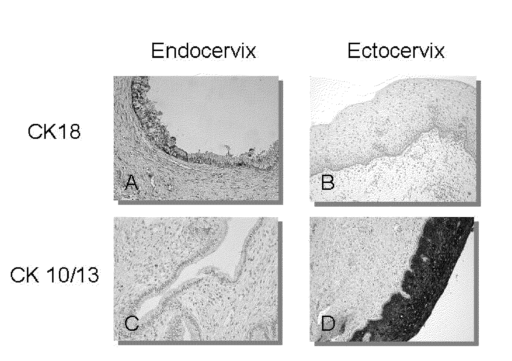
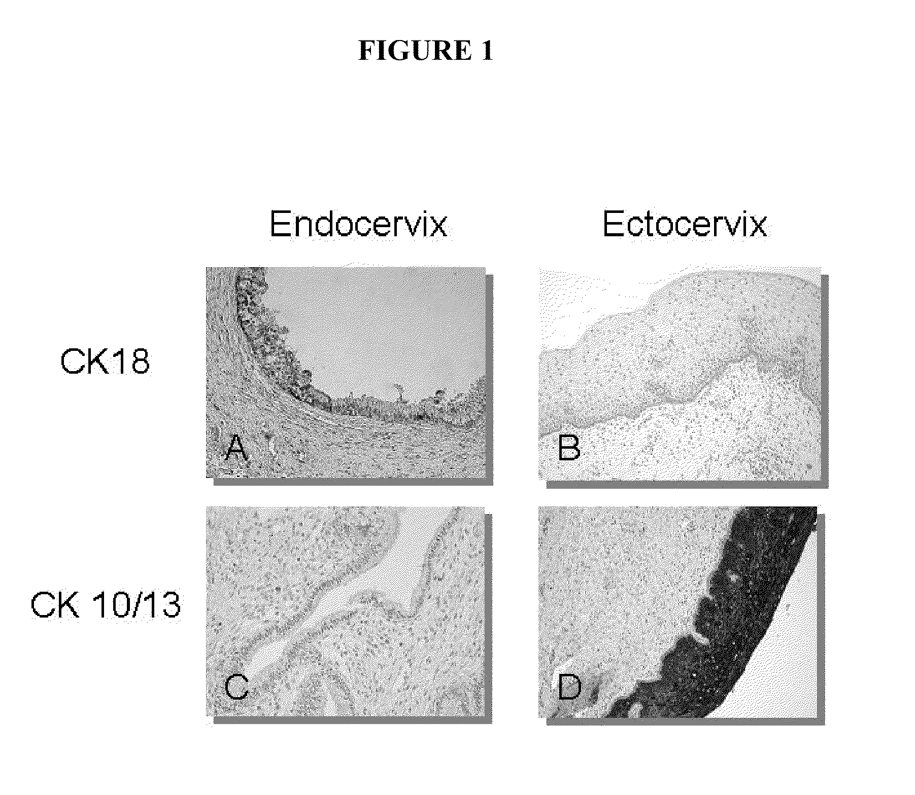
[0100]In order to evaluate markers indicating the adequacy of cervical swabs, cervical sections (fixed in 4% formaldehyde solution and paraffin-embedded) were stained with antibodies directed against Cytokeratin 18 (marker for endocervical columnar epithelia) and Cytokeratin 10 / 13 (marker for ectocervical squamous epithelia). FIG. 1 shows specific staining of endocervical epithelia with anti-Cytokeratin 18 antibody and specific staining of ectocervical epithelia with anti-Cytokeratin 10 / 13 antibody. The experiment was performed as follows:

[0101]Formalin-fixed, paraffin-embedded sections were deparaffinized in xylene bath for 5 min (step was repeated once), excess liquid was tapped off and slides were placed in 95-96% ethanol for 3 (±1) min, in 70% ethanol for 3 (±1) min (step was repeated once) and finally in distilled water for a minimum of 30 sec. For epitope retrieval, sli...

example 2

Western Blot Analysis of Solubilized Samples from Cervical Swabs

[0103]In order to evaluate, whether western blot analysis of solubilized samples allows assessing diagnosis of cervical lesions, clinical samples with known diagnosis were subjected to an immuno-chemical analysis on the basis of marker molecules after lysis of the sample material.

[0104]The clinical material (cervical swabs) samples were analyzed by Standard Western Analysis as follows.

[0105]In brief, the clinical material was in a first step solubilzed by boiling (5 min, 95° C.) in Lämmli Protein Sample buffer (100 mM Tris pH.6.8, 2% SDS, 200 mM DTT, 0.05% BpB) prior to sonification. In a second step, protein samples were resolved on a SDS-PAGE (12% Acrylamide) and subsequently transferred on a nitrocellulose membrane by tank blotting (Towbin et al., 1979, Proc Natl Acad Sci: 76:4350-4354). In a further step, the membranes were: blocked to prevent unspecific antibody binding (10% non fat dry milk in PBS) and subsequentl...

example 3

Western Blot and ELISA Analysis to Demonstrate Sample Adequacy

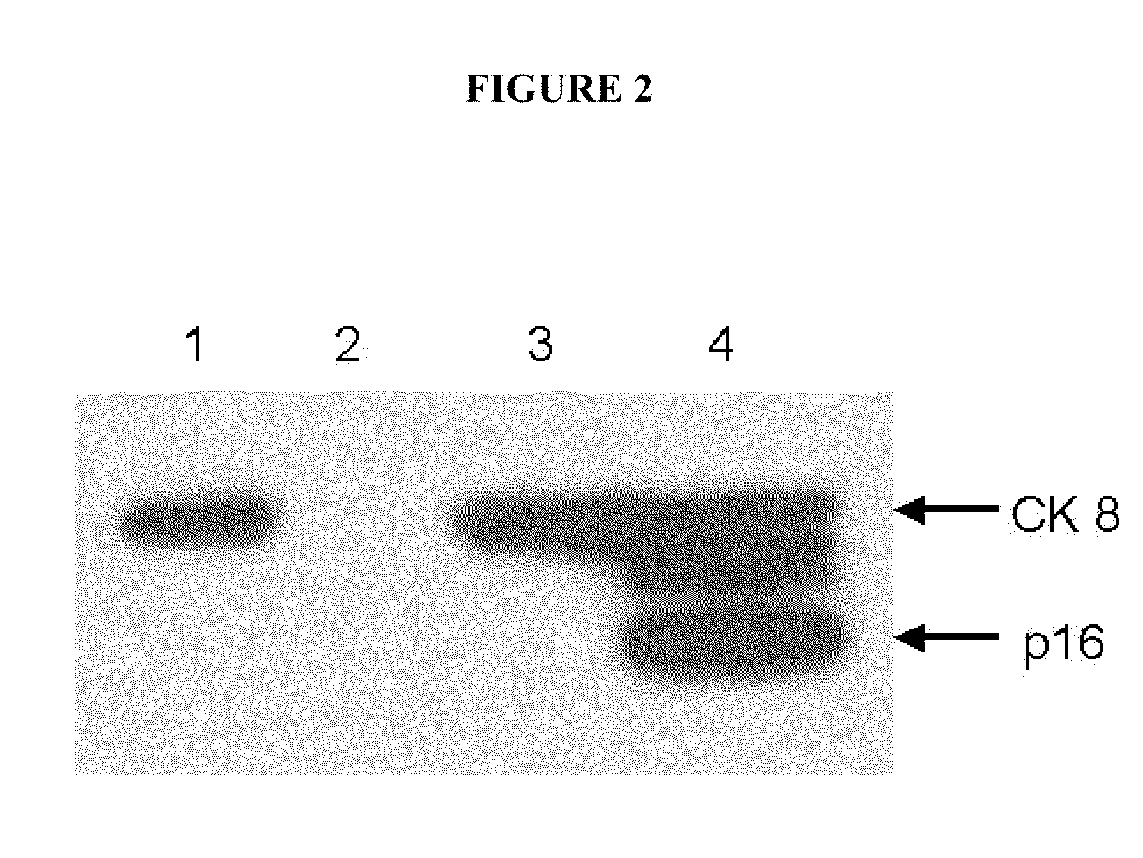
[0108]To evaluate, whether results of solution based analysis differing from diagnosis of samples may be due to inadequacy of sample, Western blot analysis of cervical swabs of four different patients with ascertained diagnosis (high-grade cervical intraepithelial neoplasia according to the cytological diagnosis of Pap IVa and Pap IVb) was performed. Antibody against p16INK4a was used to indicate presence of dysplastic cells, whereas antibodies against CK18 and CK10 / 13 were used to demonstrate adequacy of the sample.

[0109]Western blot analysis was performed as follows: Patient samples were collected with a cervical brush and directly lysed in Laemmli Sample Buffer (2% SDS, 60 mM Tris pH.6.8, 0.01%, 100 mM DTT) for 5 min at 95° C. (1×10⁷ cells / ml) with subsequent sonification (5×5 sec pulses, maximum intensity). Lysates were centrifuged for 12 min at 16,600×g) in a microcentrifuge and supernatant was transferred into a new...


PUM

PropertyMeasurementUnit
concentrationsaaaaaaaaaa

Abstract

This invention provides methods and kits for improved diagnosis of medically relevant conditions by solution based biochemical testing procedures performed in solutions of test samples. The invention provides a method to substitute the cell based morphological information contained within the cytological and / or histological data of the test sample by molecular information obtainable from the solution, wherein the original test sample is dissolved and thus enables for accurate and reproducible assessment of medically relevant diagnosis from dissolved test samples. The method according to the invention comprises the steps of determining the levels of one or more disease markers associated with the condition to be diagnosed, determining the level of one or more normalization markers suitable to substitute the information related to morphological aspects of the sample, comparing and / or combining the data of the disease and normalization markers, and assessing diagnosis of a medically relevant condition.

Description

[0001]This application is a divisional of U.S. application Ser. No. 10 / 633,484, filed Jul. 31, 2003, which claims benefit to a foreign application, EP 02017313.4, filed Aug. 1, 2002. The contents of the above applications are incorporated herein by reference in their entirety.REFERENCE TO ELECTRONIC SEQUENCE LISTING FILE[0002]This application includes a sequence listing submitted electronically herewith as an ASCII text file named “sequence.txt”, which is 53 kB in size and was created Mar. 9, 2009; the electronic sequence listing is incorporated herein by reference in its entirety.FIELD OF THE INVENTION[0003]This invention relates to methods for performing diagnosis of medically relevant conditions by detecting the levels of relevant markers characteristic for the medically relevant condition and the levels of normalization markers. The methods pertain to characterization of the sample in a solution phase, without relying on morphological cell based information.BACKGROUND OF THE INV...

Claims


Application Information

Patent Timeline
Patent Type & Authority Applications(United States)
IPC IPC(8): G01N33/53C12Q1/02C12Q1/04C12Q1/68G01N33/50G01N33/566G01N33/569G01N33/574
CPCG01N33/5076G01N33/5091G01N2333/82G01N33/57411G01N33/574
Inventor RIDDER, RUDIGERRUDY, WOLFGANGHERKERT, MATTHIASTRUNK-GEHMACHER, MARCUSREICHERT, ANJADOEBERITZ, MAGNUS VON KNEBEL
Owner VENTANA MEDICAL SYST INC
Why Eureka
Try Eureka