
System and Method for Intracranial Implantation of Therapeutic or Diagnostic Agents

a technology of intracranial implantation and therapeutic agents, which is applied in the field of system and method for intracranial implantation of therapeutic or diagnostic agents, can solve problems such as obviating the need, and achieve the effects of simple and intuitive manipulation, accurate positioning of wafers into tumors, and accurate positioning of wafers

Active Publication Date: 2009-07-30
UNIV OF VIRGINIA ALUMNI PATENTS FOUND +1
View PDF13 Cites 27 Cited by
  • Summary
  • Abstract
  • Description
  • Claims
  • Application Information

AI Technical Summary

Benefits of technology

[0008]Design of a minimally invasive instrumentation and methods for delivering such a treatment would address these limitations. Patients could thus begin to receive the benefits of chemotherapy earlier and this regimen could be re-administered with minor surgery to provide more than a single 3 week course. It is important to remember that a successful design will not guarantee realization of an ideal treatment; other limitations will exist such as choice of active agent but the limitations of physical delivery initially discussed would certainly be addressed by such a design. A successful design may thus be defined as one which causes minimal trauma to the patient compared to trauma caused by an open craniotomy (or procedure or treatment to other location or portion of the subject), allows accurate positioning of the wafer into the tumor, maintains desired wafer placement upon retraction of delivery instrument, provides for simple and intuitive manipulation by the surgeon, and is biocompatible / sterilizable.
[0009]In order to enable accurate placement of the carmustine wafers within the brain (or other location or portion of the subject) without necessitating open craniotomy, stereotactic techniques would have to be employed. Stereotaxis is a standard minimally invasive neurosurgical technique which allows surgeons to accurately guide instruments to parts of the brain through a small burr hole in the skull. One method involves the use of a rigid frame affixed to the patient's skull. Such a frame allows much more precise manipulation of the placement and angle of the instrument trajectory than would be possible freehand. However, a comparison was performed and a frameless stereotaxis system was instead chosen as the platform for this design. Frameless stereotaxis uses real time image guidance to ensure accurate placement of instruments and thus obviates the need for a rigid frame which causes further trauma by necessitating attachment to the skull. However, it should be appreciated that frame-based system may be used instead or in addition to.

Problems solved by technology

Frameless stereotaxis uses real time image guidance to ensure accurate placement of instruments and thus obviates the need for a rigid frame which causes further trauma by necessitating attachment to the skull.

Method used


Image

  • System and Method for Intracranial Implantation of Therapeutic or Diagnostic Agents
  • System and Method for Intracranial Implantation of Therapeutic or Diagnostic Agents
  • System and Method for Intracranial Implantation of Therapeutic or Diagnostic Agents

Examples


Embodiment Construction

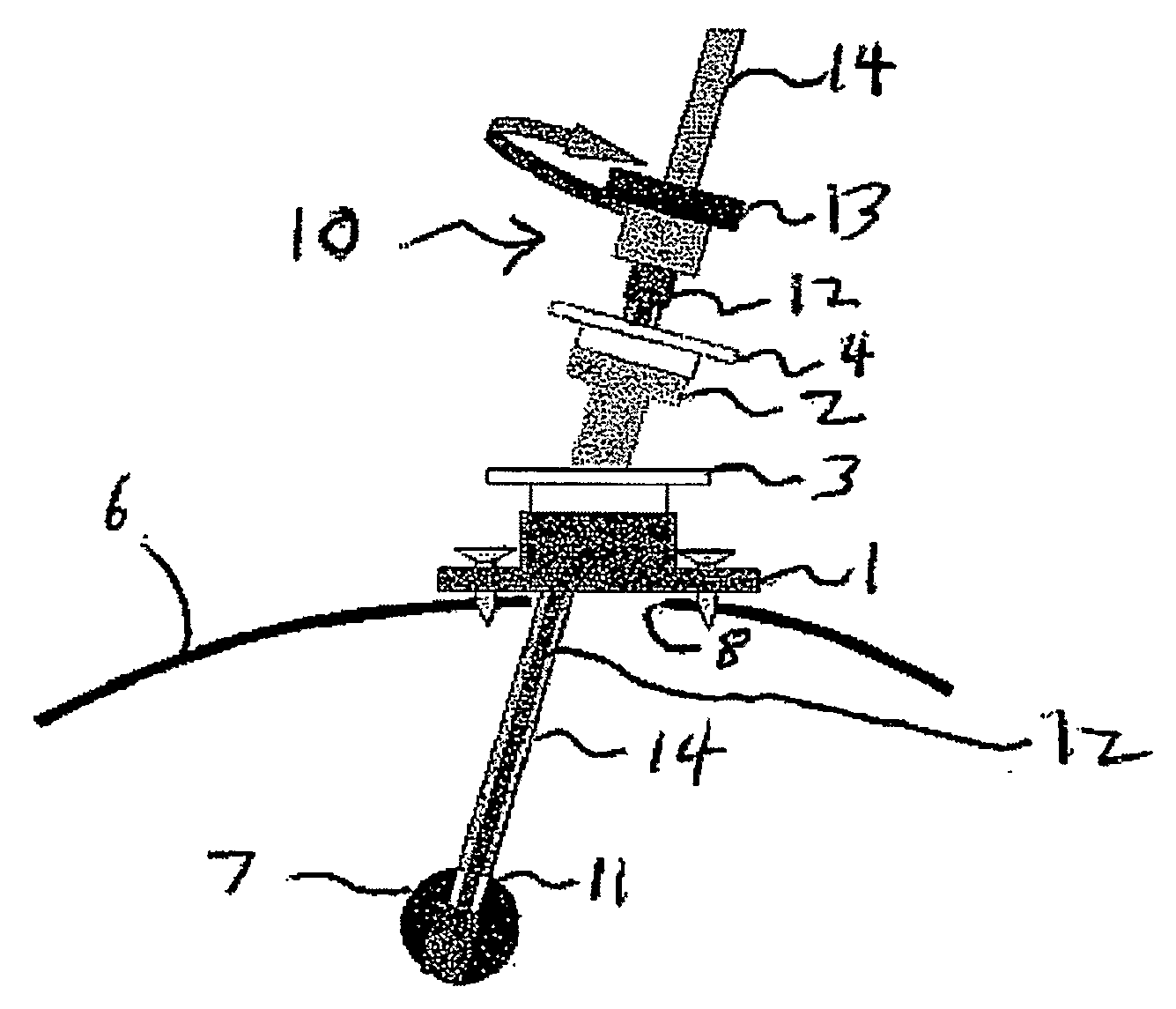
[0055]In frameless stereotaxis, the patient's head is immobilized and fiducials or markers which are visible to an infrared image guidance system are placed at reference points on the patient's skull and on the instruments entering the brain. Thus the guidance system can track the position of the instruments relative to the immobilized head. Surgeons can then visualize this information as it is coupled with preoperative MRI or CAT scan images of the brain and displayed on a monitor, as shown in FIG. 4(A)-(B). FIG. 4(A)-(B) are photographic depiction of screenshots from the StealthStation® Treatment Guidance System by Medtronic. In FIG. 4(A), reference points on the skull are visible. Also, the dot 16 on the skull shows the point of entry with the trajectory pointed directly towards the screen. Upper panes show MRI cross section images in planes which intersect to form the line containing the desired trajectory. FIG. 4(B) is a close up of the upper left pane from FIG. 4(A). The progr...


PUM


Abstract

A system and related method for delivering the anti-tumoral agent carmustine or other types of diagnostic or therapeutic agents into the brain of a patient with a brain tumor includes an insertion device, a skull mount, and a reformulated geometry of the carmustine compound (or other material) optimized for use in the insertion device and for maximized biodegradation time. The insertion device may be front loaded with the carmustine material (or other material) and inserted through the mount on a skull, to the location of the brain tumor, where the carmustine (or other material) is then released. It should be appreciated that the diagnostic and / or therapeutic system and related method thereof are not necessarily limited to the brain of a subject. It may also be used in the organ structures or tubular structures, as well as portions and locations thereof.

Description

CROSS-REFERENCE TO RELATED APPLICATIONS[0001]The present application claims benefit of priority from U.S. Provisional Application Ser. No. 60 / 833,834, filed Jul. 27, 2006, entitled “Method and Means for Intracranial Implantation of Carmustine Anti-Tumoral Agents,” the disclosure of which is hereby incorporated by reference herein in its entirety.BACKGROUND OF THE INVENTION[0002]Approximately 13,000 people develop new cases of brain tumors every year [1]. Of all central nervous system (CNS) tumors, nearly 42% are specifically diagnosed as Glioblastoma Multiforme (GBM). Conventional treatment consists of surgical resection and external beam radiation. The median survival for patients undergoing surgical resection alone is 6 months while those who undergo a more complete regimen including radiation is 9 months [2]. Thus the fact remains that even aggressive treatment with established methods of surgery, radiation, and chemotherapy leads to a median survival rate of less than one year f...

Claims


Application Information

Patent Timeline
Patent Type & Authority Applications(United States)
IPC IPC(8): A61B6/12A61M5/00
CPCA61B19/201A61B6/508A61M31/007A61B90/11A61P35/00
Inventor BROADDUS, WILLIAM C.MAHAJAN, RAHULGILLIES, GEORGE T.
Owner UNIV OF VIRGINIA ALUMNI PATENTS FOUND
Learn More