Image based quantitation of molecular translocation

a molecular translocation and quantitation technology, applied in the field of molecular translocation quantitation based on image, can solve the problems of limited microscopic applications and the inability to produce the best measurement method

Active Publication Date: 2009-08-13
AMNIS CORP
View PDF64 Cites 59 Cited by
  • Summary
  • Abstract
  • Description
  • Claims
  • Application Information

AI Technical Summary

Problems solved by technology

Until recently, microscopic applications have been limited due to the subjective nature and the lack of means to quantitate imagery.
However, this method is unlikely to produce the best measurement because it relies on an accurate nuclear mask, subjective erosion and dilation routines that determine the nuclear and cytoplasmic boundaries, an additional subjective dilation of the cytoplasm mask to create an annular volume, and both the cytoplasm and the nucleus have areas that are not represented in the calculation.

Method used


Image

  • Image based quantitation of molecular translocation
  • Image based quantitation of molecular translocation
  • Image based quantitation of molecular translocation

Examples


example 1

Induction of Translocation in Adherent Cells

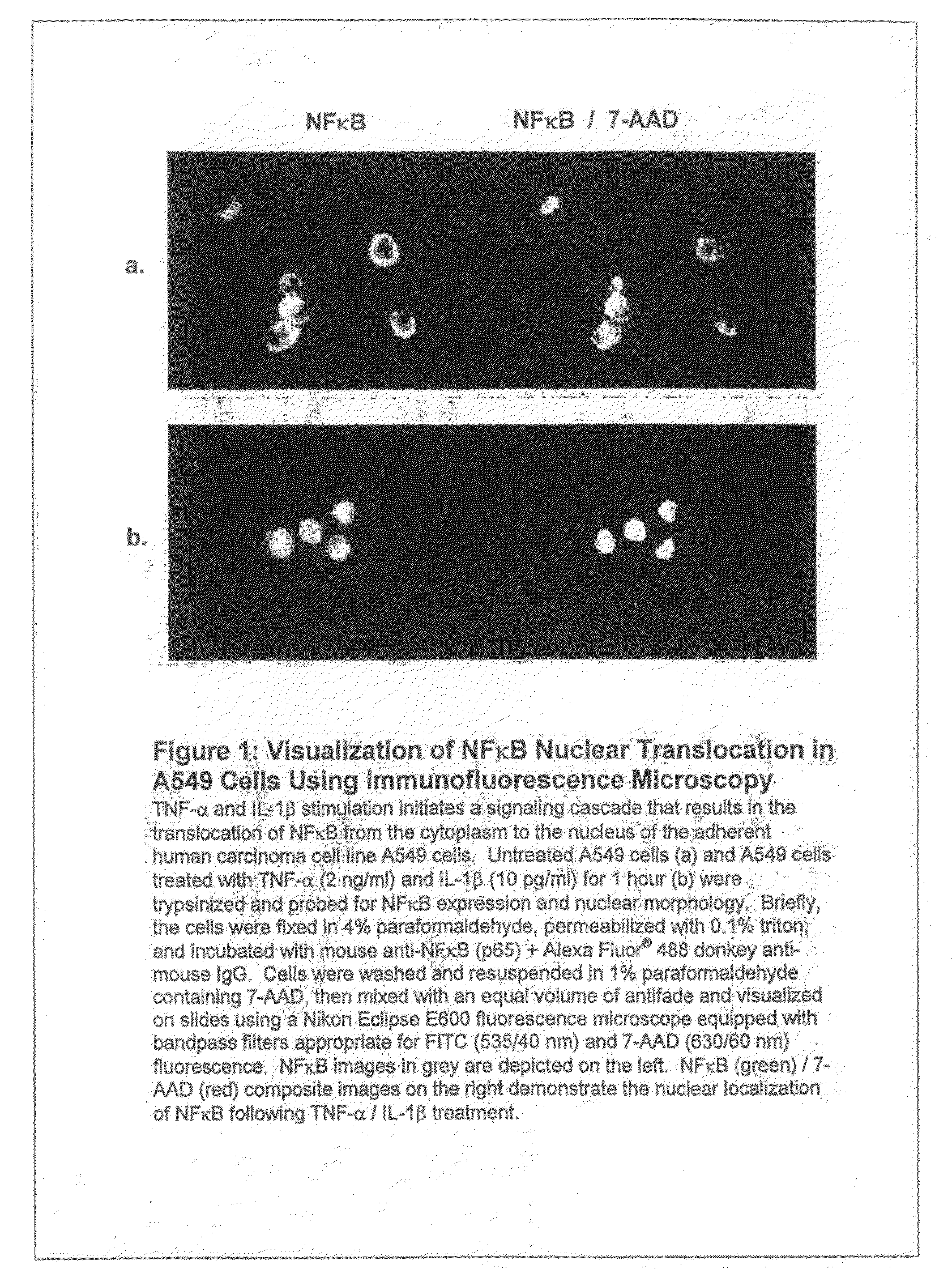
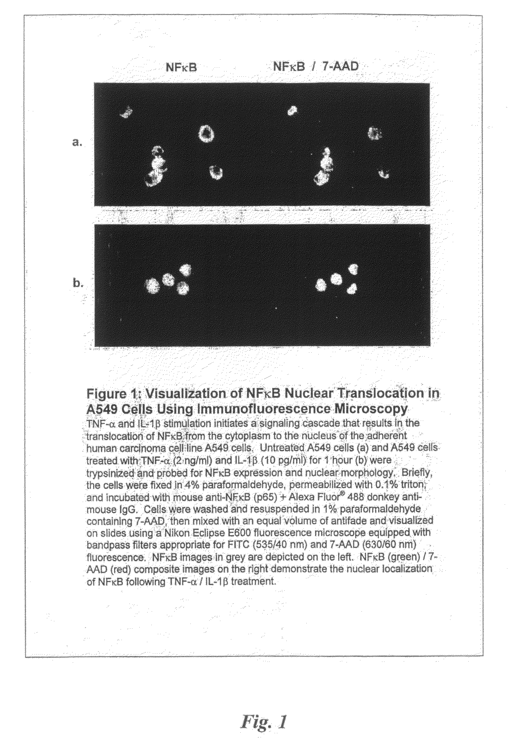
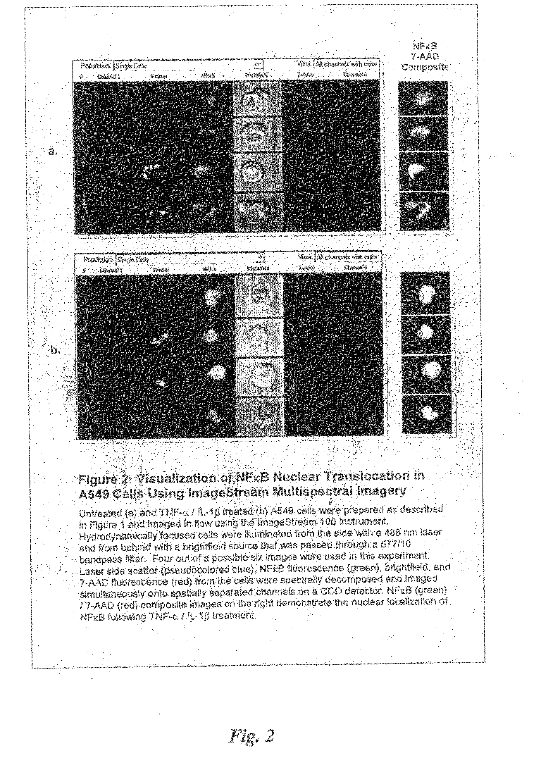
[0043]Human lung carcinoma cell line A-549, obtained from ATCC (Rockville, Md.), was maintained in RPMI 1640 (Gibco, Grand Island, N.Y.) containing 5% fetal bovine serum, 1 mM sodium pyruvate (Mediatech, Herndon, Va.), 100 μM nonessential amino acids, 100 U / ml penicillin, 100 μg / ml streptomycin, and 2 mM L-glutamine (BioWhittaker, Walkersville, Md.) in 5% CO2 atmosphere at 37° C. The density of exponentially growing cells was less than 3×10⁵ cells per ml at the time of all treatments. To induce NF-κB translocation into the nucleus from the cytoplasm, cells were treated for 1 hr with IL-1β and TNF-α.

[0044]The following is the experimental procedure for TNF-α / IL-1β induced Nuclear Translocation of NF-κB in A-549 cells

Samples:

[0045]1) Unstained and single fluorescent color control samples—start with 3.0×10⁶ total cells each. In this experiment, controls are:[0046]unstained[0047]NFκB Alexa Fluor488[0048]7-AAD

At the end, resuspend in 100 μl 0.1...

example 2

Induction of Translocation in Non-Adherent Cells

[0078]Human monocyte cell line THP-1, obtained from ATCC (Rockville, Md.), were maintained in RPMI 1640 (Gibco, Grand Island, N.Y.) containing 5% fetal bovine serum, 1 mM sodium pyruvate (Mediatech, Herndon, Va.), 100 μM nonessential amino acids, 100 U / ml penicillin, 100 μg / ml streptomycin, and 2 mM L-glutamine (BioWhittaker, Walkersville, Md.) in 5% CO2 atmosphere at 37° C. The density of exponentially growing cells was less than 3×10⁵ cells per ml at the time of all treatments. To induce NF-κB translocation into the nucleus from the cytoplasm, cells were treated for 1 hr with LPS.

[0079]The following is the experimental procedure for LPS-induced Nuclear Translocation of NF-κB in THP-1 cells

Samples:

[0080]1) Unstained and single fluorescent color control samples—start with 3.0×10⁶ total cells each. In this experiment, controls are:[0081]unstained[0082]NFκB Alexa Fluor488[0083]7-AAD

At the end, resuspend in 100 μl 0.1% triton X-100 / PBS.

[0...

example 3

Nuclear Staining and NF-κB Staining

[0108]Control (untreated) cell and LPS or IL-1β / TNF-α treated cells were independently counted and washed once in phosphate buffered saline (PBS, Fair Lawn, N.J.). Each cell group was resuspended at 107 cells / ml in 10 μM 7-aminoactinomycin D (7-AAD, Molecular Probes) for 10 minutes at room temperature. Cells were additionally stained with anti-NF-κB mAb-AF488 donkey anti-mouse IgG. Each cell group was washed, fixed in 2% paraformaldehyde (Sigma), and analyzed by flow cytometry and immunofluorescence microscopy.


PUM

PropertyMeasurementUnit
molecular movementaaaaaaaaaa
fluorescentaaaaaaaaaa
time delayaaaaaaaaaa

Abstract

The use of an imaging system, cell compartment markers, and molecular markers in methods for correlating the movement of molecules within a cell to a particular compartment are provided, including measuring and correlating molecule movement in adherent and non-adherent cells.

Description

BACKGROUND OF THE INVENTION[0001]1. Field of the Invention[0002]The present disclosure relates generally to methods for detecting specific molecules in cells, and more specifically, to the use of imagery in methods for quantitating the movement of molecules within a cell, including adherent and non-adherent cells.[0003]2. Description of the Related Art[0004]Signal transduction pathways regulate most cellular biological processes and have a critical influence on cellular responses to external stimuli. Typically, cell surface receptors that bind to a specific extracellular mediator trigger a cascade of intracellular signaling events that alter cellular metabolism or gene expression, and such changes contribute to the cellular response. The intracellular signaling cascade often involves the translocation of transcription factors or second messengers from the cytoplasm to the nucleus.[0005]Historically, nuclear translocation events have been studied microscopically by observing the sub-...

Claims


Application Information

Patent Timeline
Patent Type & Authority Applications(United States)
IPC IPC(8): G06K9/00G01N21/64G01N33/50G01N33/569G01N33/58
CPCG01N33/56966
Inventor GEORGE, THADDEUS C.BASIJI, DAVID A.FROST, KEITHHALL, BRIAN E.ORTYN, WILLIAM E.SEO, MICHAEL J.
Owner AMNIS CORP
Why Eureka
Try Eureka