Human corneal endothelial cell-derived precursor cells, cellular aggregates, methods for manufacturing the same, and methods for transplanting precursor cells and cellular aggregates

a technology of corneal endothelial cells and precursor cells, which is applied in the field of human corneal endothelial cellderived precursor cells, cellular aggregates, methods for manufacturing the same, and methods for transplanting precursor cells and cellular aggregates. it can solve the problems of corneal transplantation, tissue rejection with allotransplants, and inability to transplant, so as to enhance the ability to proliferate during culturing

Inactive Publication Date: 2009-09-17
AMANO SHIRO +2
View PDF2 Cites 23 Cited by
  • Summary
  • Abstract
  • Description
  • Claims
  • Application Information

AI Technical Summary

Benefits of technology

[0011]The present invention provides human corneal endothelial precursor cells derived from human corneal endothelium tissue. The present invention further provides a cellular aggregate in the form of stem cell-like cells derived from corneal endothelial cells. When the precursor cells or cellular aggregate is transplanted onto the parenchyma of cornea (Descemet's membrane, which is the base membrane of corneal endothelial cells), it adheres readily, forming a membrane functioning as a corneal endothelium. Thus, the corneal endothelium can be regenerated without a corneal transplant.
[0012]The present invention relates to human corneal endothelial precursor cells derived from human corneal endothelial tissue. The term “derived from human corneal endothelial tissue” means obtained by culturing using human corneal endothelial tissue as the starting material. Human corneal endothelial tissue includes the monolayer of human corneal endothelial cells and Descemet's membrane.
[0013]The human corneal endothelial precursor cells of the present invention are desirably positive for ne

Problems solved by technology

However, at least in Japan, cornea donors, and thus the availability of transplants, are currently in extremely short supply.
Further, there is an issue of tissue rejection with allotransplants.
Thus, trea

Method used


Image

  • Human corneal endothelial cell-derived precursor cells, cellular aggregates, methods for manufacturing the same, and methods for transplanting precursor cells and cellular aggregates
  • Human corneal endothelial cell-derived precursor cells, cellular aggregates, methods for manufacturing the same, and methods for transplanting precursor cells and cellular aggregates
  • Human corneal endothelial cell-derived precursor cells, cellular aggregates, methods for manufacturing the same, and methods for transplanting precursor cells and cellular aggregates

Examples


embodiment 1

Preparation of the Precursor Cells of Human Corneal Endothelial Cells

[0057]Corneal endothelial cells were collected from the Descemet membrane of strong sections of cornea following a full cornea transplant and primarily cultured. The cells were cultured in DMEM supplemented with 15 percent FBS at 100 percent humidity in the presence of 5 percent CO2. Cultured endothelial cells were separated from cell plates when the cell density reached saturation and subcultured for three generations, yielding precursor cells.

embodiment 2

Preparation of Cellular Aggregate from Cultured Corneal Endothelial Cells

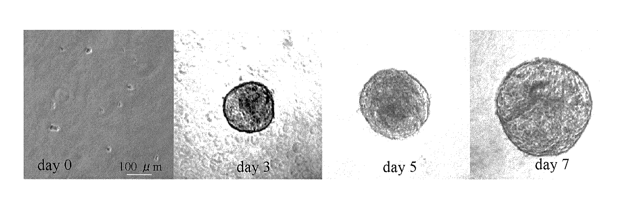
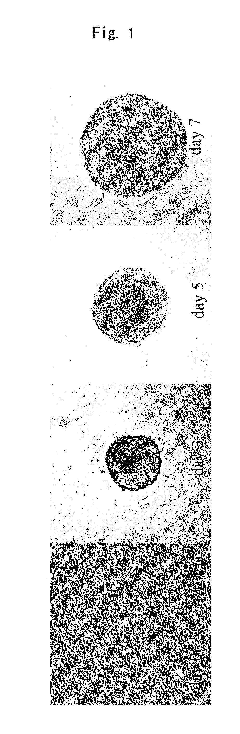
[0058]The neurosphere method was employed as the cell culture technique. A culture solution to which 8 g / mL of methyl cellulose gel matrix had been added to prevent cellular reaggregation was employed. The base medium employed was DMEM / F12 (1:1, Sigma) to which were added 20 ng / mL of B27 (Invitrogen, San Diego, Calif.), 20 ng / mL of epidermal growth factor (EGF, Sigma), and 40 ng / mL of basic fibroblast growth factor (bFGF, Sigma). Stock cells (precursor cells) were inoculated to 50 cells / microliter (50,000 cells per well) into 24-well culture plates without coatings. Cell reaggregation did not occur under these conditions. The CO2 concentration was 5 percent.

[0059]After 7 days of culturing, the cells had grown and formed cellular aggregates. FIG. 1 shows the status of the cellular aggregates at the outset of culturing, on the first day (day 0), and on days 3, 5, and 7. The diameter of the cellular aggregates on ...

embodiment 3

Transplantation of Stem Cell-Like Cells from Cultured Corneal Endothelial Cells

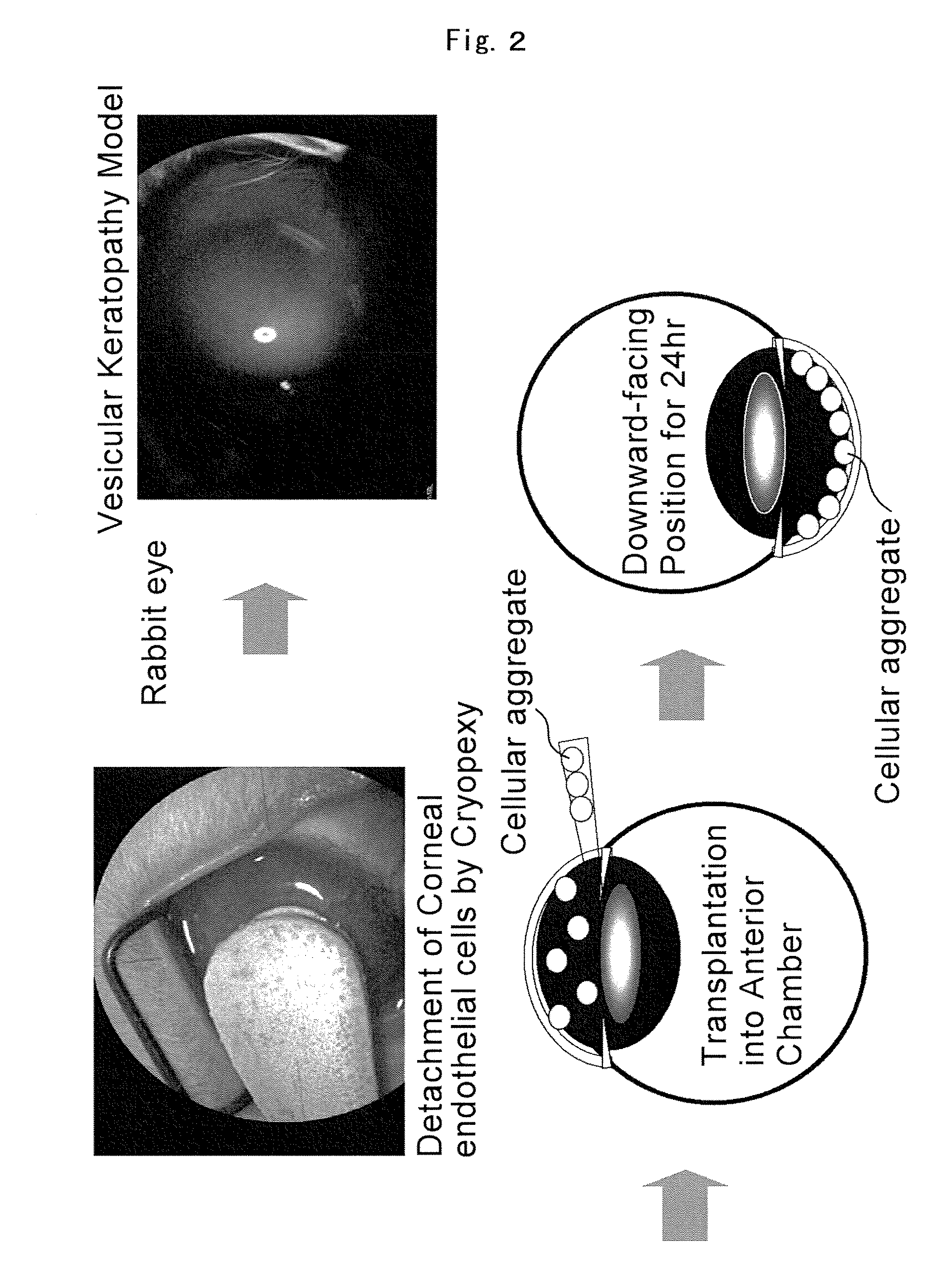
[0062]The cellular aggregate obtained in Embodiment 2 was transplanted with an injector into the anterior chamber of a rabbit eye in which corneal endothelial cells had been detached by cryopexy. That is, all of the cellular aggregates in the 30 to 500 micrometer range that had been obtained by the above-described method were collected in uncoated 60 millimeter cell plates containing PBS so that the cells did not adhere. The cellular aggregates were then centrifuged and, without replacing the solution, those cells that were spherical under a stereoscope were recovered. The cellular aggregates were then washed several times with PBS by replacement of the PBS. A 300 microliter quantity of solution containing 30 to 200 cellular aggregates was introduced into the anterior chamber of each eye. A 27 or 30 G gauge was employed in this process. A downward-facing position was maintained for 24 hours following the ...


PUM


Abstract

Providing is cellular aggregates derived from corneal endothelial cells that, when transplanted, readily adhere to the parenchyma of cornea and function in a manner equivalent to corneal endothelial cells, and a method of transplantation of the cellular aggregates. Cellular aggregates derived from corneal endothelial cells. The cellular aggregates derived from corneal endothelial cells is prepared by culturing human corneal endothelial cells in a medium containing fetal bovine serum, growth factor and glucose; and then float culturing the cells obtained in a medium containing growth factor. A method of transplantation into the anterior chamber the cellular aggregate or the cellular aggregate prepared by the above method, comprising inserting a tube into the parenchyma of cornea, introducing the cellular aggregate into the anterior chamber through the inserted tube, and causing the cellular aggregate that has been introduced to adhere to Descemet's membrane by assuming in a downward-facing position.

Description

TECHNICAL FIELD[0001]The present invention relates to human corneal endothelial precursor cells derived from human corneal endothelial tissue, cellular aggregates in the form of stem cell-like cells derived from human corneal endothelial cells, and methods for manufacturing the same. The present invention further relates to methods for transplanting these precursor cells or cellular aggregates into the anterior chamber to regenerate the endothelial cell layer.BACKGROUND ART[0002]The conventional method for treating severe corneal disease is to perform a cornea transplant. However, at least in Japan, cornea donors, and thus the availability of transplants, are currently in extremely short supply. Further, there is an issue of tissue rejection with allotransplants. Thus, treatment by cornea transplant is far from being an ideal solution.[0003]As shown in FIG. 5, the cornea 1 is comprised of a multilayered structure in the form of the anterior corneal epithelium 2, Bowman's membrane 3,...

Claims


Application Information

Patent Timeline
IPC IPC(8): A61K45/00C12N5/08A61P27/02A61K35/12A61K35/44C12N5/071
CPCA61K35/12A61K35/44C12N5/0621A61L27/3839A61L2430/16A61L27/3808A61P27/02
Inventor AMANO, SHIROYAMAGAMI, SATORUMIMURA, TATSUYAOSAKABE, YASUHIRO
Owner AMANO SHIRO
Why Eureka
Try Eureka