Bioprinting Three-Dimensional Structure Onto Microscale Tissue Analog Devices for Pharmacokinetic Study and Other Uses

a tissue analog and three-dimensional structure technology, applied in the field of microfluidic systems, can solve the problems of inability to create high-fidelity three-dimensional tissue analogs, current practice does not permit the control of spatiotemporal placement of cells within a biomaterial matrix, residue formation and subsequent channel occlusion, etc., to achieve the effect of improving hydrophilicity

Inactive Publication Date: 2009-10-22
DREXEL UNIV
View PDF11 Cites 62 Cited by
  • Summary
  • Abstract
  • Description
  • Claims
  • Application Information

AI Technical Summary

Benefits of technology

[0021]In certain embodiments, at least one of the cover platform or the substrate platform comprises a surface with an improved hydrophilicity.

Problems solved by technology

The existing technologies available and biomaterials employed are not currently amenable to creating 3-dimensional tissue analogs with high fidelity.
Current practice also does not permit control of spatiotemporal placement of cells within a biomaterial matrix.
Furthermore, current use of chemical coatings and modifications for cell / matrix attachment of microfluidic channels leads to residue formation and subsequent channel occlusions.
Published biological data show that existing in vitro micro devices do not demonstrate good cell viability or preservation of normal in vivo cell-specific physiological function necessary to accurately perform pharmacokinetic studies on a long-term basis.
This system is very expensive to operate and requires a large amount of space in which to operate.
It is impossible to accurately generate physiologically realistic conditions at such a large scale.
A disadvantage of this system is that it still relies upon cell migration for cell seeding.
There is no possibility for direct positional control of cell placement.
This method does not have the ability for 3-D positional control and patterning of cells.
This reference does not describe creating an artificial three dimensional tissue incorporated into a microfluidic device and therefore, it is limited to interactions of cells seeded on the surfaces of the chamber.

Method used


Image

  • Bioprinting Three-Dimensional Structure Onto Microscale Tissue Analog Devices for Pharmacokinetic Study and Other Uses
  • Bioprinting Three-Dimensional Structure Onto Microscale Tissue Analog Devices for Pharmacokinetic Study and Other Uses
  • Bioprinting Three-Dimensional Structure Onto Microscale Tissue Analog Devices for Pharmacokinetic Study and Other Uses

Examples


Embodiment Construction

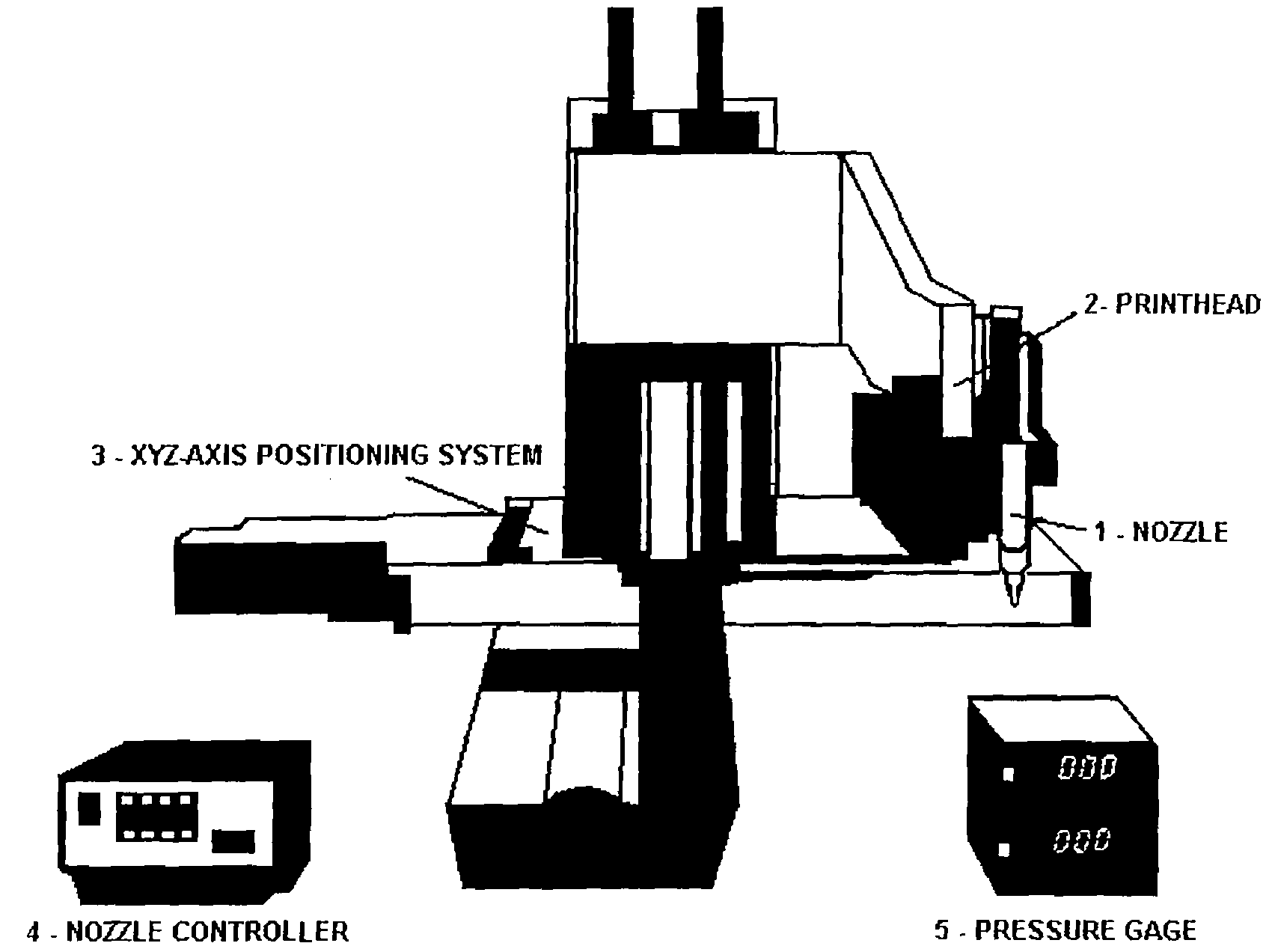
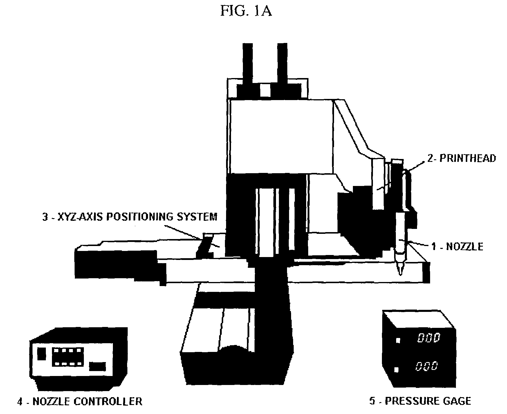
[0064]The object of the invention is a new device and process for manufacturing such devices that reliably aids in the drug screening and drug discovery process. Additionally, the device will be able to perform metabolic and cytotoxicity studies on a microscale that is comparable to human physiologic scales. Faster drug screening methods with high-throughput capability and portability can lead to significant cost reductions attributed to reduced time and effort in the number of animal and human trial studies conducted. A suitable in vitro drug screening processes can aid in new drug discovery processes.

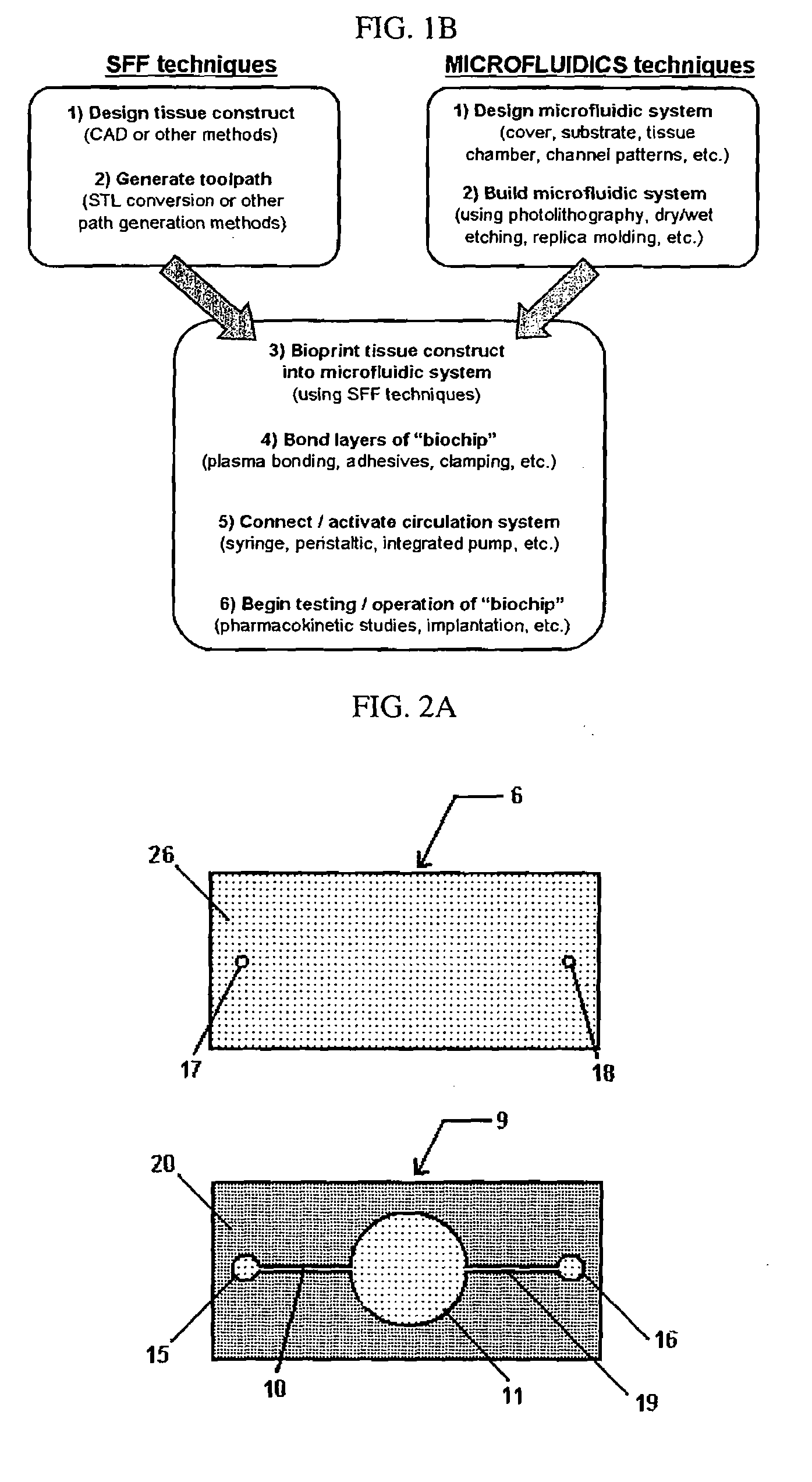
[0065]The invention further includes a method of making the microfluidic system. Furthermore, the fabrication process of bioprinting has been developed to build a 3-dimensional heterogeneous cell-encapsulated hydrogel-based construct within a microfluidic device which serves as a fluid circulator and as a platform for experimental drug / chemical analysis and toxicology.

[0066]The presen...


PUM

PropertyMeasurementUnit
diameteraaaaaaaaaa
volumeaaaaaaaaaa
hydrophilicityaaaaaaaaaa

Abstract

A microfluidic system for monitoring or detecting a change in a parameter of an input substance, which includes a microfluidic device having a tissue chamber and a tissue analog placed in the tissue chamber, wherein the tissue analog has a vessel structure mimicking naturally occurring vessel network incorporated in the tissue analog.

Description

BACKGROUND OF THE INVENTION[0001]1. Field of Invention[0002]This invention relates to a microfluidic system for monitoring or detecting a change in a parameter of an input substance. Specifically, the invention relates to a model for in vitro pharmacokinetic study and other pharmaceutical applications, as well as other uses such as computing, sensing, filtration, detoxification, production of chemicals and biomolecules, testing cell / tissue behavior, and implantation into a subject.[0003]2. Description of Related Art[0004]The existing technologies available and biomaterials employed are not currently amenable to creating 3-dimensional tissue analogs with high fidelity. Current practice also does not permit control of spatiotemporal placement of cells within a biomaterial matrix. Furthermore, current use of chemical coatings and modifications for cell / matrix attachment of microfluidic channels leads to residue formation and subsequent channel occlusions. Published biological data show...

Claims


Application Information

Patent Timeline
Patent Type & Authority Applications(United States)
IPC IPC(8): C12Q1/02C12M1/34
CPCB01L3/502707B01L3/502746B01L2300/10B01L2300/0874B01L2300/0816
Inventor SUN, WEICHANG, ROBERT C.STARLY, BINILNAM, JAE
Owner DREXEL UNIV
Why Eureka
Try Eureka