However, according to a hand-held, mechanical scanning ultrasonic probe that performs scanning by mechanically oscillating an ultrasonic element, since it is impossible for the oscillating ultrasonic element to directly contact an organism, an acoustic
coupling liquid must be sealed by a window and a probe case, and the ultrasonic element must be oscillated or rotated inside the liquid to perform scanning.
Therefore, it is difficult for an
ultrasonic transmission / reception area near the surface of an organism to be reduced, or for the oscillating center of the ultrasonic element to be located either in the vicinity of the
body surface or at a location near the
body surface.
Furthermore, this apparatus is also not one with which a three-dimensional ultrasonic probe can be employed for diagnosing an additional area, such as the carotid
artery or the
thyroid.
Thus, diagnosis of an additional area, such as the carotid
artery or the
thyroid, can not be easily performed.
Thus, this arrangement is not preferable for a hand-held three-dimensional ultrasonic probe.
And especially in a case for performing a diagnosis for the carotid artery or the thyroid, a problem is that a hand-held three-dimensional ultrasonic probe can not contact a desired location for a target portion of an organism because a jaw, etc., obstructs the location.
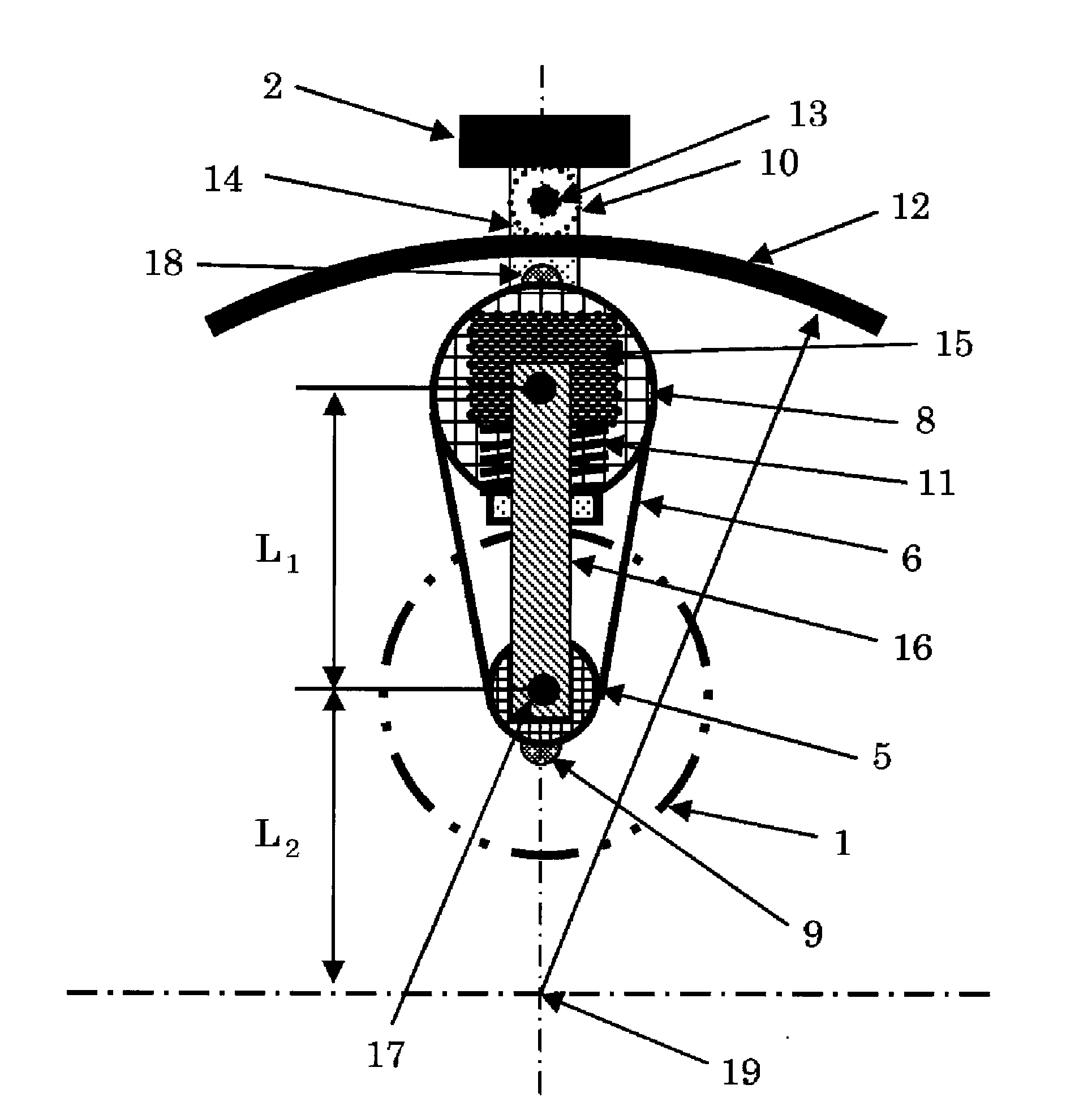
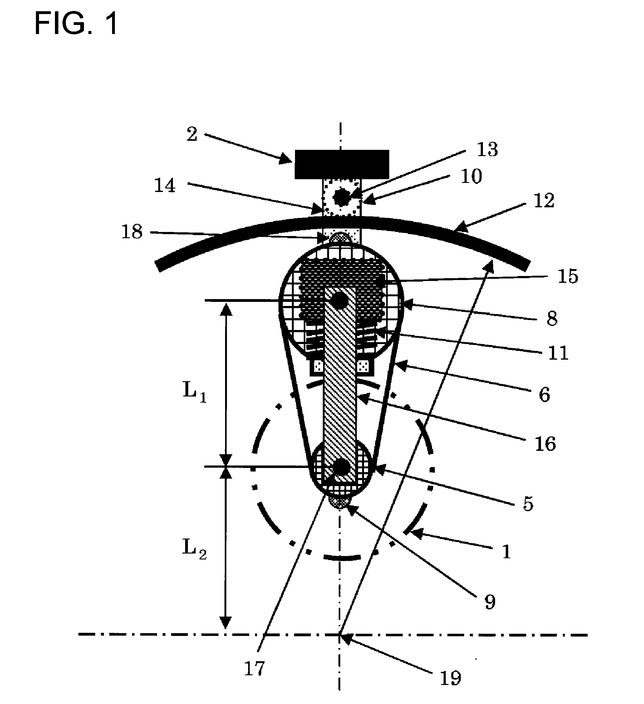
Thus, in proportion to the distance from the rotation center, the
pitch of two-dimensional slices is increased, and when a three-dimensional image is formed based on image data for a position separated from the rotation center, a problem is that the resolution for the portion separated from the rotation center is lower.
Thus, in a case wherein a diagnosis for a portion such as the carotid artery or the thyroid is to be performed, a problem is that an organism contact portion longer than the element may strike the jaw portion, making it difficult for an ultrasonic probe to contact a desired location.
In addition, when the distance from the mechanical oscillation center to the tip of the array type element is extended, the size of a hand-held, three-dimensional ultrasonic probe is increased, and this presents a problem in that the increase in the size and the weight of the hand-held three-dimensional ultrasonic probe make handling the probe difficult when performing a diagnosis.
Therefore, a problem encountered is that the oscillation angle of the element is not proportional to the rotational angle of the motor, and in a case wherein the velocity at which the motor rotates is a constant, the
ultrasonic beam can not be oscillated at the same angle.
Furthermore, since an inverse proportion is established between the scanning line density and the angular oscillation velocity, a problem is that, for the above described mechanism, the angular oscillation velocity is high in the middle of the oscillation angle that is especially important, i.e., the scanning line density is lowered.
Thus, a problem is that, when an organ, such as the ribs, that has a large
acoustic impedance and that greatly reflects an
ultrasonic beam, is present near the organism, an ultrasonic beam is interrupted by the organ, such as the ribs, and can not reach the organ present below, inside the organism.
Thus, there is a problem in that, when an organ, such as a rib, that has a large
acoustic impedance and that greatly reflects an ultrasonic beam, is present near the organism, an ultrasonic beam will be interrupted by the organ, such as the rib, and can not reach an organ located below, inside the organism.
In addition, as in
patent document 6, since it is difficult to provide an improvement for time-transient tracking relative to an organ that moves rapidly, the oscillation velocity is increased by reducing the oscillation angle, and it is also difficult for the scanning line density and the oscillation velocity to be arbitrarily set in consonance with a diagnosis portion.
Therefore, this is a problem related to a reduction in the size and the weight of the probe.
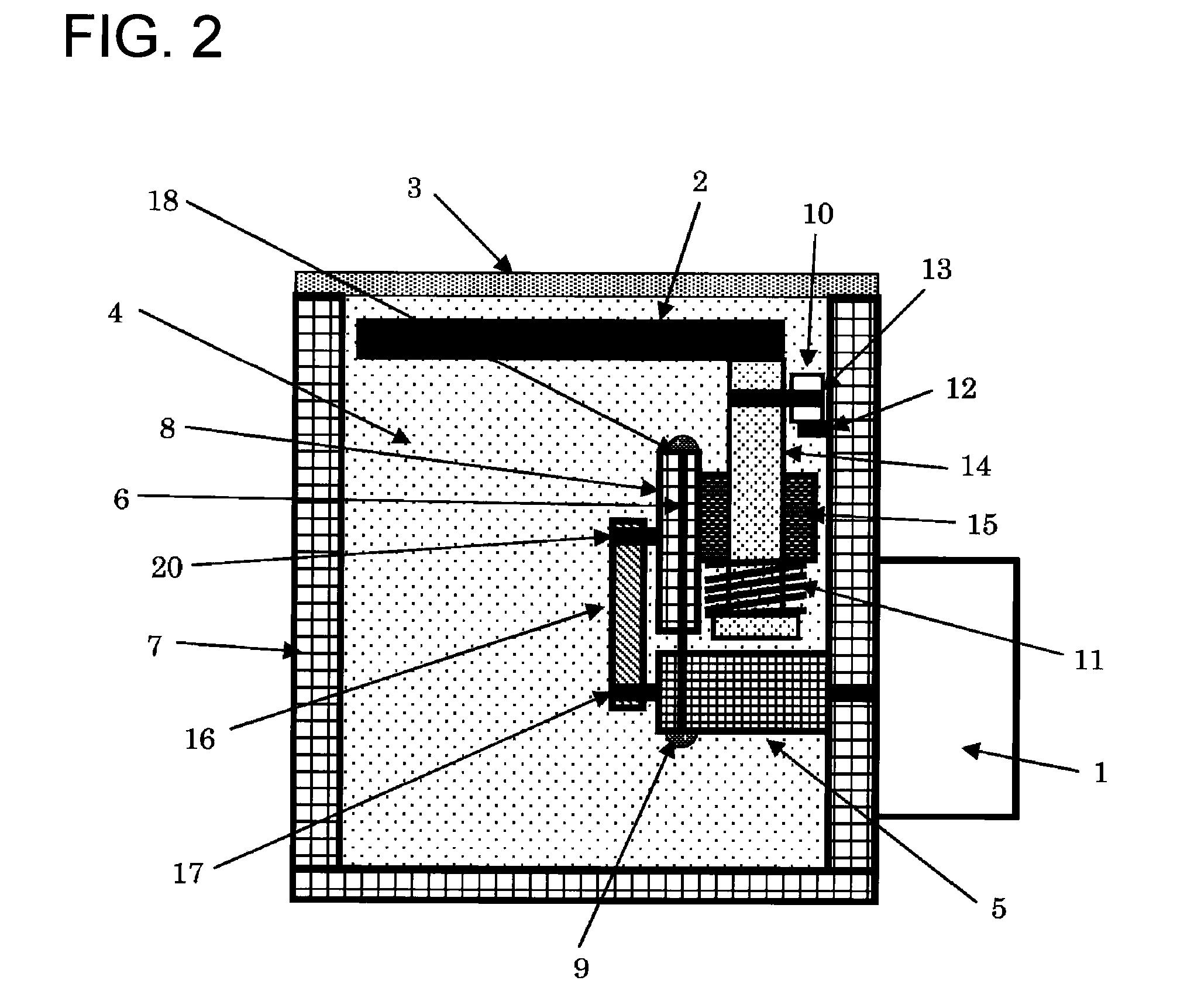
Further, when an organ, such as the heart, is to be scanned along the lower edge of the costal arch, i.e., from below the ribs, the probe must be moved from below the ribs to substantially parallel to the
body surface, and the roundness of the probe due to the presence of the roller interferes with the scanning operation.
In addition, since multiple parts, such as a guide rail, a plurality of rollers and a belt, are required for the mechanism, the total cost for these parts and the complicated
assembly process are also problems.

