Apparatus and method for external fluorescence imaging of internal regions of interest in a small animal using an endoscope for internal illumination

an endoscope and fluorescence imaging technology, applied in the field of apparatus and method for external fluorescence imaging of internal regions of interest in a small animal using an endoscope for internal illumination, can solve the problems of large fluoresced parts of the body of the subject, too deep near infrared imaging probes to be excited by illumination applied externally and non-invasively, and limited detection of the region of interes

Inactive Publication Date: 2009-11-12
BRUKER BIOSPIN
View PDF94 Cites 7 Cited by
  • Summary
  • Abstract
  • Description
  • Claims
  • Application Information

AI Technical Summary

Benefits of technology

[0015]A method according to the invention is useful for capturing an optical molecular image of one or more internal regions of interest of a small animal having a cranio-caudal axis. The method may include steps of immobilizing the animal on a support member; inserting an endoscope into the body of the animal to allow the endoscope to be positioned selectively proximate such internal regions of interest within the body; connecting a light

Problems solved by technology

An often-encountered problem is that light applied from the exterior of the subject to excite the imaging probe dye to fluoresce may be of a wavelength that cannot readily pass through the tissue of the body, or is impeded by the endogenous tissue absorption, either of which may result in limited detection of the region of interest.
In some cases even near infrared

Method used


Image

  • Apparatus and method for external fluorescence imaging of internal regions of interest in a small animal using an endoscope for internal illumination
  • Apparatus and method for external fluorescence imaging of internal regions of interest in a small animal using an endoscope for internal illumination
  • Apparatus and method for external fluorescence imaging of internal regions of interest in a small animal using an endoscope for internal illumination

Examples


Embodiment Construction

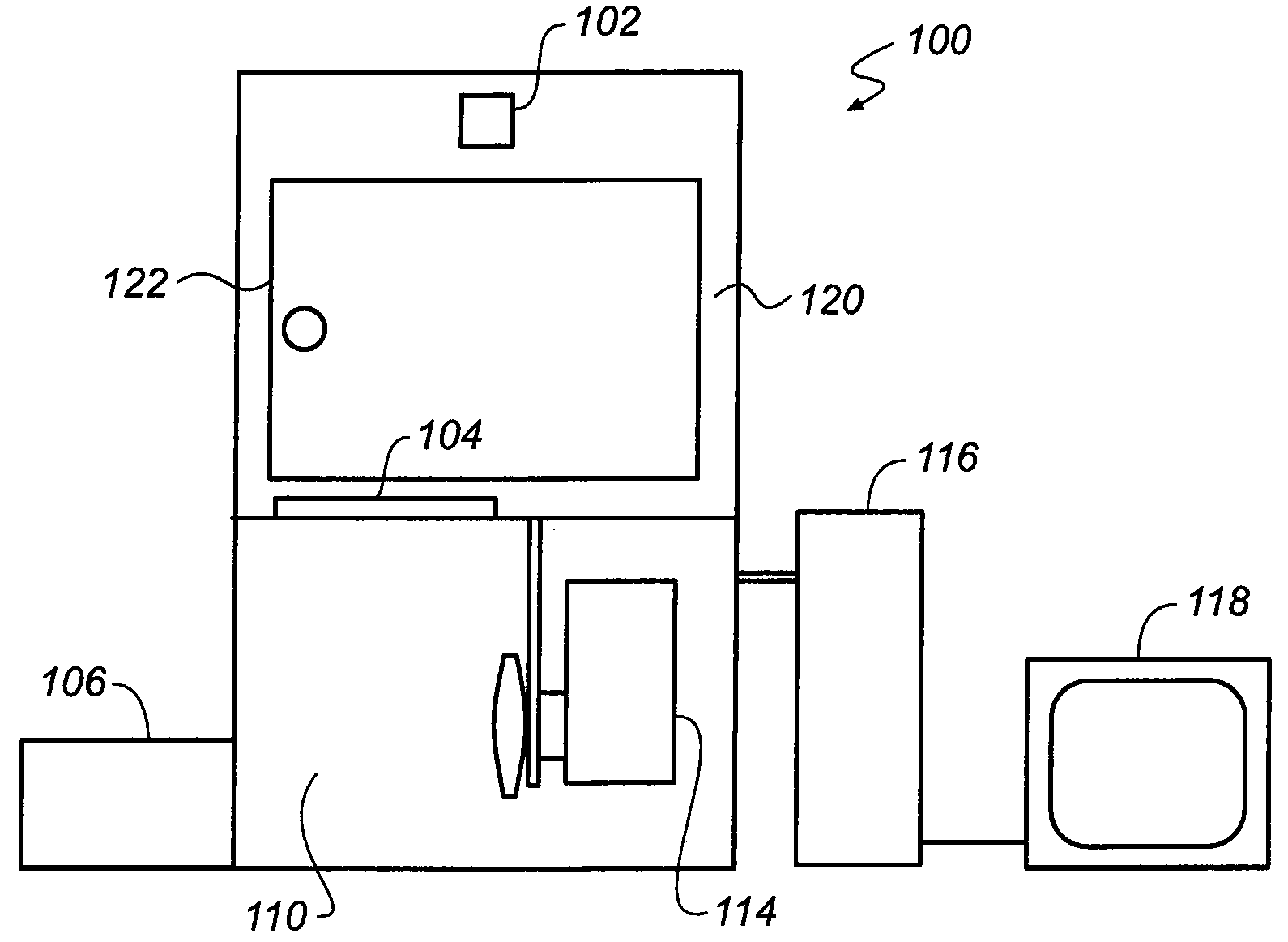
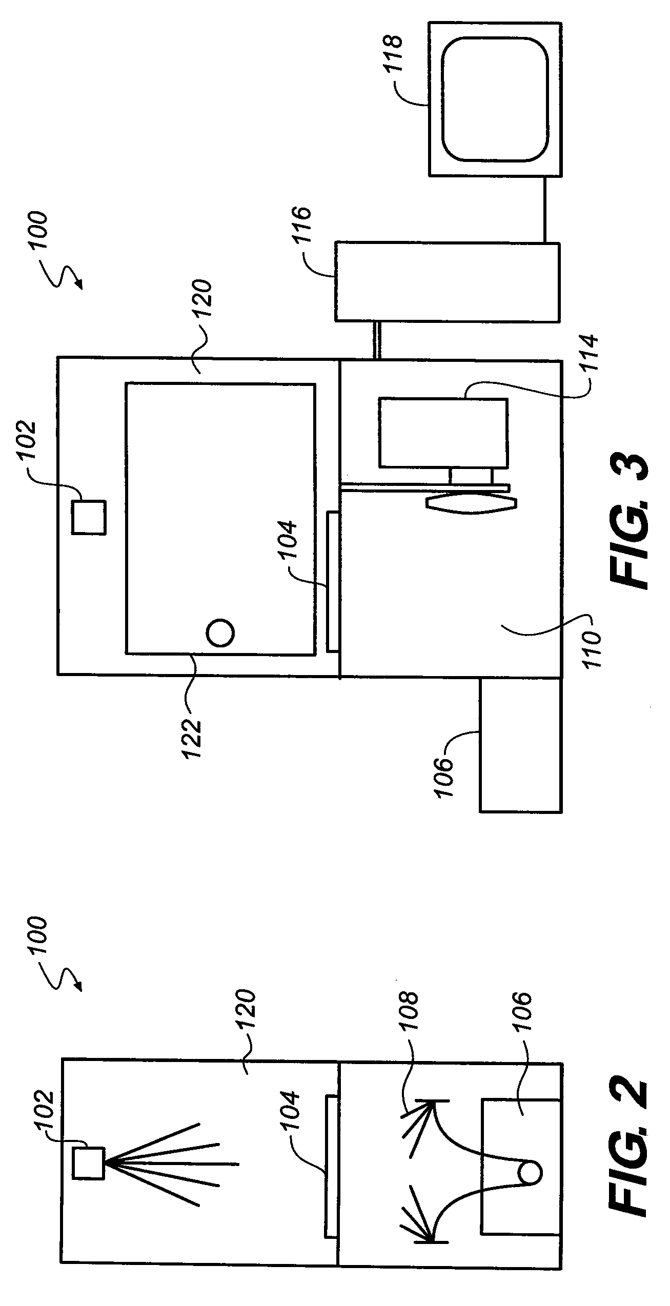
[0031]The invention will be described in detail with particular reference to certain embodiments thereof, but it will be understood that variations and modifications can be effected within the spirit and scope of the invention. The following is a detailed description of various embodiments of the invention, reference being made to the drawings in which the same reference numerals identify the same elements of structure in each of the several figures.

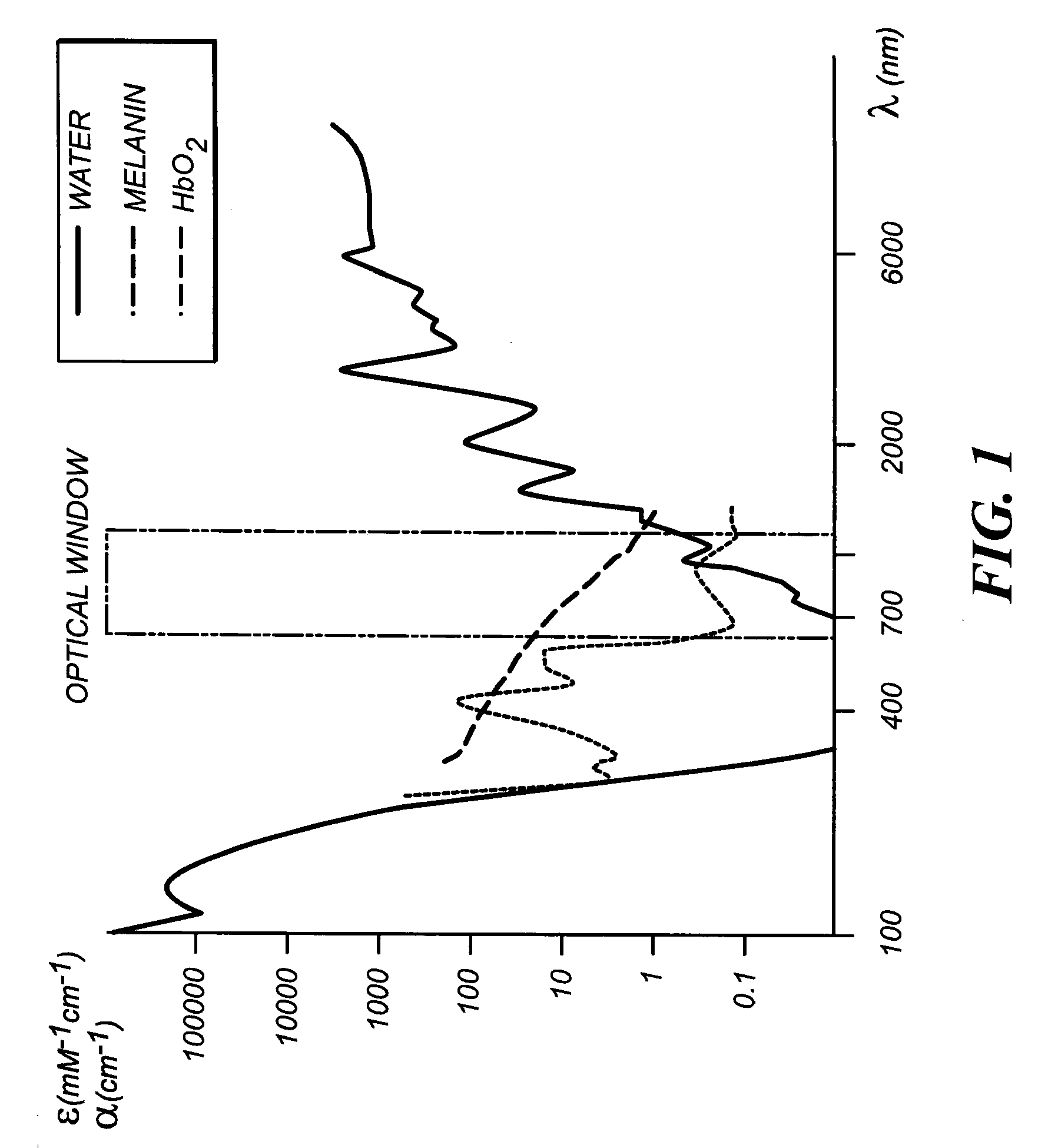
[0032]The present inventors have recognized a need for a more efficient apparatus and method for exciting optical imaging probes internally located in regions of interest of a subject such as a small animal for the purpose of molecular imaging. This invention provides a method to enhance and maximize the depth of tissue penetration of light from an excitation source. As shown in the graphical representation of FIG. 1, endogenous chromophores like hemoglobin (oxy-hemoglobin) and melanin absorb light and interfere by autofluorescence below...


PUM


Abstract

An apparatus for capturing an optical molecular image of one or more internal regions of interest of a small animal having a cranio-caudal axis. The apparatus may include a support member for the animal in an immobilized state; an endoscope for insertion into and withdrawal from the body of the animal to allow the endoscope to be positioned selectively proximate the internal regions of interest within the body; a light source connected to the endoscope to provide illumination within the body at the regions of interest; a mechanism for rotating the animal about its cranio-caudal axis to enable image capture from different angles; and a device for capturing images of the one or more internal regions of interest from the different angles, due to light emitted from the one or more internal regions of interest through an external surface of the body in response to illumination from the light source. A corresponding method is disclosed.

Description

CROSS REFERENCE TO RELATED APPLICATIONS[0001]Priority is claimed from commonly assigned, copending U.S. provisional patent application Serial No. 61 / 075,033 filed Jun. 24, 2008 by Papineni et al and entitled SMALL ANIMAL OPTICAL IMAGING ENDOSCOPES—GAVAGE SCOPES AND RECTAL SCOPES, the contents of which are incorporated by reference into the present specification.[0002]This application is a continuation in part of the following commonly assigned, copending U.S. patent applications, the contents of each of which also are incorporated by reference into the present specification:[0003]U.S. Ser. No. 11 / 221,530 filed Sep. 9, 2005 by Vizard et al., entitled APPARATUS AND METHOD FOR MULTI-MODAL IMAGING;[0004]U.S. Ser. No. 12 / 381,599 filed Mar. 13, 2009 by Feke et al., entitled METHOD FOR REPRODUCING THE SPATIAL ORIENTATION OF AN IMMOBILIZED SUBJECT IN A MULTI-MODAL IMAGING SYSTEM; and[0005]U.S. Ser. No. 12 / 475,623 filed Jun. 1, 2009 by Feke et al, entitled TORSIONAL SUPPORT APPARATUS AND MET...

Claims


Application Information

Patent Timeline
IPC IPC(8): A61B1/04
CPCA61B5/0059A61B6/00A61B2503/40A61B6/5247A61B6/4417A61B5/0035
Inventor PAPINENI, RAOFEKE, GILBERT
Owner BRUKER BIOSPIN
Why Eureka
Try Eureka