Medical valve and method to monitor intra-abdominal pressure

a technology of intra-abdominal pressure and medical valve, which is applied in the direction of diaphragm valve, catheter, instruments, etc., can solve the problems of organ damage and patient death, tissue edema in the body, and increased risk of infection for both patient and health practitioner, so as to achieve sufficient flow rate of bleed-down fluid

Inactive Publication Date: 2009-12-24
CONVATEC TECH INC
View PDF45 Cites 17 Cited by
  • Summary
  • Abstract
  • Description
  • Claims
  • Application Information

AI Technical Summary

Benefits of technology

[0022]One preferred embodiment of a hydraulically actuated valve may be characterized by providing a plurality of fluid paths. In such valve, a first path may be defined between an entrance port and a drain port. A portion of the first fluid path may be provided by structure associated with the valve's housing and disposed between a control port and the entrance port. A second fluid path can be defined between the control port and the drain port. A bleed-down port of the valve is arranged to discharge bleed-down fluid along a path that overlaps a portion of that second fluid path. Bleed-down fluid can be directed along a third fluid path between the bleed-down port and a fluid-flow restriction valve. Desirably, a bleed-down valve is associated with the bleed-down port to cause the rate of flow of bleed-down fluid to be sufficiently low that it does not generate a significant back-pressure when passing through the flow-restriction valve. At certain times during valve operation, a fluid path followed by the bleed-down fluid overlaps a portion of the first fluid path.

Problems solved by technology

Elevated intra-abdominal pressure leads to major changes in the body's physiology that, if undetected and untreated, can result in organ damage and patient death.
When patients become critically ill, they may develop a capillary leak phenomenon that causes the tissues in their body to become edematous with extra fluid that seeps out of the capillaries.
Unfortunately, opening the closed drainage system plumbing places both the patient and the health practitioner at increased risk of infection.
It is possible to use a three-way Foley catheter, but such catheters are more expensive and are not routinely used.
The former option increases costs, and the latter would increase both costs and risk of patient infection.
Undesirably, approaches involving use of needles, particularly in the vicinity of the patient's legs to assemble the pressure measuring apparatus, place both the patient and the health practitioner at risk of needle sticks.
However, operable medical grade valves that are commercially available, such as two-way or three-way stopcocks 110 and 112, typically introduce undesired complications.
One complication is that the available medical grade stopcocks typically provide drainage passageways that are too small in diameter for use in a urinary catheter drain.
Clogging of the urine drain bore would be a serious problem.
Another complication substantially precluding direct inclusion of commercially available medical grade two-way or three-way valves or stopcocks is that such devices route fluid conduits in orthogonal directions at the valve connection locations, thereby creating protruding and invasive plumbing that is uncomfortable to the patient.
Furthermore, currently available valves and stopcocks also have protrusions (such as valve actuators or handles), and sharp corners or abrupt changes in shape, that place a patient at risk of injury should such protrusion or corner be impressed into a patient's skin.
It is undesirable for the anesthesiologist to move from a traditional station, at the patient's head, repeatedly to make periodic IAP measurements.
Further, it might not be political to have an anesthesiologist to invade the “turf” of the surgeon to make the IAP measurement.
In any case, periodic manual activation of a urine valve by the anesthesiologist, or other personnel present in the operatory, also may cause an interference with the surgeon.
The procedures for measuring trends in a patient's IAP described above undesirably place a patient at risk of infection, or require tiresome manual adjusting of a plurality of plumbing devices, such as two-way valves or stopcocks.

Method used


Image

  • Medical valve and method to monitor intra-abdominal pressure
  • Medical valve and method to monitor intra-abdominal pressure
  • Medical valve and method to monitor intra-abdominal pressure

Examples


Embodiment Construction

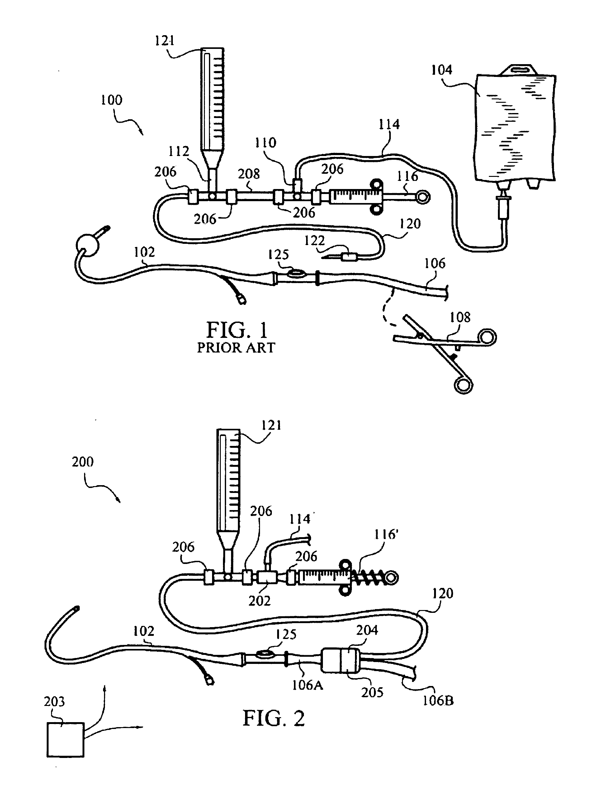
[0051]FIG. 2 illustrates an exemplary embodiment, generally indicated at 200, of an apparatus for measuring trends in a patient's intra-abdominal pressure. The apparatus 200 includes a fluid supply tube or conduit 114 with one end in fluid communication with a sterile saline or other fluid source (not illustrated). Fluid supply tube or conduit 114 desirably is connected at a second end for fluid communication with an automatic, direction-of-flow control device 202 to urge fluid flow through tubing 120 in a direction toward a patient. A hydraulic pressure in tubing 120 is measured by a pressure transducer, such as pressure measuring device 121.

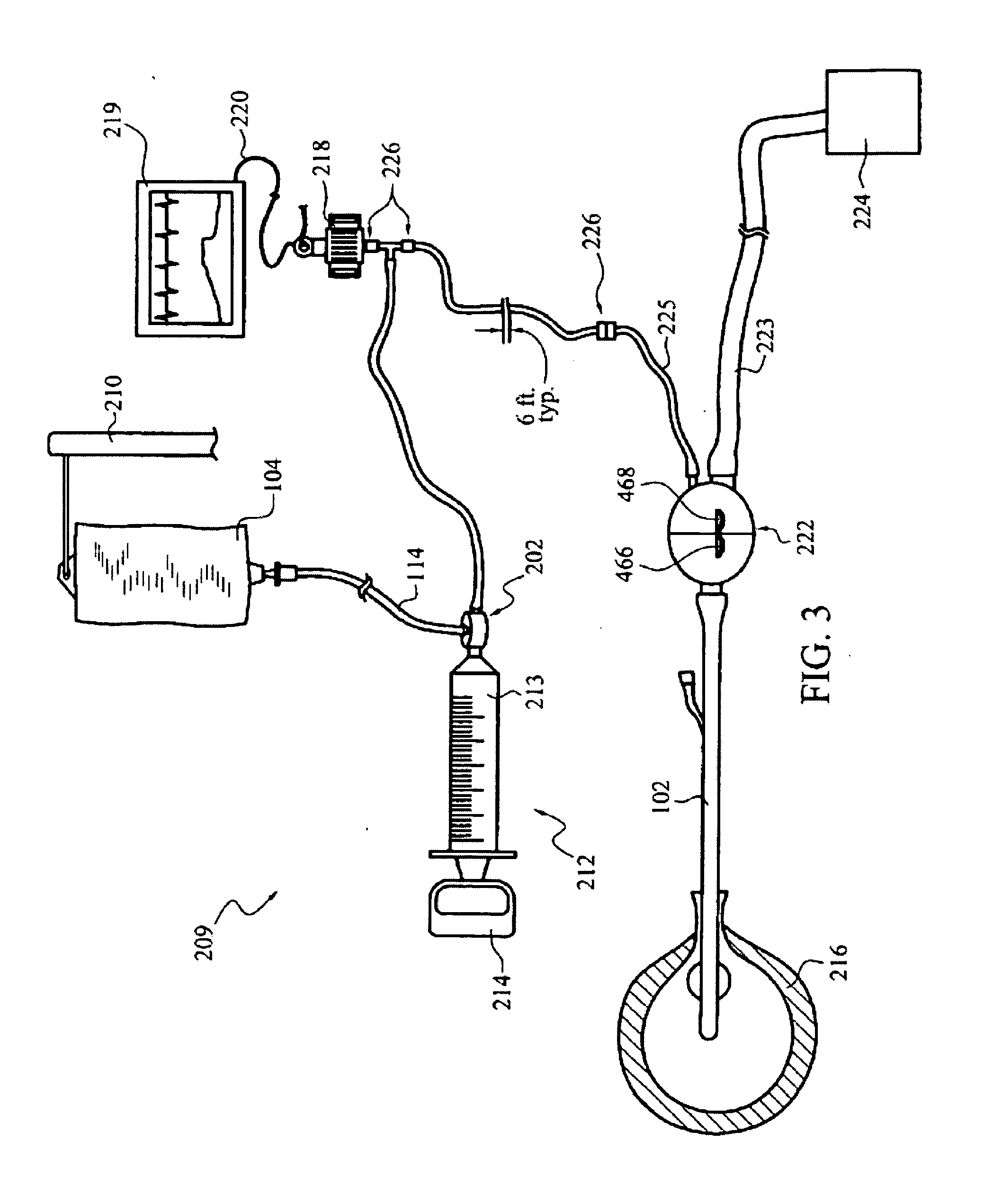
[0052]As illustrated in FIG. 3, it is sometimes preferred to arrange the pressure transducer in a dead-ended conduit, compared to the flow-through arrangements illustrated in FIGS. 1 and 2. The illustrated arrangement requires a clinician to make only one attachment at the pressure transducer area. However, it should be realized that additional...


PUM


Abstract

An apparatus for monitoring the intra-abdominal pressure of a hospitalized patient includes a urinary catheter connected to a urine valve providing selectable communication between a discharge end of the urinary catheter and either a drain or a fluid source. Preferably, the urine valve is adapted for remote actuation and has a housing adapted to resist patient discomfort from leg-valve contact. Plumbing structure desirably maintains fluid supply and drain conduits in a substantially parallel arrangement to assist routing those conduits between a patient's legs. When the urine valve is oriented to permit communication with the fluid source, an infusion pump may be used to infuse a known quantity of fluid through the urine valve and into the patient's bladder. A pressure transducer desirably is connected in-circuit to indicate the fluid's pressure. To facilitate the infusion process, an automatic flow control device may be included in a fluid supply path and arranged to permit repetitive operation of a syringe to inject a bolus of fluid into the patient's bladder. Subsequent to a period of time in which to make a pressure measurement, preferred embodiments of the urine valve automatically return to a bladder draining position.

Description

CROSS-REFERENCE TO RELATED APPLICATIONS[0001]This patent application is a continuation of U.S. patent application Ser. No. 11 / 199,790, filed Aug. 9, 2005, now U.S. Pat. No. ______, which application is a continuation-in-part of co-owned PCT International Patent Application PCT / US04 / 006409 filed Mar. 1, 2004, designating the United States of America, and published in English as WO 2004 / 078235, which is a continuation-in-part of U.S. patent application Ser. No. 10 / 379,222, filed on Mar. 4, 2003, now U.S. Pat. No. 7,112,177, issued Sep. 26, 2006, the contents of each of which are incorporated herein by this reference in their entirety.TECHNICAL FIELD[0002]The invention relates generally to medical equipment including valves and conduits, and to pressure measurement equipment. The invention relates particularly to apparatus configured as an assembly to infer intra-abdominal pressure of a medical patient by measuring the pressure of fluid in the patient's bladder.BACKGROUND[0003]Elevated...

Claims


Application Information

Patent Timeline
Patent Type & Authority Applications(United States)
IPC IPC(8): F16K21/00
CPCA61B5/036A61B5/205F16K7/126A61M25/0017A61M39/223A61B5/412Y10T137/2557Y10T137/7722
Inventor CHRISTENSEN, MARK A.WOLFE, TIMOTHY R.CROLL, PERRY W.DENTON, MARSHALL T.KIMBALL, EDWARD J.
Owner CONVATEC TECH INC
Why Eureka
Try Eureka