
Apparatus for real-time 3D biopsy

a technology of 3d biopsy and apex, which is applied in the field of surface registration of images, can solve the problems of no ideal standard, no ideal standard, and sample collection by urologists may produce false negatives, etc., and achieve the effect of reducing the time of biopsy and facilitating the visualization of the registration surface on the screen

Inactive Publication Date: 2009-12-31
EIGEN INC
View PDF58 Cites 25 Cited by
  • Summary
  • Abstract
  • Description
  • Claims
  • Application Information

AI Technical Summary

Benefits of technology

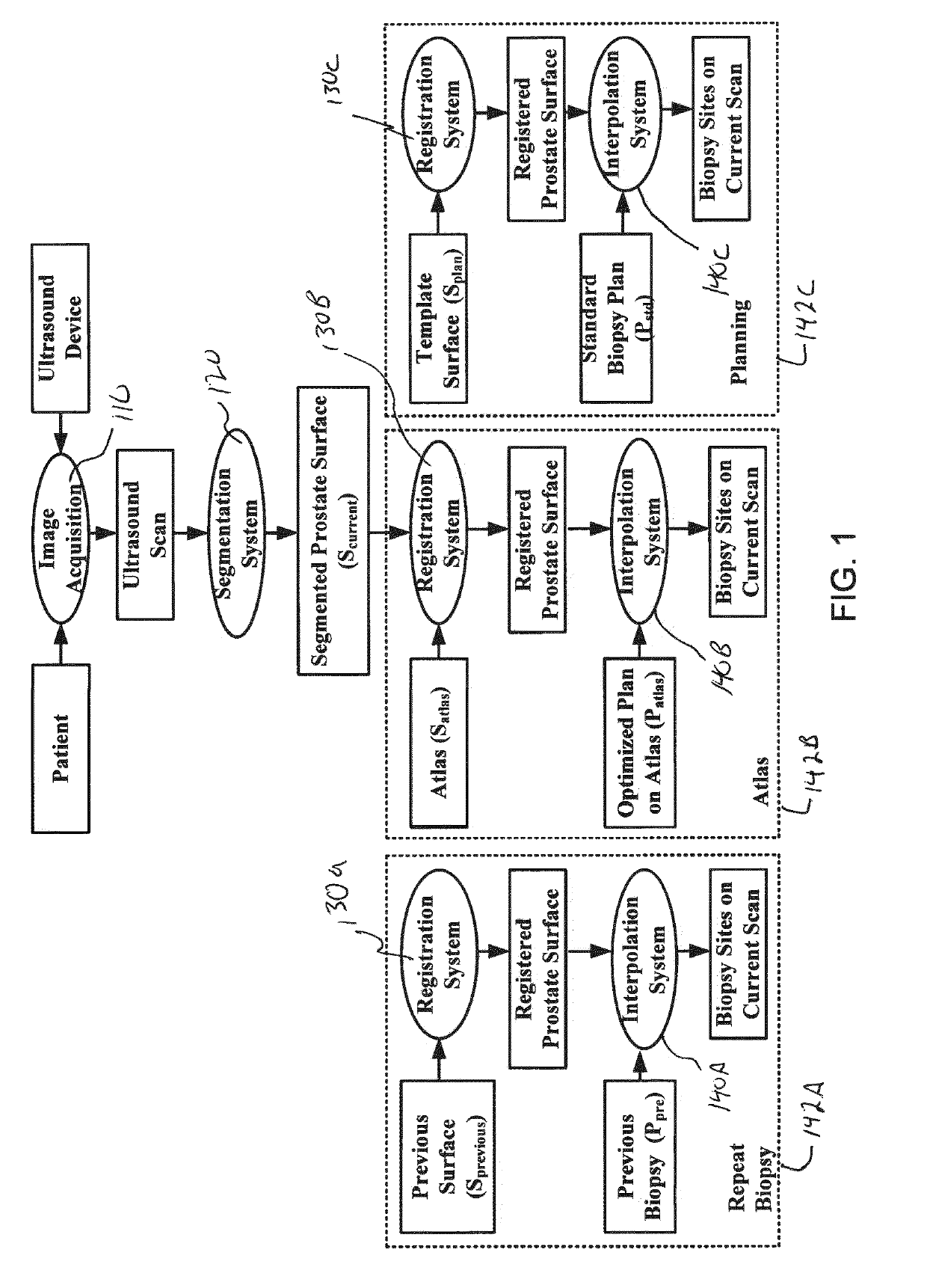
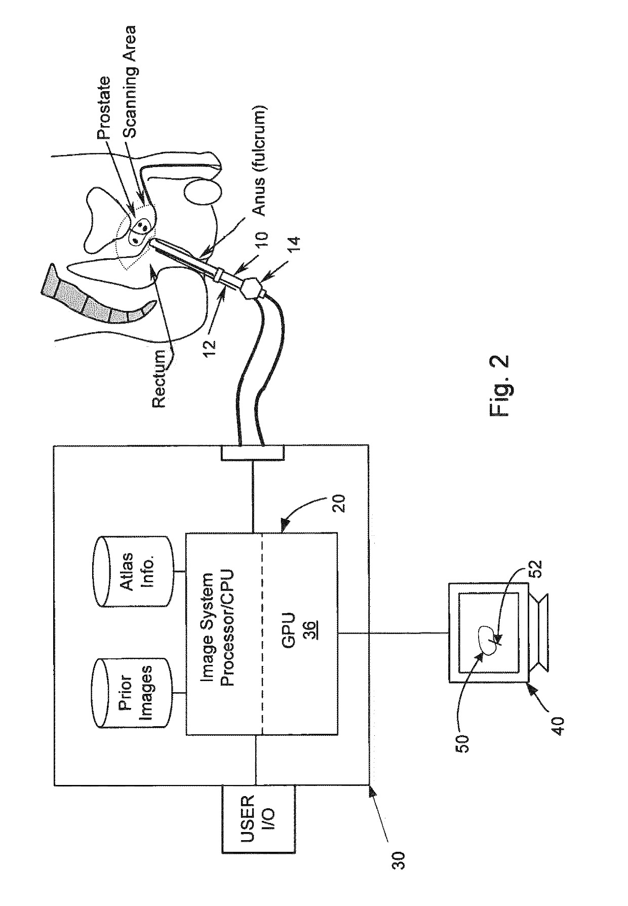
[0008]There are typically four stages involved in an imaged biopsy procedure: image acquisition, image segmentation, image registration, and biopsy navigation. The first stage has a constant operating time; the fourth stage depends on the number of biopsy sites planned. It is therefore important to make the segmentation and registration procedure real-time or near real-time. Graphics processing units (GPU) have now evolved into a computing powerhouse for parallel computation. The numerous multiprocessors, and fast memory units within the GPU may be favorably exploited to run large data parallel tasks simultaneously, and with high arithmetic intensity allowing the creation of several hundreds or thousands of data parallel threads.
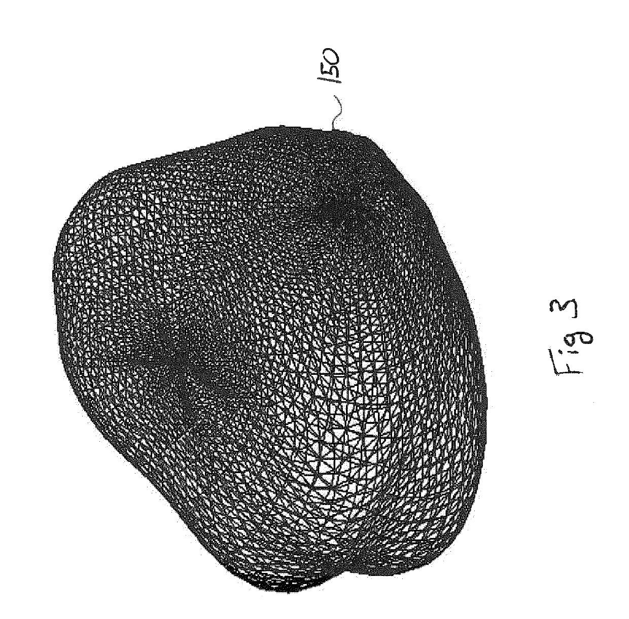
[0010]In this application, the inventors present an implementation of surface-based registration algorithm using parallel computation that may be performed on a GPU. Also presented is an elastic warping solver in 3D to warp 3D volumes based on surface correspondences. The inputs are a current segmented prostate surface and a model surface (e.g., previous prostate surface, atlas surface etc). The inputs surfaces have vertices and facets defined (e.g., mesh surfaces). The output is a deformed mesh surface after registration. The deformation information (i.e, correspondences) generated from deforming the model mesh surface to the current mesh surface is used to warp 3D volumes after surface registration. That is, initially only the surfaces of 3D volumes (e.g., current prostate volume and a previous prostate volume) surfaces are used to determine correspondences. After determining these correspondences they are applied to elastically deform, for example, a previous volume to a current volume. In the procedure of registration, both local and global features are used by adjusting the searching resolution and step size. A parallel computation implementation not only makes the registration near real-time, but also facilitates the visualization of registration surface on screen.
[0011]The systems and methods (utilities) presented in this patent allow for reducing biopsy procedure times. Three procedures which may benefit from the presented utilities includes: statistical Atlas based warping to map optimal biopsy sites defined on the atlas space to the current anatomical volume; mapping of biopsy locations of the same patient from a previous visit to the current anatomical volume; and mapping of planned biopsy sites, e.g. sextant, extended 12 core systematic biopsy, etc on to the current anatomical volume. All three methods provide useful information to help guide biopsy target selection. They require the registration of surfaces followed by interpolating needle locations from one image to another.

Problems solved by technology

The samples collected by the urologist may produce a false negative if the biopsy does not detect malignant tissues despite high PSA levels or other indicators of PCa (e.g. transurethral resection, digital rectal examination).
Traditional biopsy protocols most notably the systematic sextant biopsy protocol has a poor positive predictive value.
Several studies have suggested the use of more optimal biopsy protocols based on the non uniform occurrence of cancers within the prostate but no ideal standard exists at this time.
The image processing involved to accomplish these 3D registration tasks are very computationally intensive making target selection guided by a) atlas, b) previously visited sites and / or c) standard plans difficult to achieve in real time by processing data on a CPU.
Long registration times can lead to patient anxiety, risk of motion that may invalidate the relevance of the TRUS image acquired and reconstructed to 3D, and longer biopsy procedures.

Method used


Image

  • Apparatus for real-time 3D biopsy
  • Apparatus for real-time 3D biopsy
  • Apparatus for real-time 3D biopsy

Examples


Embodiment Construction

[0025]Reference will now be made to the accompanying drawings, which assist in illustrating the various pertinent features of the various novel aspects of the present disclosure. Although the invention is described primarily with respect to an ultrasound imaging embodiment, the invention is applicable to a broad range of imaging modalities and biopsy techniques, including MRI, CT, and PET, which are applicable to organs and / or internal body parts of humans and animals. In this regard, the following description is presented for purposes of illustration and description. Furthermore, the description is not intended to limit the invention to the form disclosed herein. Consequently, variations and modifications commensurate with the following teachings, and skill and knowledge of the relevant art, are within the scope of the present invention.

[0026]In one embodiment of the present invention, a needle positioning system to aid a urologist in rapidly finding biopsy target sites is presente...


PUM


Abstract

A method and apparatus are disclosed for performing software guided prostate biopsy to extract cancerous tissue. The method significantly improves on the current system by accelerating all computations using a graphical processing unit (GPU) keeping the accuracy of biopsy target locations within tolerance. The result is the computation of target locations to guide biopsy using statistical priors of cancers from a large population, as well as based on previous biopsy locations for the same patient, and finally via mapping protocols with predefined needle configurations onto the patient's current ultrasound image.

Description

CROSS-REFERENCE TO RELATED APPLICATION[0001]This application claims priority and the benefit of the filing date under 35 U.S.C. 119 to U.S. Provisional Application No. 61 / 062,009, entitled, “AN APPARATUS FOR REAL-TIME 3D BIOPSY,” filed on Jan. 23, 2008, the entire contents of which are incorporated herein as if set forth in full.FIELD OF INVENTION[0002]The present invention is directed to the registration of medical images. More specifically, the present invention is directed to surface registration of images in a parallel processing system that reduces the computational time requiredBACKGROUND OF THE INVENTION[0003]A biopsy is recommended when a patient shows high levels of prostate specific antigen (PSA), which is often an indicator of prostate cancer (PCa). 3-D Transrectal Ultrasound (TRUS) guided prostate biopsy is one method to test for prostate cancer. The samples collected by the urologist may produce a false negative if the biopsy does not detect malignant tissues despite hi...

Claims


Application Information

Patent Timeline
IPC IPC(8): G06K9/00G06T15/00
CPCG06K9/00986G06K9/6206G06T7/0032G06T2207/30081G06T2207/10068G06T2207/10136G06T2207/20128G06T17/20G06T7/344G06V10/955G06V10/754
Inventor NARAYANAN, RAMKRISHNANGUO, YUJUNSURI, JASJIT S.
Owner EIGEN INC
Learn More