Dissolution of amyloid fibrils by flavonoids and other compounds
a technology of flavonoids and amyloid fibrils, which is applied in the direction of biocide, tetracycline active ingredients, drug compositions, etc., can solve the problems of systemic amyloidosis that can be deadly, amyloid deposits may develop in the heart, and patients can di
- Summary
- Abstract
- Description
- Claims
- Application Information
AI Technical Summary
Problems solved by technology
Method used
Image
Examples
example 2
Thioflavin T Fluorescence Assay for Detecting the Presence of Amyloid Fibrils
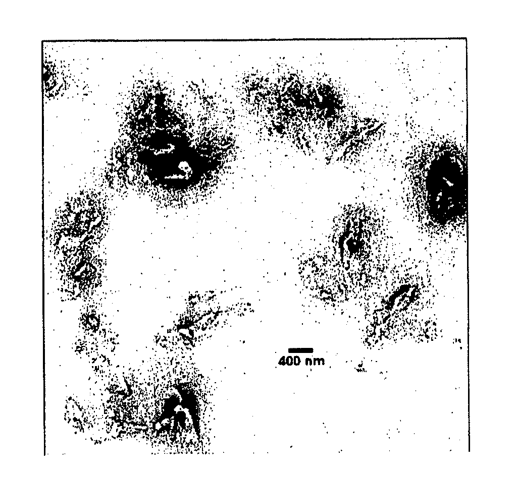
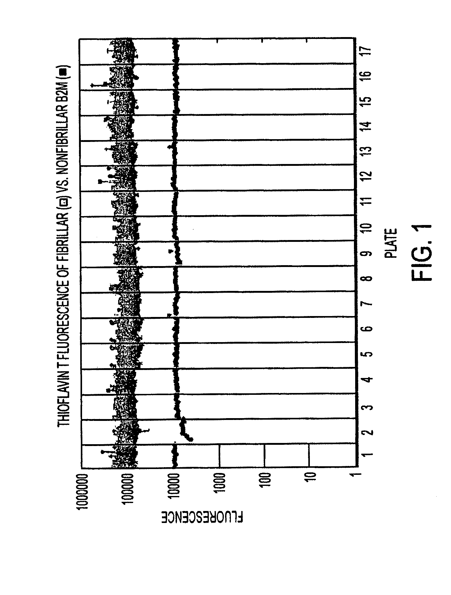
[0069]In our experiments, disruption of the fibrillar structure of preformed β-2 microglobulin fibrils was monitored by fluorescence. The dye used, thioflavin T, serves as a probe for the presence or absence of amyloid fibrils. Thioflavin T binds specifically to human β-2 microglobulin fibrils, causing a red shift in the fluorescence excitation spectra of the dye to produce a peak in the fluorescence emission spectrum at 482 nm when the excitation wavelength is set at 444 nm. Nonfibrillar β-2 microglobulin does not cause such a red shift (FIG. 1) (LeVine, H. 1999 Methods in Enzymology; 309:274-84.). Because aliquotting of fibrils has an inherent variability, we aimed for a high signal / noise ratio of ˜10. FIG. 1 shows that this ratio is sufficient to distinguish fibrillar and nonfibrillar samples.
[0070]β-2 microglobulin fibrils prepared in vitro retain the characteristic amyloid properties, including binding...
example 3
[0071]In our experiments, we screened 5200 compounds to determine whether a compound could disrupt preformed human β-2 microglobulin fibrils. Preformed β-2 microglobulin fibrils and ThT were mixed in the ratios described above in Example 2 and divided among seventeen 384-well glass-bottom plates (Costar #6569) using 40 μL / well. Each of the sixteen commercial plates contained four columns of control wells: columns 1, 2, and 23 contained preformed fibrils+dimethylsulfoxide (herein, DMSO), but no compound; column 24 contained nonfibrillar β2M+DMSO (no compound). The final concentrations of compound and DMSO in the other wells were 10 μM and 1%, respectively. Compounds were obtained from the following chemical libraries: the MicroSource Discovery Systems, Inc. Spectrum Collection library (J Virology 77: 10288 (2003); Ann Rev Med 56: 321 (2005)), the first four plates from the Prestwick Chemical Inc. library, two BIOMOL plates (phosphatase / kinase inhibitor librar...
example 4
Dose-Response Testing
[0076]Twelve compounds were selected for testing their dose-response effects. Dose-response curves were derived as follows. Preformed β-2 microglobulin fibrils were mixed with ThT in the ratios described above and divided into portions of 40 μL per well across 8 wells. The final concentrations were the same as used in the high-throughput screen: 4.1 μM ThT, 41.2 mM glycine, 0.018 mg / mL B2M, 35 mM NaCl, 4.4 mM phosphate. The first and last wells of each row were controls (B2M fibrils+ThT+solvent (water or DMSO)). To wells 2-7 of a given row, test compound was added to the β2M fibril / ThT mix such that the following final concentrations in the well were reached: 10 mM, 1 mM, 100 μM, 10 μM, 1 μM, and 100 nM. Each row was set up in duplicate. The wells were incubated for 99 hours and data collected at 2.5 hour intervals. Compound 22 was tested using both water and DMSO as solvent.
[0077]The dose-response curves for compounds 22 and L18 are shown in FIG. 3. It is visua...
PUM
| Property | Measurement | Unit |
|---|---|---|
| molar concentration | aaaaa | aaaaa |
| molar concentration | aaaaa | aaaaa |
| molar concentration | aaaaa | aaaaa |
Abstract
Description
Claims
Application Information
 - R&D
- Intellectual Property
- Life Sciences
- Materials
- Tech Scout
- Unparalleled Data Quality
- Higher Quality Content
- 60% Fewer Hallucinations
Browse by: Latest US Patents, China's latest patents, Technical Efficacy Thesaurus, Application Domain, Technology Topic, Popular Technical Reports.
© 2025 PatSnap. All rights reserved.Legal|Privacy policy|Modern Slavery Act Transparency Statement|Sitemap|About US| Contact US: help@patsnap.com


