
Methods and apparatus for providing and processing sliced thin tissue

a tissue and tissue technology, applied in the field of methods and apparatus for providing and processing sliced thin tissue, can solve the problems of inability to address the needs of the larger community of neuroscientists, the inability to integrate the deluge of thousands of individual in vivo tracing experiments into a coherent whole, and the inability to achieve the goal of facilitating subsequent imaging of sliced thin tissue, facilitating fully automated production, collection, handling, imaging and storag

Inactive Publication Date: 2010-04-15
PRESIDENT & FELLOWS OF HARVARD COLLEGE
View PDF19 Cites 42 Cited by
  • Summary
  • Abstract
  • Description
  • Claims
  • Application Information

AI Technical Summary

Benefits of technology

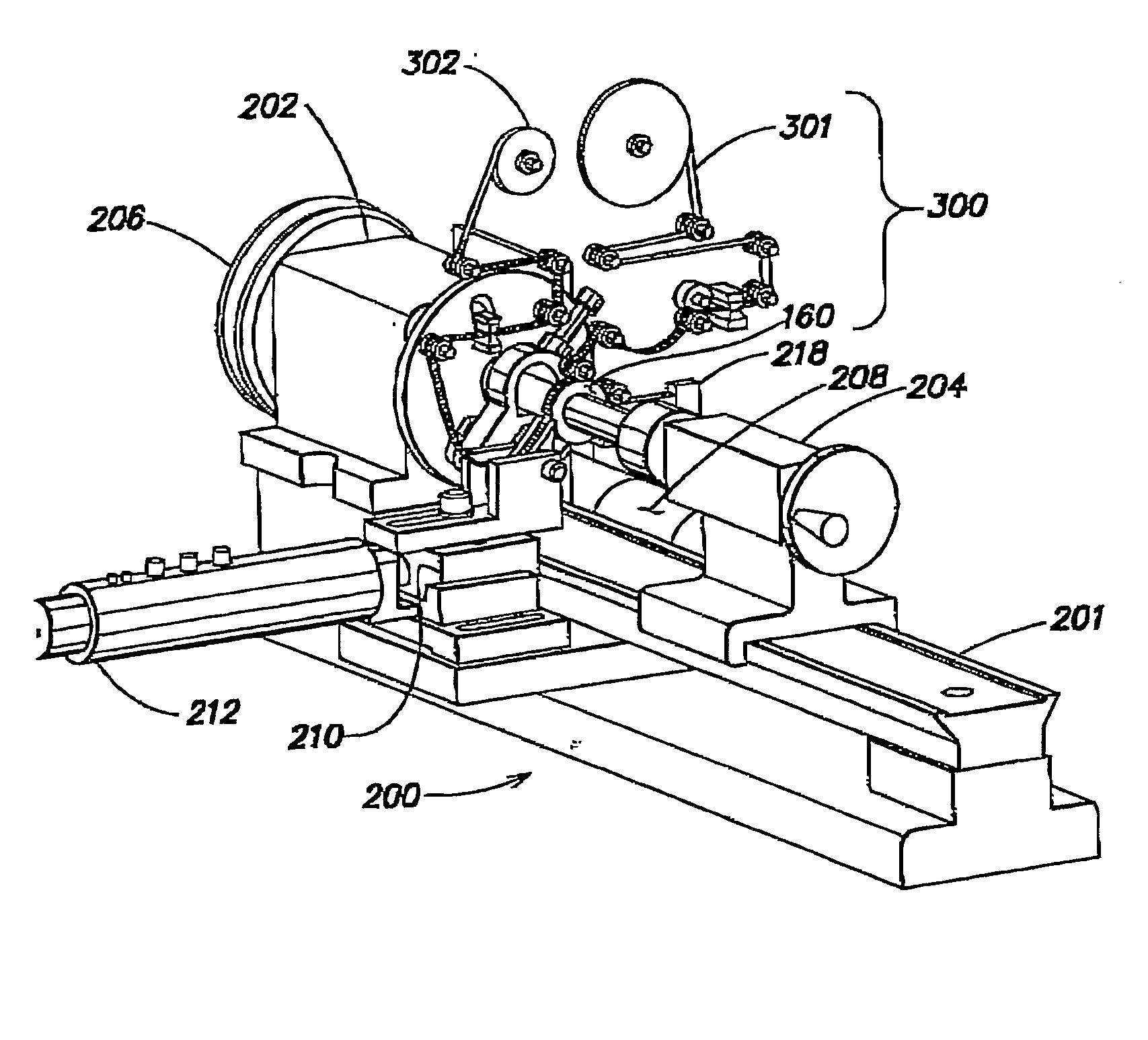
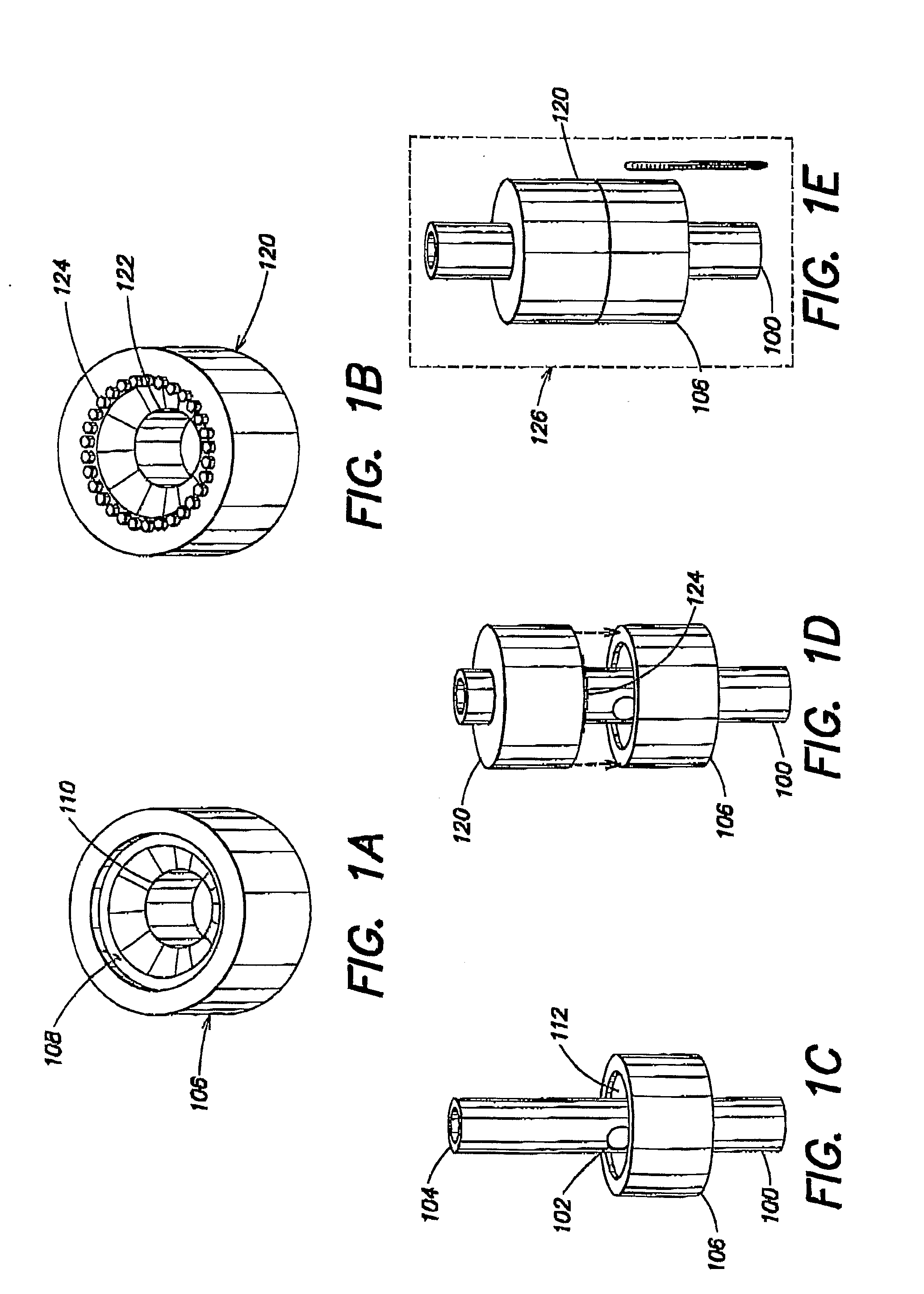
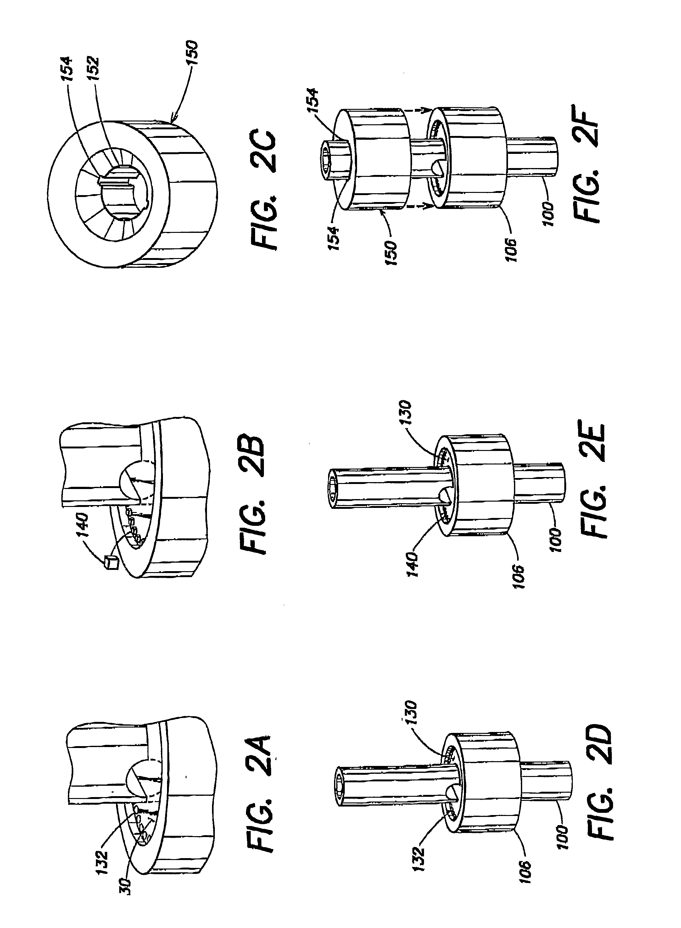
[0010]Various embodiments of the present invention are directed to methods and apparatus for providing and processing serial tissue sections. In exemplary embodiments, an “automatic tape collecting lathe ultramicrotome” (ATLUM) is disclosed, in which the basic ratcheting motion of the microtome is redesigned, replacing the conventional discontinuous ratcheting motion with a continuous rotary Motion of a lathe. Using an ATLUM, a block of tissue sample having various geometries may be sliced into a continuous ribbon Of thin tissue; or multiple thin tissue sections, and disposed on an appropriate substrate to facilitate subsequent imaging of the sliced thin tissue. As will be described in greater detail below, the continuous lathe cutting design makes possible continuous taping and slice collection. The result is a mechanically more stable, more reliable, faster, and more easily constructed design that facilitates fully automated production, collection, handling, imaging, and storage of thousands of semi-thin and ultra-thin tissue sections.
[0011]In some inventive embodiments disclosed herein, closed-loop control of section thickness of thin tissue sections or ribbons sliced from a tissue sample is implemented to produce thinner sliced tissue sections or ribbons having tightly controlled thickness. Thinner samples with predictable thickness in turn facilitate high quality volume reconstructions of biological samples. In one exemplary implementation, one or more capacitive sensors are employed in an ATLUM to facilitate regulation of a distance between a slicing knife and a tissue sample to be sliced, thereby controlling sliced tissue thickness with improved precision. Other types of distance sensing techniques may be employed in other implementations to control and regulate sliced tissue thickness.
[0012]In yet other inventive embodiments disclosed herein, thin tissue sections or ribbons are particularly processed / prepared to facilitate imaging with a scanning electron microscope (SEM) (e.g., in electron backscatter mode). Imaging via an SEM is generally a significantly simplified process as compared to imaging via a TEM (transmission electron microsope), and images obtained via SEM of sufficient quality, and in many instances equivalent quality, to conventional TEM images. In one aspect of these embodiments, collected tapes of thin tissue sections or ribbons sliced from a tissue sample are used to create UltraThin Section Libraries (UTSLs) that allow for fully automated, time-efficient imaging in the SEM.
[0013]In view of the foregoing, aspects of the current invention provide for thinner tissue sections having tightly controlled thickness produced in a fully automated fashion. In exemplary applications made possible by embodiments of the present invention, tens of meters of ultrathin sections may be automatically sliced and collected on a tape that is subsequently stained with heavy metals and mounted onto plates for any appropriate imaging mode in a scanning electron microscope (SEM), such as, but not limited to, electron backscatter imaging. ATLUM-collected sections may provide images equivalent to TEM images, showing detail down to individual synaptic vesicles within synapses. The ATLUM can also quickly create a UTSL of many cubic millimeters of tissue, enough to encompass multiple brain regions and their interconnecting axonal tracts. The UTSL can also be swiftly SEM imaged, and this can be used to intelligently direct subsequent time intensive high-resolution imaging forays. In this manner, researchers may efficiently map out specific neural circuits spanning several millimeters with a resolution in the nanometer range.

Problems solved by technology

Unfortunately, these experiments and models involve brain systems where incomplete information regarding the system's underlying neural circuitry presents one of the largest barriers to research success.
Unfortunately, the current approach of attempting to integrate the deluge of thousands of individual in vivo tracing experiments into a coherent whole is proving to be a virtually impossible task.
Because of the manual nature of this current process, this technique is totally impractical to apply to larger brain structures and so it is currently unable to address the needs of the larger community of neuroscientists who require a map of the brain connectivity of rodent and primate brains.
We are unaware of any current microtome design (either in production or disclosed in the open literature) that adequately addresses this need for automating the production, collection, handling, and imaging of large numbers of thin tissue sections suitable for use in light and transmission electron microscopic 3D reconstruction work.
Although there is a vast number of patents pertaining to microtomes and their automation, these designs are targeted toward automating the slicing process only, and do not address the tissue collection and handling processes.
Thus, current “automated microtome” designs still require manual slice retrieval and manual slide or grid mounting for imaging.
Such manual slice retrieval necessitates skilled, delicate, and incredibly time-consuming work be expended on each tissue slice (or small series of slices) as it involves “fishing” each tissue slice out of a water boat attached to the knife of the ultramicrotome instrument and onto a TEM grid.
That design also makes no modification to the current standard microtome design, and thus also suffers from the discontinuous ratcheting action.
The tape in Bolles' design and the glass slide in the Voneiff and Gibson design are much too thick for this.

Method used


Image

  • Methods and apparatus for providing and processing sliced thin tissue
  • Methods and apparatus for providing and processing sliced thin tissue
  • Methods and apparatus for providing and processing sliced thin tissue

Examples


Embodiment Construction

[0062]Following below are more detailed descriptions of various concepts related to, and inventive embodiments of, methods and apparatus according to the present disclosure for providing and processing sliced thin tissue. It should be appreciated that various aspects of the subject matter introduced above and discussed in greater detail below may be implemented in any of numerous ways, as the subject matter is not limited to any particular manner of implementation. Examples of specific implementations and applications are provided primarily for illustrative purposes.

[0063]One embodiment of the present invention discloses a device, an automated tape collecting lathe-ultramicrotome (“ATLUM”), and associated methods and apparatus for fully automating the collection, handling, and imaging of large numbers of serial tissue sections.

[0064]Classical TEM tissue processing and imaging methods begin by embedding an approximately 1 mm3 piece of biological tissue that has been fixed with mixed ...


PUM

PropertyMeasurementUnit
thicknessaaaaaaaaaa
thicknessaaaaaaaaaa
thicknessaaaaaaaaaa

Abstract

Methods and apparatus for providing and processing serial tissue sections. In one example, an “automatic tape collecting lathe ultramicrotome” (ATLUM) slices a block of tissue sample having various geometries into a continuous ribbon of thin tissue, or multiple thin tissue sections, and disposes the sliced thin tissue on an appropriate substrate to facilitate subsequent imaging of the sliced thin tissue. Closed-loop control of section thickness of the sliced thin tissue sections or ribbons is implemented to produce thinner sliced tissue sections or ribbons and tightly regulate thickness. Thin tissue sections or ribbons may be particularly processed / prepared to facilitate imaging with a scanning electron microscope (SEM). Collected thin tissue sections or ribbons may be used to create UltraThin Section Libraries (UTSLs) that allow for fully automated, time-efficient imaging in the SEM to facilitate expansive tissue studies.

Description

FIELD OF THE INVENTION[0001]The present invention relates generally to automating the process of producing sliced thin tissue from a block of sample tissue block so as to allow the thin tissue to be reliably collected, handled, stored, and digitally imaged (via automated retrieval).BACKGROUND[0002]Today neuroscientists are routinely carrying out evermore-advanced physiological experiments and cognitive scientists are proposing and testing evermore-comprehensive models of brain function. Unfortunately, these experiments and models involve brain systems where incomplete information regarding the system's underlying neural circuitry presents one of the largest barriers to research success. It is widely accepted within the neuroscience community that what is needed is a comprehensive and reliable wiring diagram of the brain that will provide a neuroanatomical scaffolding (and a set of foundational constraints) for the rest of experimental and theoretical work in the neuro- and cognitive...

Claims


Application Information

Patent Timeline
Patent Type & Authority Applications(United States)
IPC IPC(8): G01N1/30B26D5/08H01J37/28G05B13/02
CPCG01N1/06G01N1/312G01N2001/066H01J2237/26H01J2237/201H01J2237/202G01N2001/362Y10T83/141Y10T83/04
Inventor HAYWORTH, KENNETHKASTHURI, NARAYANANLICHTMAN, JEFFHARTWIEG, ERIKA
Owner PRESIDENT & FELLOWS OF HARVARD COLLEGE
Learn More