Spatial characterization of a structure located within an object by identifying 2d representations of the structure within section planes

Inactive Publication Date: 2010-04-22
KONINKLIJKE PHILIPS ELECTRONICS NV
View PDF4 Cites 66 Cited by
  • Summary
  • Abstract
  • Description
  • Claims
  • Application Information

AI Technical Summary

Benefits of technology

[0047]According to a further embodiment of the invention at least one known property of the stent or vessel lumen is used in order to identify the stent within the 2D representations. This may provide the advantage that a comparison of an identified structure within the measured lumen with e.g. the maximal possible expanded stent lumen may be carried out. Thereby, one may carry out an automatic consistency check whether it is really possible that the identified structure is an inserted stent. The reliability of a corresponding algorithm of such a consistency check will depend on such an input information.
[0048]According to a further embodiment of the invention the step of acquiring a volumetric dataset of the object under examination is carried out with contrast agent being inserted into the vessel lumen. This may provide the advantage that the morphology of at least a portion of the vascular system can be clearly visualized.
[0049]According to a further embodiment of the invention the method further comprises the step of acquiring a further volumetric dataset of the object under examination in the absence of a contrast agent. This

Problems solved by technology

However, determining and monitoring the stent expansion on images

Method used


Image

  • Spatial characterization of a structure located within an object by identifying 2d representations of the structure within section planes
  • Spatial characterization of a structure located within an object by identifying 2d representations of the structure within section planes
  • Spatial characterization of a structure located within an object by identifying 2d representations of the structure within section planes

Examples


Embodiment Construction

[0069]The illustration in the drawing is schematically. It is noted that in different figures, similar or identical elements are provided with the same reference signs or with reference signs, which are different from the corresponding reference signs only within the first digit.

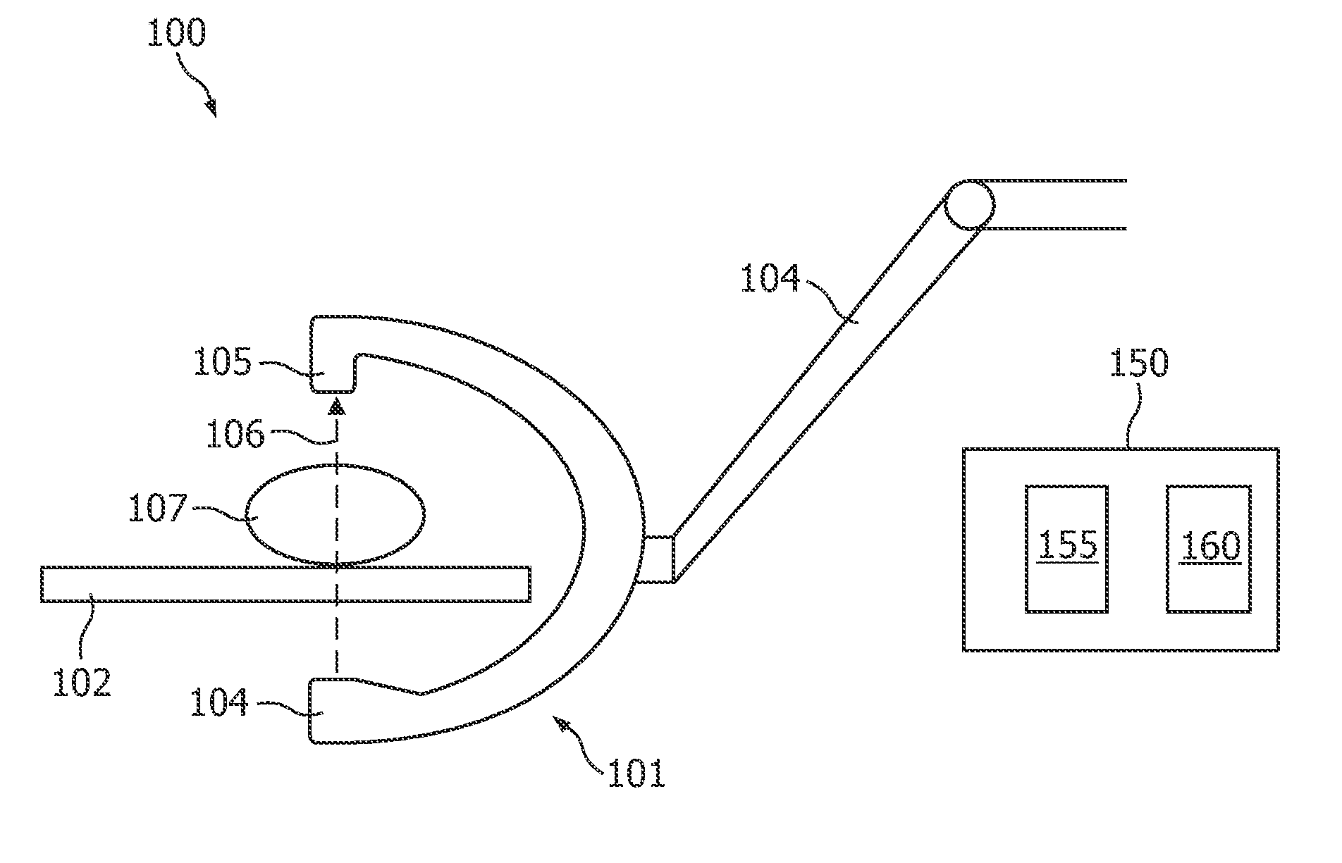
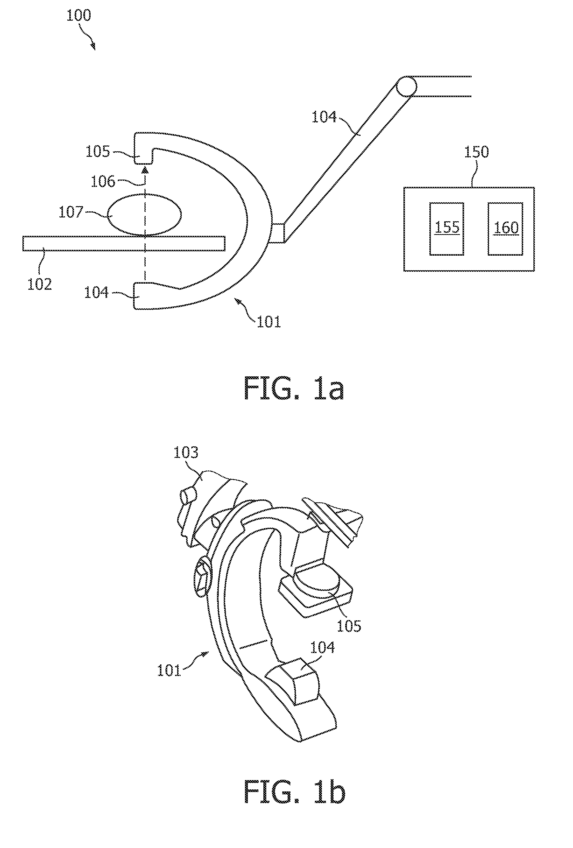
[0070]Referring to FIG. 1a and 1b of the drawing, a medical X-ray imaging system 100 according to an embodiment of the invention comprises a swing arm scanning system (C-arm) 101 supported proximal a patient table 102 by a robotic arm 103. Housed within the swing arm 101, there is provided an X-ray tube 104 and an X-ray detector 105. The X-ray detector 105 is arranged and configured to receive X-rays 106, which have passed through a patient 107 representing the object under examination. Further, the X-ray detector 105 is adapted to generate an electrical signal representative of the intensity distribution thereof. By moving the swing arm 101, the X-ray tube 104 and the detector 105 can be placed at any desir...


PUM


Abstract

It is described a virtual pullback as a visualization and quantification tool that allows an interventional cardiologist to easily assess stent expansion. The virtual pullback visualizes the stent and/or the vessel lumen similar to an Intravascular Ultrasound (IVUS) pullback. The virtual pullback is performed in volumetric data along a reference line. The volumetric data can be a reconstruction of rotational 2D X-ray attenuation data. Planes perpendicular to the reference line are visualized as the position along the reference line changes. This view is for interventional cardiologists a very familiar view as they resemble IVUS data and may show a section plane through a vessel lumen or a stent. In these perpendicular section planes automatic measurements, such as minimum and maximum diameter, and cross sectional area of the stent can be calculated and displayed. Combining these 2D measurements allows also volumetric measurements to be calculated and displayed.

Description

FIELD OF INVENTION[0001]The present invention generally relates to the field of digital image processing, in particular for medical purposes in order to provide for a visualization and for a quantitative analysis of an object being inserted within a body of a patient.[0002]Specifically, the present invention relates to a method for spatially characterizing a structure located within an object under examination, in particular for spatially characterizing a medical device being inserted into the body of a patient.[0003]Further, the present invention relates to a data processing device and to a medical X-ray examination apparatus, in particular a C-arm system or a computed tomography system, comprising the described data processing device, wherein the data processing device is adapted for spatially characterizing a structure located within an object under examination, in particular for spatially characterizing a medical device being inserted into the body of a patient.[0004]Furthermore...

Claims


Application Information

Patent Timeline
IPC IPC(8): A61B5/05G06K9/62G01N23/04
CPCG06T7/0012G06T2219/008G06T2207/30101G06T19/00
Inventor SCHOONENBERG, GERTWINK, ONNOMOVASSAGHI, BABAK
Owner KONINKLIJKE PHILIPS ELECTRONICS NV
Why Eureka
Try Eureka