

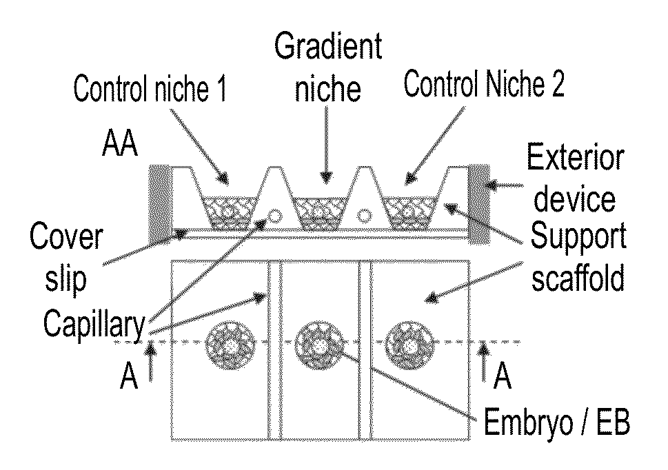
Niche system for biological culturing
a niche system and niche technology, applied in the field of culturing niche system, can solve the problems of inability to achieve the goal using standard tissue culture techniques, the inability of culturing systems of this kind to be constructed sophisticated (typically microfluidics-based) and the inability to construct sophisticated devices to control the micro-environment. the effect of implantation chance and improving human fertility
- Summary
- Abstract
- Description
- Claims
- Application Information
AI Technical Summary
Benefits of technology
Problems solved by technology
Method used
Image

Examples
example 1
Diffusion Gradients
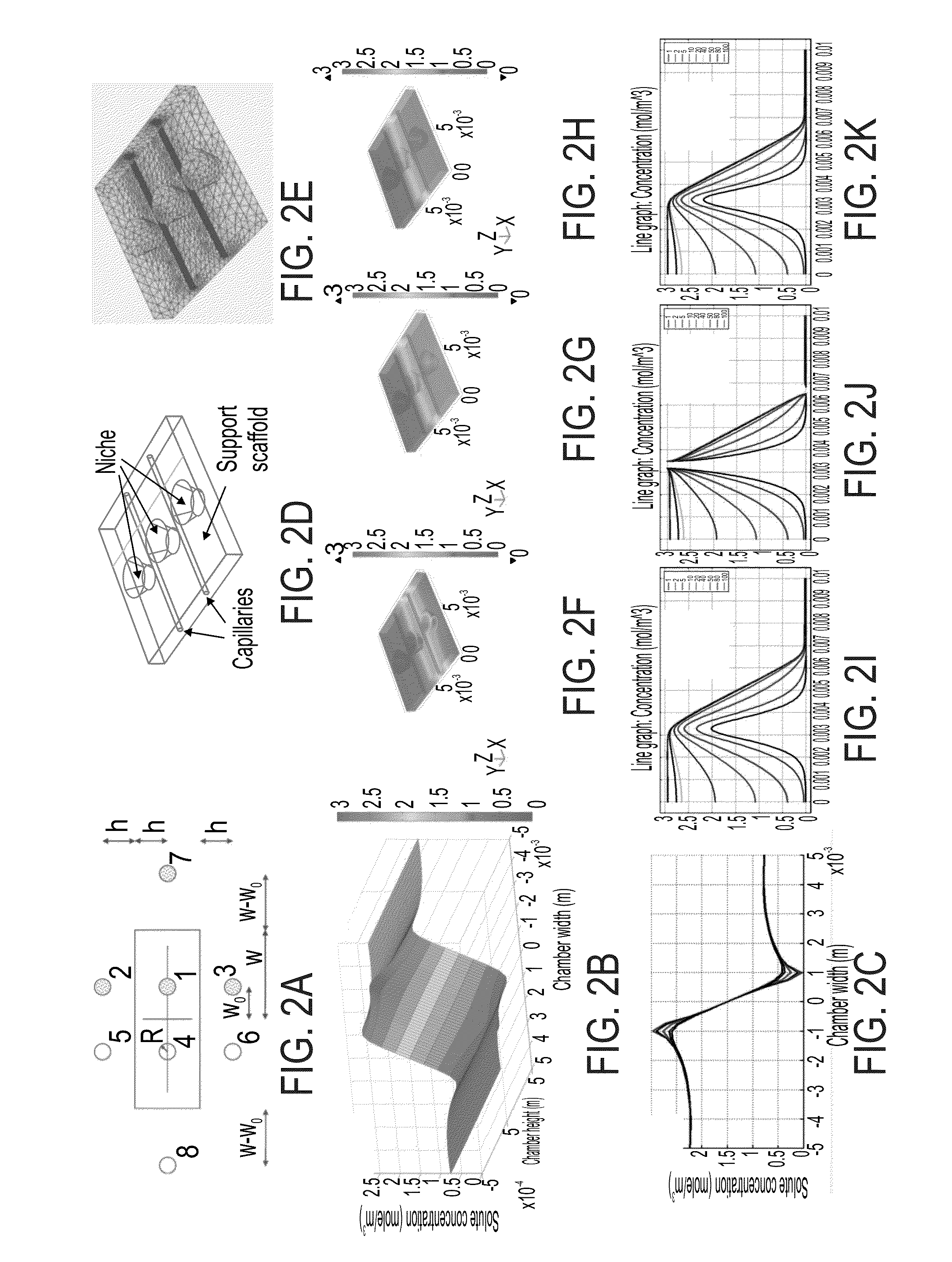
[0102]The present system was tested with small and large molecules in order to determine the diffusion gradients for these molecules.
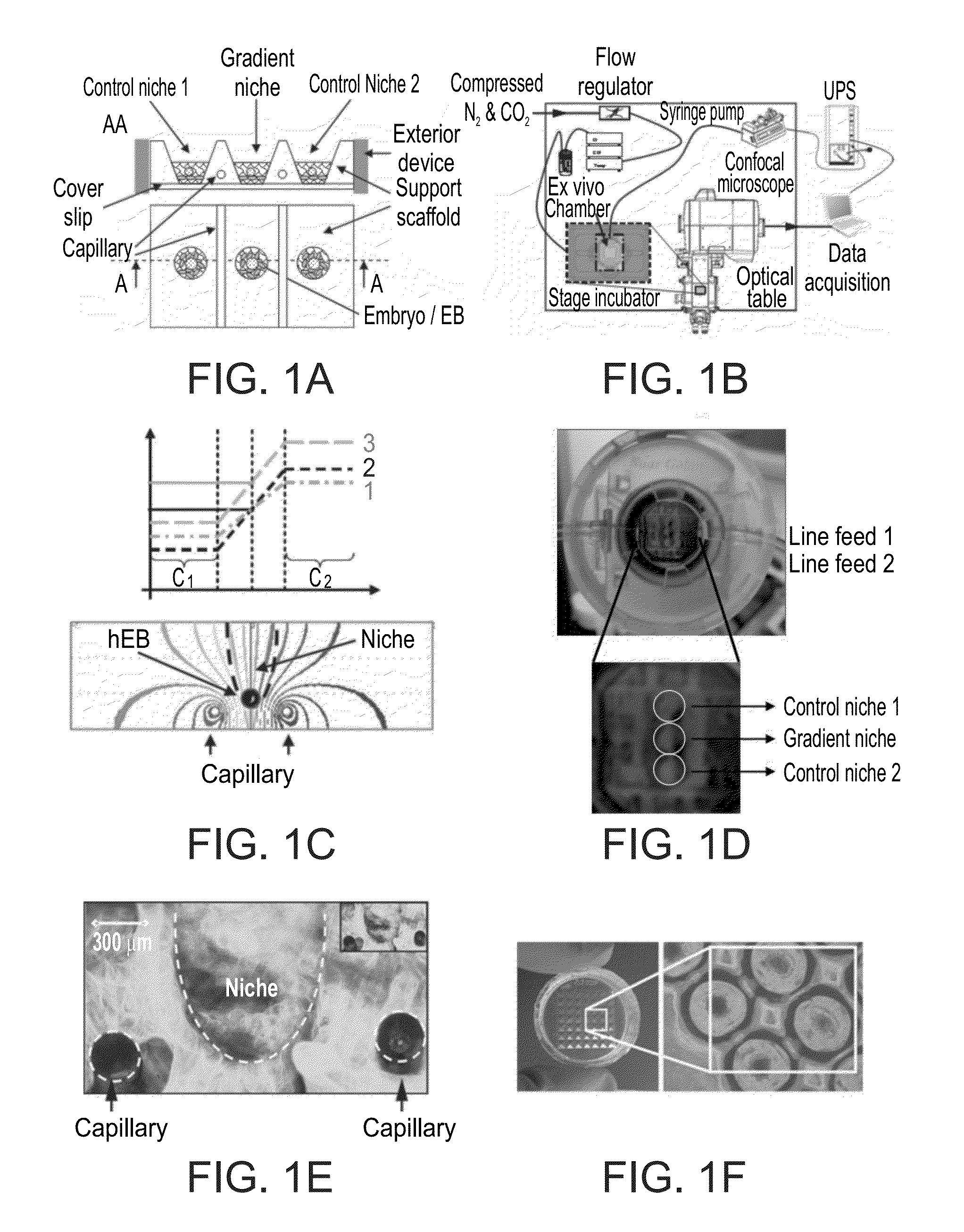
[0103]The Food colorants Green-mix (mixture of E133 / Brilliant Blue FCF, Mw=792.84 Da, and E110 / Sunset Yellow FCF, Mw=452.37 Da) and Red-Azorubine (E122, Mw=502.4 Da) and the fluorescent molecule Calcein (Sigma-Aldrich C0875, Mw=622.53 Da) were used as models for small molecules (FIGS. 2N-R). The kinetics and spatial distribution of the Calcein was investigated using confocal microscopy time lapse (Zeiss LSM 710) and is presented in FIG. 2R. In this Figure, a simple moving average (SMA, fifty elements) data smoothing algorithm is used to filter the noise. FIG. 2R demonstrates the chamber ability to maintain a stable small molecule gradient over an extended period of hours. This Figure also illusatrtes that the gradient rapidly stabilizes within less than two hours.
[0104]To demonstrate the ability of the present system to maintain a st...
example 2
Culturing of EBs
[0106]Several cell lines (293T, ECC-1, Ishikawa, JAR, H1 / H9 / Hues hESCs), fruit fly embryos (D. melanogaster) and EBs (hESCs, constitutively labeled with CFP or genetically marked for the expression of the pluripotency gene OCT4 with GFP—a knock-in line kindly provided by the Thomson lab, FIG. 4D) were used in order to demonstrate the ability of the present system to support culturing of embryonic models. The present system was tested for the ability to induce organization of EBs in a gradient niche (vs. control niches). The effects of in vivo-like signals (e.g., BMP-4 and FGF) on the polarization were tested. The simple readout for these experiments is spatially organized differentiation responses within the EBs.
[0107]Two approaches were tested for establishment of an implantation model. The first approach relied on generating human EBs consisting of both trophoblast and ESC lines and co-culturing them with receptive endometrial lines (FIG. 4C). The second approach u...
PUM
| Property | Measurement | Unit |
|---|---|---|
| time- | aaaaa | aaaaa |
| height | aaaaa | aaaaa |
| radius | aaaaa | aaaaa |
Abstract
Description
Claims
Application Information