Preparation of tissue for meniscal implantation

a tissue and meniscus technology, applied in the field of tissue preparation for meniscal implantation, can solve the problems of severe damage or extensive tear of the meniscus, difficulty in preparing an immunologically inert or decellularised tissue for transplantation, swelling and/or loss of knee joint function,

Active Publication Date: 2010-06-17
TISSUE REGENIX
View PDF4 Cites 10 Cited by
  • Summary
  • Abstract
  • Description
  • Claims
  • Application Information

AI Technical Summary

Benefits of technology

[0066]It is believed that the methods and products of the present invention advantageously provides natural meniscal tissue that is truly biocompatible with a host with minimal risk of rejection by the host by virtue of the improved method of decellularisation.
[0067]The meth...

Problems solved by technology

When menisci are damaged through injury, disease or inflammation arthritic changes can develop in the knee joint with consequent swelling, pain and/or loss of knee joint function in the affected individual.
Whilst it is possible to repair a torn meniscus, a meniscus that is severely damaged or has an extensive tear may have to be removed.
A problem associated with the use of donor tissue for meniscal replacement is that the meniscus is a dense fibrocartilagenous tissue impregnated throughout with fibrochondrocytes which are the cells responsible for synthesis, maintenance and repair of the extracellular matrix.
Because the meniscus is a thick and dense tissue with cells located throughout and es...

Method used


Image

  • Preparation of tissue for meniscal implantation
  • Preparation of tissue for meniscal implantation
  • Preparation of tissue for meniscal implantation

Examples


example 1

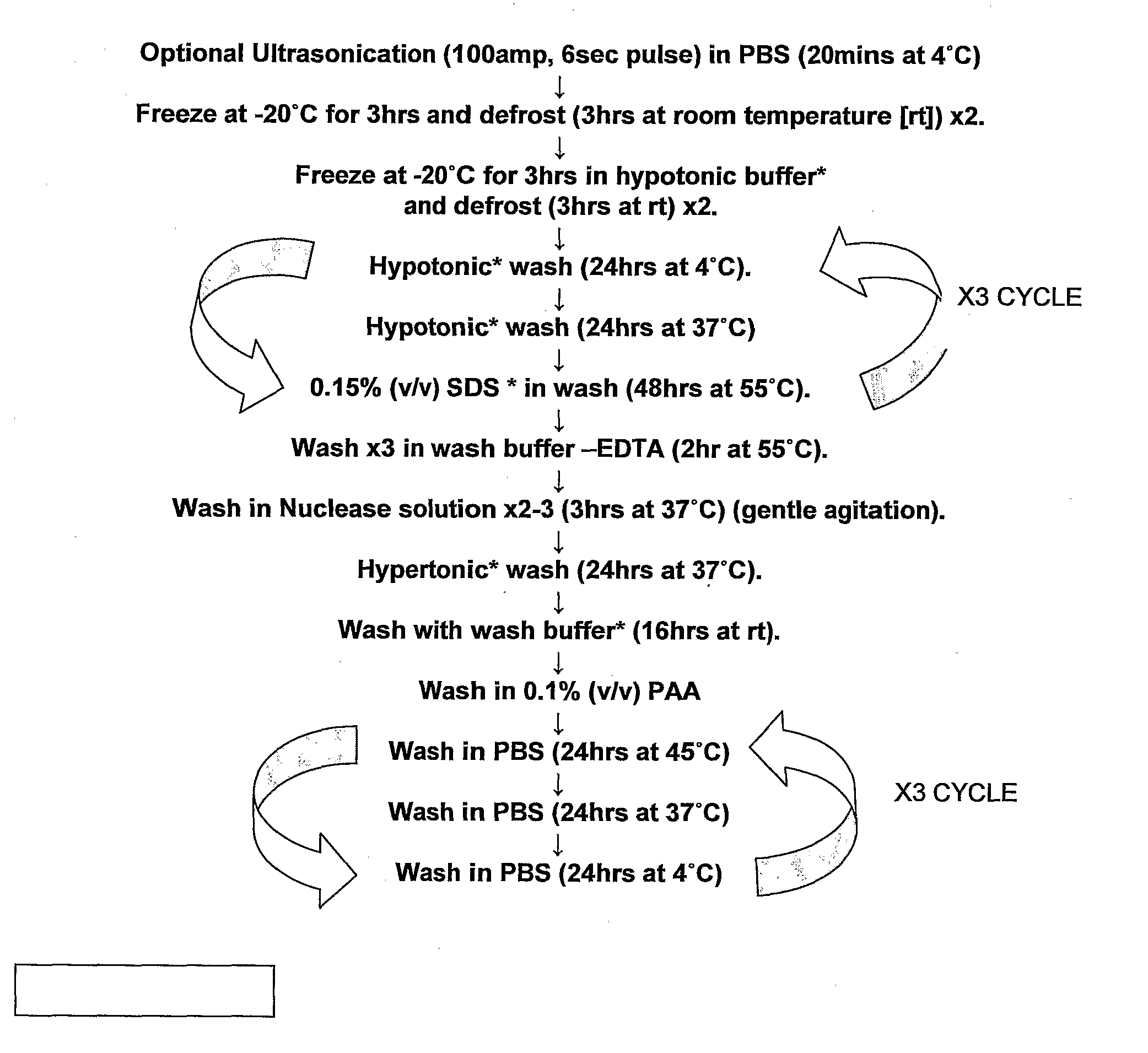
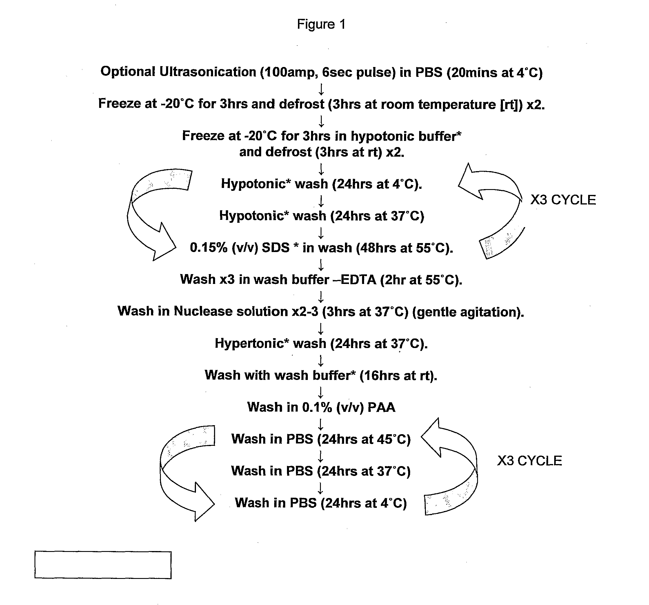
[0085]With reference to FIG. 1 there is shown a typical flow chart of one embodiment of the method of the present invention. With regard to the protocol of FIG. 1 the order of ultrasonication and freeze / thawing may be reversed, however as will be demonstrated hereinafter the steps of ultrasonication, freeze / thawing and treatment with PAA are essential in order to effect total decellularisation of meniscal tissue.

example 2

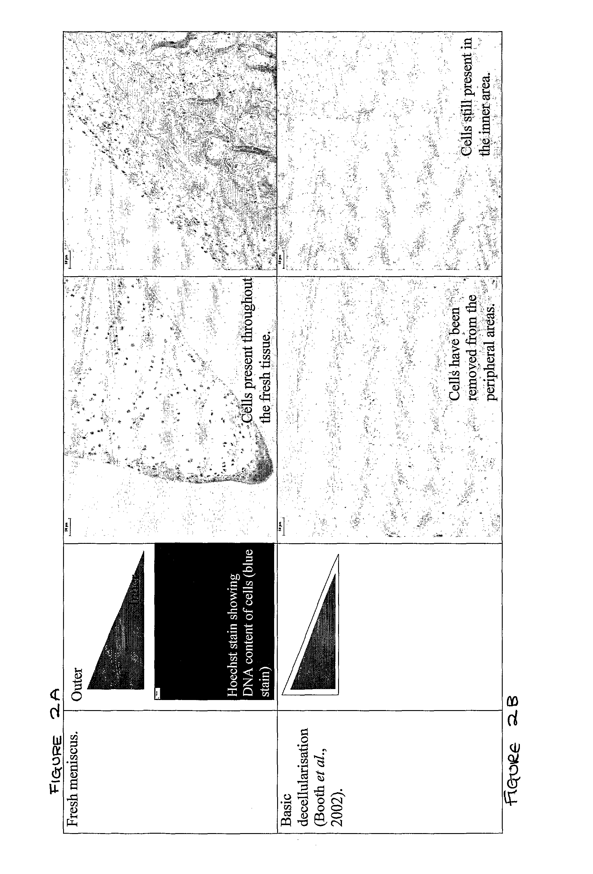
[0086]With reference to FIG. 2A-G, there is shown a schematic representation of the meniscus as a right-angled triangle to approximate the overall cross sectional shape of the meniscus and a Hoechst stain showing the DNA content of cells which is an indication of cell density. In addition histology slides are provided to the right. FIGS. 2A-G shows the DNA content and histology of the medial porcine meniscus after decellularisation using various protocols.

[0087]FIG. 2A shows fresh porcine medial meniscus and the presence of cells throughout the tissue with a uniform distribution of DNA content of cells or cell density from the outer to inner regions of the meniscus.

[0088]The protocol followed for decellularisation of porcine medial meniscus in FIG. 2B is described in Booth, C et al “Tissue engineering of cardiac valve prostheses I: Development and histological characterisation of an acellular porcine scaffold”The Journal of Heart Valve Disease 11, pp. 457-462, (2002). This process d...

example 3

[0090]With reference to FIG. 3A there is shown cell distribution within fresh medial porcine meniscus. A cross-sectional area of the meniscus approximates to a right-angled triangle (see FIGS. 2A-G) and shows that the areas problematic to decellularisation include the outer and central areas of the meniscus, especially around microvascularisation deep within the centrally located tissue. As shown in Example 2A-E the areas which could be decellularised using an incomplete method according to the present invention were the superior and inferior peripheral meniscus and the inner meniscus. With reference to FIG. 3B there is shown a comparable section of a porcine medial meniscus decellularised according to the method of the present invention wherein the meniscus, including the problematic outer and central areas, is completely devoid of cells, in other words the meniscus is completely decellularised and provides a tissue that is suitable for transplantation into a host.


PUM


Abstract

The present invention relates to a method of preparing a tissue matrix and its subsequent use in the replacement and/or repair of a damaged or defective meniscus. The invention also provides meniscal tissue that is substantially decellularised.

Description

[0001]The present invention relates to a method of preparing a tissue matrix, especially a soft tissue matrix, for the replacement and / or repair of a damaged or defective meniscus. The invention also provides a meniscus tissue that is substantially decellularised for subsequent transplantation / implantation.BACKGROUND[0002]The human knee is an important and complex joint comprising three spatially inter-related bones (the femur, tibia and patella), ligaments and cartilaginous structures all of which interact to create a variety of motions. The surfaces of the knee bones within the joint are covered with articular cartilage. This important surface allows the bones to smoothly glide against each other without causing damage to the bone. The meniscus, a C-shaped cartilage cushion sits between the articular cartilage surfaces of the bone and acts as a shock absorber by distributing weight and so improves the overall stability of the knee joint. Each knee joint has a medial and lateral me...

Claims


Application Information

Patent Timeline
IPC IPC(8): A61F2/38A01N1/00C12N5/071
CPCA61F2/3872A61L2430/40A61L27/3683A61L27/3612A61P19/02A61L2430/06A61L27/36
Inventor INGHAM, EILEENFISHER, JOHNSTAPLETON, THOMASINGRAM, JOANNE
Owner TISSUE REGENIX
Why Eureka
Try Eureka