
Ultrasoft atomic force microscopy device and method

a microscopy and ultrasonic technology, applied in the field ofatomic force microscopy, can solve the problems of sample damage, ineffective feedback technique for very fragile samples, and the limitations of afm, and achieve the effect of accurately and directly measuring vibration and deflection, low mass and low mass

Inactive Publication Date: 2010-10-07
PURDUE RES FOUND INC +1
View PDF6 Cites 22 Cited by
  • Summary
  • Abstract
  • Description
  • Claims
  • Application Information

AI Technical Summary

Benefits of technology

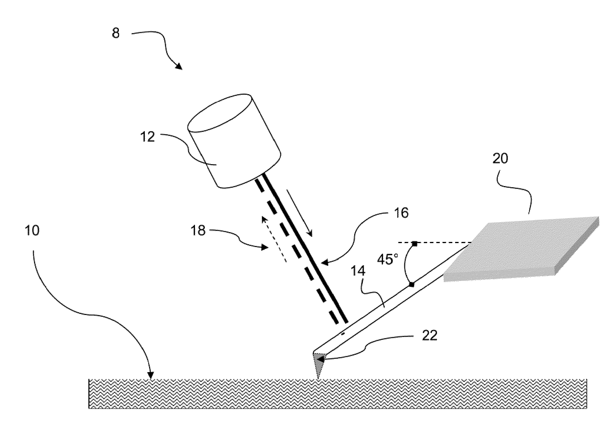
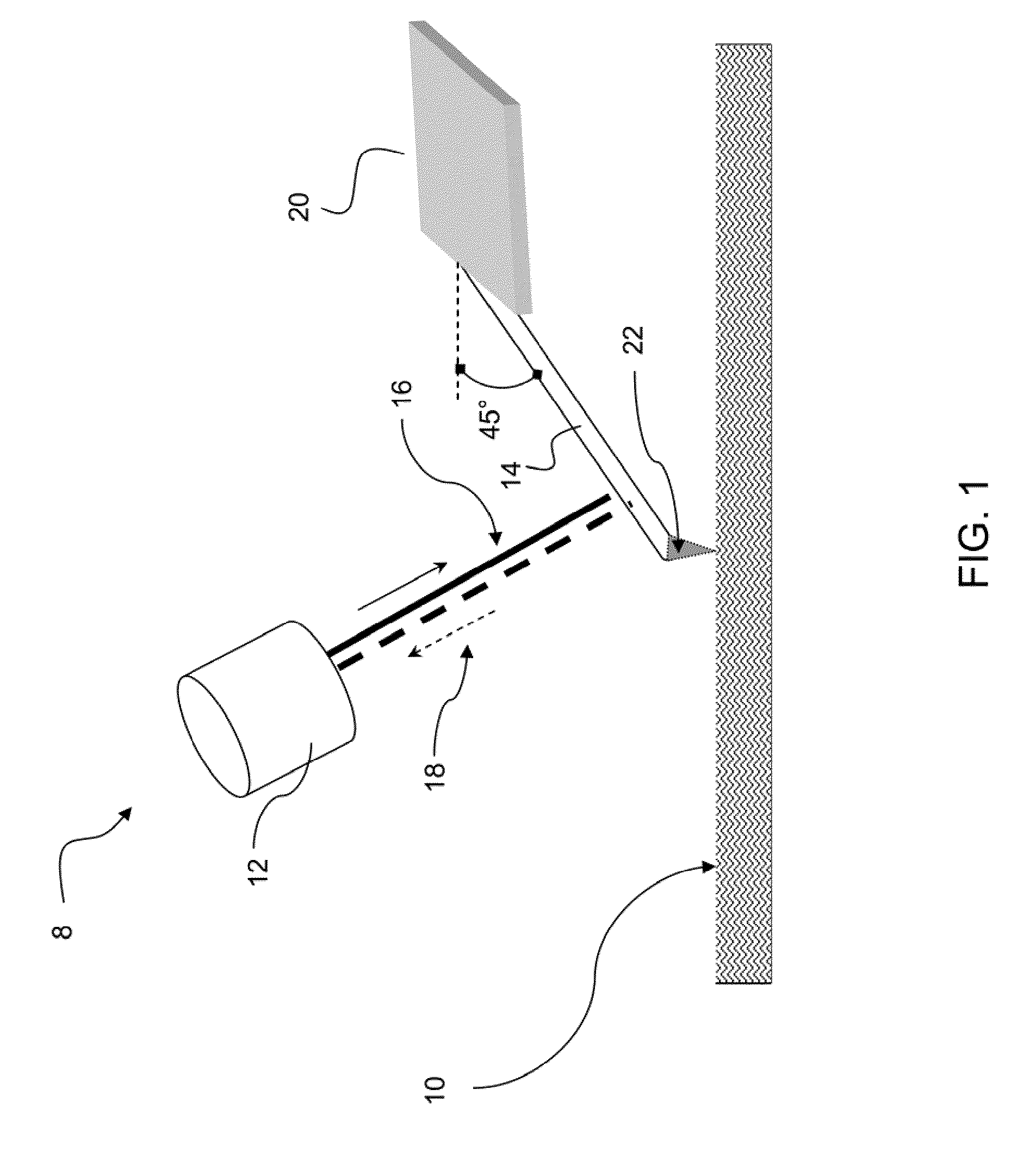
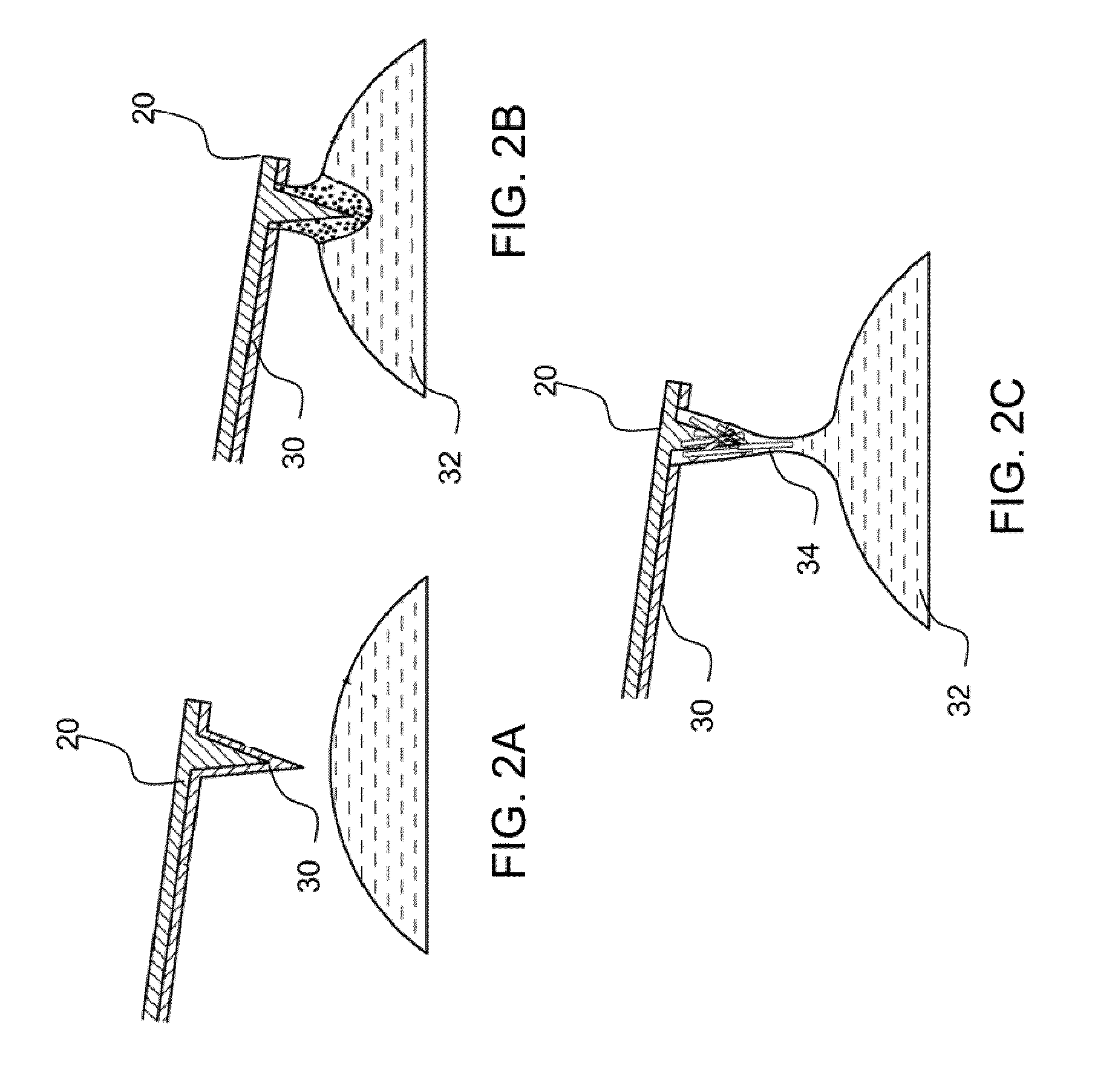
[0007]A preferred embodiment of the invention provides an ultra-soft atomic force microscope device that has a nanoneedle cantilever that terminates in a smaller diameter nanofiber tip. Deflection of the nanoneedle cantilever is detected directly by a laser Doppler vibrometer. The invention simultaneously provides a very low mass nanoneedle cantilever arm with a very small diameter nanofiber tip, while being able to image the vibration and displacement. An AFM device of the invention simultaneously provides a ultra low mass and soft cantilever, the ability to accurately and directly measure vibration and deflection of the very small diameter nanoneedle cantilever with the laser Doppler vibrometer, and a sharp nanofiber tip that provides sub nanometer resolution.

Problems solved by technology

The resolution attained by this type of scanning probe microscopy is orders of magnitude better than the optical diffraction limit, which is a barrier to the resolution of optical based microscopy techniques.
AFM continues to have limits however.
One problem involves sample damage.
However, the feedback technique is not effective for very fragile samples, such as soft biological macromolecule samples in liquids.
Typical AFM forces in the range of 10 picoNewtons (pN) to 1 nanoNewton (nN) can irreversibly damage such soft biomolecules.
However, the imaging forces are still sufficiently large (10 pN to 1 nN) that imaging soft biological macromolecules in their native state under liquids is very difficult.
While atomic force microscopy offers the greatest promise of achieving sub-nm resolution imaging capability, a large gap remains between its potential and the capability of current AFM technology for high-resolution imaging of soft biomolecules in their quasi-native state.

Method used


Image

  • Ultrasoft atomic force microscopy device and method
  • Ultrasoft atomic force microscopy device and method
  • Ultrasoft atomic force microscopy device and method

Examples


Embodiment Construction

[0012]The present invention provides an ultra-soft atomic force microscope device that can provide sub picoNewton forces and simultaneously provide sub nanometer resolution. Applications include high-resolution imaging and materials property characterization with sub-nm resolution of biomolecules in buffer solutions, which offers the oppourtunity to greatly advance understanding of the molecular basis of disease and of drug-cell interactions. The sub-nm resolution imaging is important in the study of biophysics in the realms of individual proteins, DNA, lipid bilayers, and viruses supported on surfaces in liquid environments. Such imaging resolution could provide, for example, high-resolution maps of protein markers expressed on a biological membrane, maps of regions on a protein with specific affinity to drug molecules or high resolution material property maps of viruses in quasi-native state.

[0013]A preferred embodiment of the invention provides an ultra-soft atomic force microsco...


PUM


Abstract

A preferred embodiment of the invention provides an ultra-soft atomic force microscope device that has a nanoneedle cantilever that terminates in a smaller diameter nanofiber tip. Deflection of the nanoneedle cantilever is measured directly by a laser Doppler vibrometer. The invention simultaneously provides a very low mass nanoneedle cantilever arm with a very small diameter nanofiber tip, while being able to image the vibration and displacement. An AFM device of the invention simultaneously provides a ultra low mass and soft cantilever, the ability to accurately and directly measure vibration and deflection of the very small diameter nanoneedle cantilever with the laser Doppler vibrometer, and a sharp nanofiber tip that provides sub nanometer resolution.

Description

PRIORITY CLAIM AND REFERENCE TO RELATED APPLICATION[0001]This application claims priority under 35 U.S.C. §119 from prior provisional application Ser. No. 61 / 153,903, which was filed Feb. 19, 2009.FIELD[0002]A field of the invention is atomic force microscopy. Example preferred applications of the invention include imaging of soft biological samples in liquid. A particular preferred application of the invention is imaging that is capable of resolving individual soft biological macromolecules, e.g., proteins.BACKGROUND[0003]Atomic force microscopy (AFM) uses a very small cantilever arm with a sharp tip, called a probe at its end. The tip can be brought proximate or into contact with a sample, and the deflection of the cantilever provides information about the sample. When a tip is brought proximate a sample, various forces can induce deflection prior to actual contact, e.g., van der Waals forces and electrostatic forces. In a typical imaging mode of operation, the tip can be moved wi...

Claims


Application Information

Patent Timeline
IPC IPC(8): G01Q60/24
CPCB82Y15/00G01Q60/38G01Q20/02B82Y35/00
Inventor REIFENBERGER, RONALDRAMAN, ARVINDBIEDERMANN, LAURA BUTLERYAZDANPANAH, MEHDI M.
Owner PURDUE RES FOUND INC
Learn More