Cancer Marker and Therapeutic Target

a cancer marker and therapeutic target technology, applied in the field of cancer markers and therapeutic targets, can solve the problems of reducing the survival rate of patients, improper treatment, and large number of false positives, and achieve the effects of improving the survival rate, enhancing the specificity, and altering the stability of the targ

Inactive Publication Date: 2010-11-04
CANCER RES TECH LTD
View PDF6 Cites 28 Cited by
  • Summary
  • Abstract
  • Description
  • Claims
  • Application Information

AI Technical Summary

Benefits of technology

[0049]The information may be used to predict whether the solid tumour or the non-haematological cell tumour of the patient will be susceptible to an anti-cancer treatment. This aspect of the invention advantageously permits stratification of cancer patients. This allows an optimal anti-cancer treatment or regime to be identified for a given individual patient.
[0100]Advantageously, the methods of the invention provide a more reliable and more accurate way of identifying, or staging cancer in an individual, or for stratifying individuals for selection of appropriate treatments, particularly in relation to solid tumours, more particularly cervical and oesophageal cancers, but also including cancers selected from the group consisting of bronchial, nasopharyngeal, laryngeal, small cell and non-small cell lung, skin (e.g. melanoma or basal cell carcinoma), brain, pancreatic, neck, lung, kidney, liver, breast, colon, bladder, oesophagus, stomach, cervical, ovarian, germ cell and prostate.
[0102]In alternative embodiments samples may be obtained from patients by other methods well known in the art, including but not limited to, samples of blood, serum, urine, sputum, ascites, intraperitoneal fluids and samples of cells taken by a ‘smear’ test. Blood samples may be taken via venipuncture, (e.g. by vacuum collection tube or syringe,) catheter, cannula, or by finger prick or heel prick as appropriate to the needs of the patient and the amount of blood required. Once a blood sample has been taken it may be treated prior to analysis (e.g. with sodium citrate, EDTA, ethanol or Heparin) for the purposes of preservation or in order to maximise the accuracy and / or reliability of the signal obtained by analysis of the sample.
[0107]The cells and / or liquid collected in a sample taken from a patient may be processed immediately or preserved in a suitable storage medium for later processing. For example, in the case of a cervical smear test the cells are often preserved in an ethanol based storage medium for later processing and analysis. The sample may be treated for the purposes of preservation or for maximising the accuracy and / or reliability of the signal obtained by analysis of the sample. Methods of processing (e.g. centrifugation and / or filtration) may be used to separate a sample into fractions each of which may be tested independently.
[0122]Fluorescence-activated cell-sorting (FACS) is a specialised type of flow cytometry. It provides a method for sorting a heterogeneous mixture of biological cells into two or more containers, one cell at a time, based upon the specific light scattering and fluorescent characteristics of each cell. It is a useful scientific instrument as it provides fast, objective and quantitative recording of fluorescent signals from individual cells as well as physical separation of cells of particular interest.
[0147]The production of recombinant monoclonal antibodies may also involve technologies, referred to as repertoire cloning or phage display / yeast display. These may involve the use of viruses or yeast to create antibodies, rather than mice. These techniques rely on rapid cloning of immunoglobulin gene segments to create libraries of antibodies with slightly different amino acid sequences from which antibodies with desired specificities can be selected. This process can be used to enhance the specificity with which antibodies recognize antigens, alter their stability in various environmental conditions, increase their therapeutic efficacy, and modulate their detectability in diagnostic applications.

Problems solved by technology

Although an abnormal Pap smear indicates possible cervical neoplasia, it is insufficient for diagnosis, which is subsequently carried out by biopsy and additional invasive procedures (‘colposcopy’).
However, due to the high level of HPV infection in the sexually active population (up to 80% infection history), this also results in the identification of a large number of false positives and makes the accuracy of the test dependent on HPV prevalence.
Thus, incorrect staging would lead to improper treatment, and material diminution of patient survivability.
Correct staging, however, can be difficult to achieve.
Pathologic staging, where a pathologist examines sections of tissue, can be particularly problematic for two specific reasons: visual discretion and random sampling of tissue.
Oversight of one cell can mean mis-staging and lead to serious, unexpected spread of cancer.
If cancerous cells present in the lymph node happen not to be present in the slices of tissue viewed, incorrect staging and improper treatment can result.

Method used


Image

  • Cancer Marker and Therapeutic Target
  • Cancer Marker and Therapeutic Target
  • Cancer Marker and Therapeutic Target

Examples


examples

Cervical Tissue Samples and Oesophageal Specimens

[0254]For the mRNA studies, fifteen tumour biopsies from patients with cervical cancer (11 squamous cell carcinoma, S1-S11, and 4 adenocarcinomas, A1-A4) and 14 samples of non-neoplastic cervical tissue (N1-N14) were obtained during surgery and snap-frozen in liquid nitrogen. Diagnosis was made by the pathology department of Barts and The London NHS Trust. Patient samples were divided according to the FIGO classification (stage I, II, III, IV) and tumour biopsies were classified according to increasing grade of nuclear atypia (1, 2, 3) or as well, moderately, or poor differentiation.

[0255]For immunohistochemistry, paraffin embedded samples (n=166) from 150 different patients were obtained from Barts and The London NHS Trust and the Clinical Centre of Serbia, Belgrade. Access to fresh and paraffin-embedded human samples satisfied the requirements of the East London and City Health Authority Research Ethics Subcommittee (LREC no. T / 02 / 0...

experiment 1

Protection Assay for Chemokine Receptors

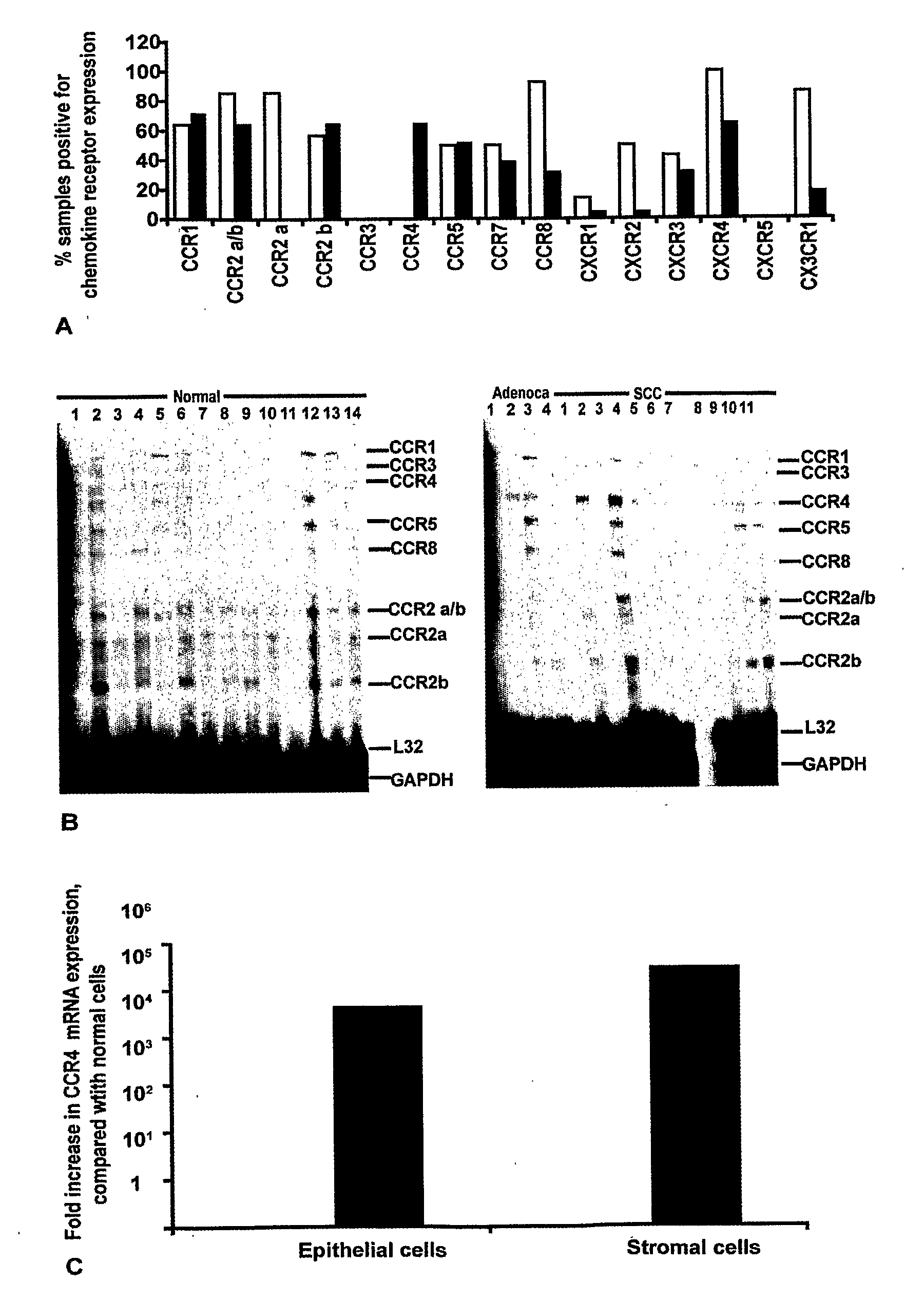
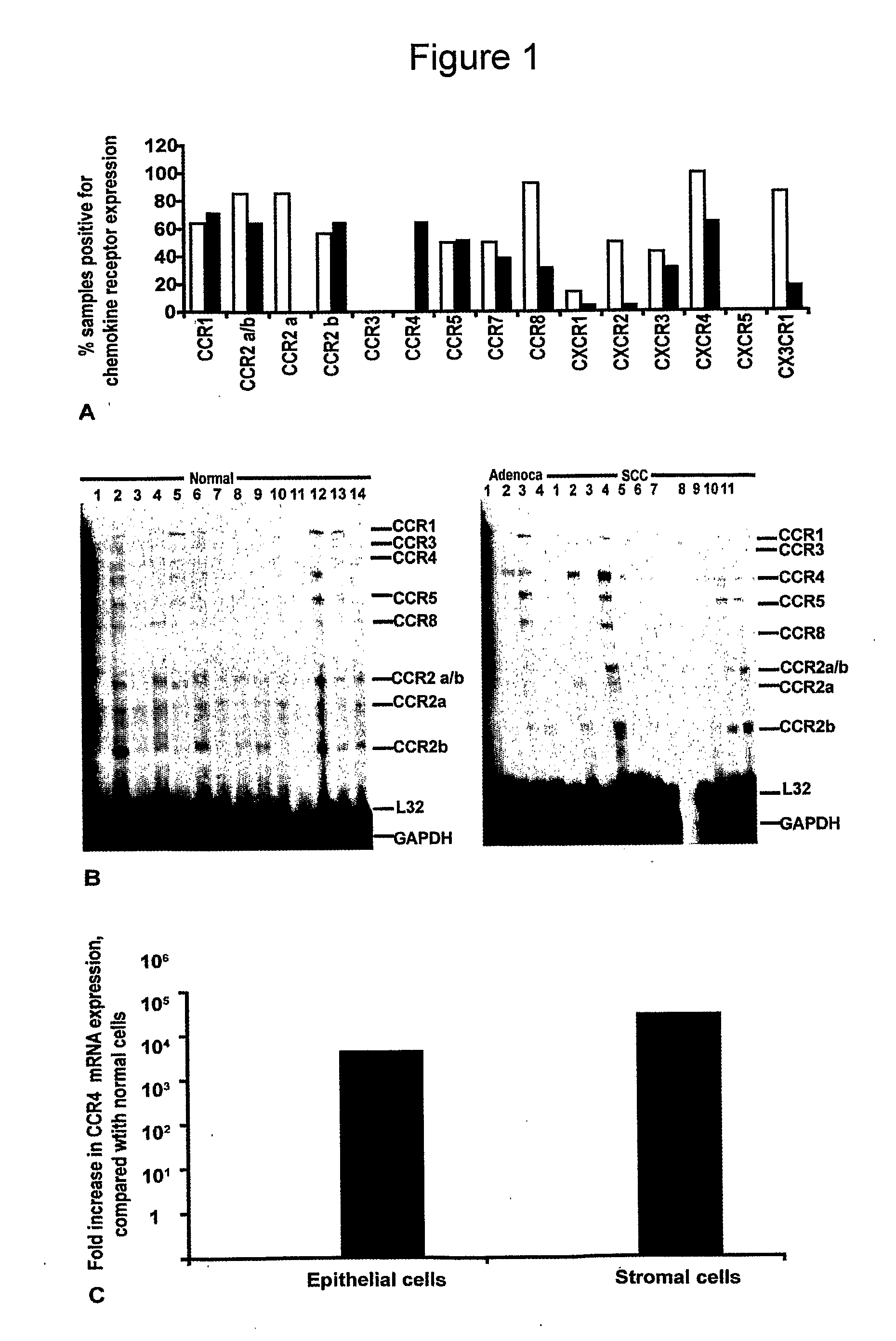
[0265]Ribonuclease protection assays (RPA) were used to screen for 13 chemokine receptor mRNAs in fresh-frozen biopsies of human cervical tissue. As can be seen from the summary graph in FIG. 1A, a range of chemokine receptor mRNAs was found in cervical tissue extracts with some discrete differences between the non-neoplastic and the malignant biopsies. Of particular interest was the chemokine receptor CCR4, which was present in the malignant cervix but not in extracts from non-neoplastic cervical biopsies (FIG. 1B).

[0266]As chemokine receptor expression was examined on whole tissue extracts containing a mixed population of stromal cells and epithelial cells, we next investigated the cellular source of the CCR4 mRNA. mRNA was extracted from laser microdissected stromal and epithelial cell areas of normal and malignant cervical biopsies, and semi-quantitative Real Time RT-PCR was used to analyse CCR4 expression with 18S rRNA as a control. As sh...

experiment 2

is Found on Infiltrating Leucocytes in Human Cervical Biopsies.

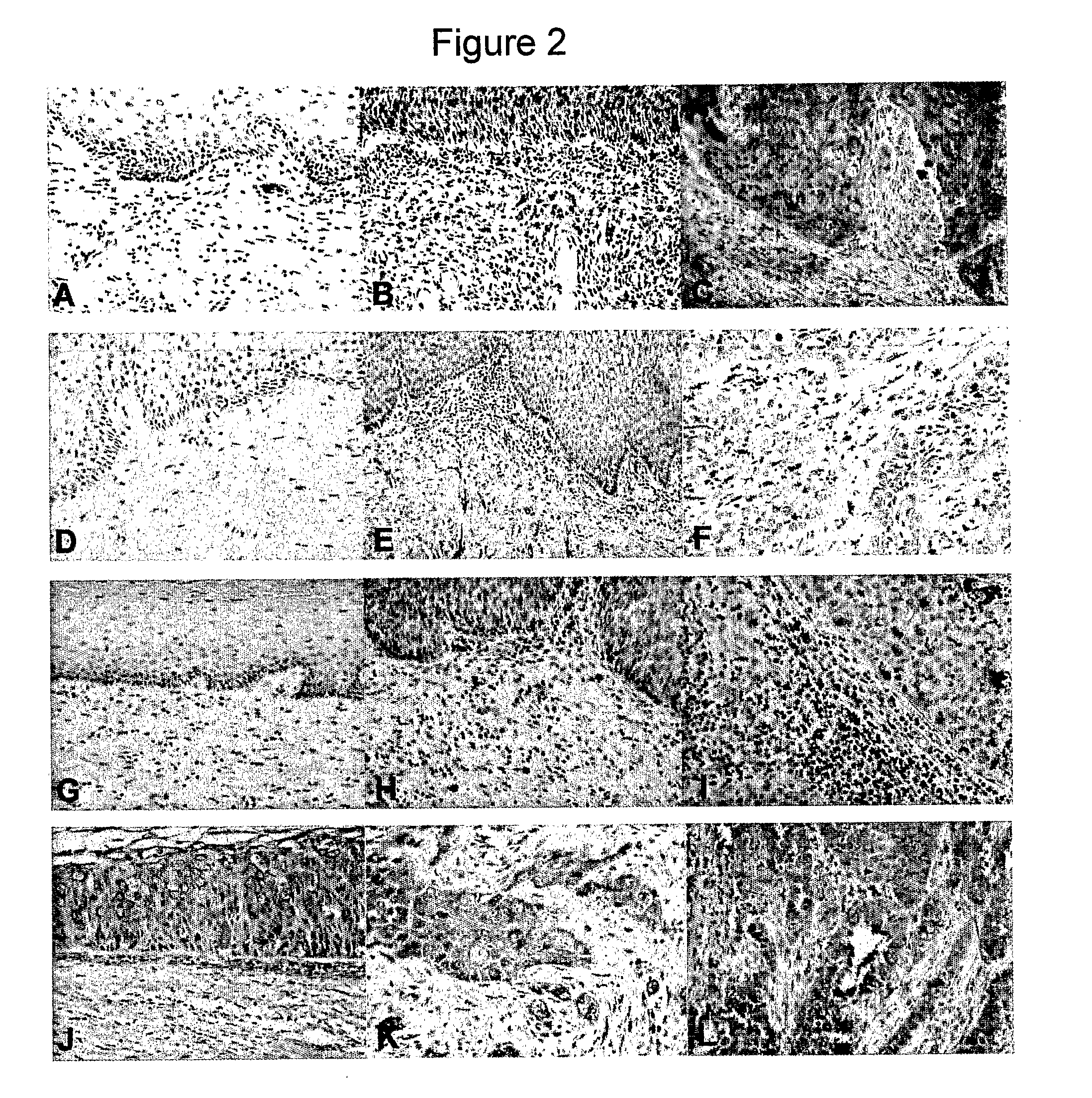
[0268]As shown in FIG. 2 (A-C), leucocytes in the stromal areas of the biopsies stained positive for CCR4. The IHC score for CCR4 positivity in the stromal areas is summarised in FIG. 3A (white bars). The non-neoplastic tissues were negative for CCR4 leucocytes (FIGS. 2A, 3A). There were more CCR4 expressing stromal cells in the CIN samples (FIG. 2 B, 3A) and this increased further in the invasive neoplastic cervical samples (FIG. 2 C, 3A). The intensity of CCR4 expression on the infiltrating leucocytes also increased with malignant progression. In non-neoplastic tissues this was mild; intensity was moderate to strong in CIN and adenocarcinomas, and intensity was strong in invasive SCC, recurrent tumours and lymph node metastases (FIG. 8).

[0269]The stroma consists of various cell types, and tests were carried out to ascertain which of the infiltrating cells contributed to CCR4 expression. As macrophages and Treg cells ex...


PUM

PropertyMeasurementUnit
amino-acid sequenceaaaaaaaaaa
sizeaaaaaaaaaa
frequencyaaaaaaaaaa

Abstract

Chemokine receptor CCR4 and its ligands CCL1 7 and CCL22 are used as markers for the identification and / or staging of cancer. The level of CCR4, CCL17 and CCL22 are found to increase during malignant tumour progression. CCR4, CCL17 and CCL22 are used as markers for the stratification of cancer patients according to their suitability for treatment with anti-cancer agents. Information of diagnostic character is provided by measuring the level of one or more of CCR4, CCL 17 and CCL22 present in a patient sample. Methods of treatment of cancer patients which agents that modulate the activity of CCR4, CCL17 and CCL22. Methods of screening for agents which modulate the biological activities of CCR4, CCL 17 and CCL22 provide anti-cancer agents.

Description

FIELD OF THE INVENTION[0001]The invention relates to oncology and methods of cancer diagnosis, stratification, disease staging and treatment. The field of the invention therefore concerns markers of predictive or clinical value in cancer diagnosis and treatment and the use of medicaments for the treatment of cancer. The invention also concerns screening assays for identifying active anti-cancer agents.BACKGROUND TO THE INVENTION[0002]Chemokine receptors and their ligands direct the trafficking of cells in normal tissue homeostasis and in disease, influencing cell motility, invasiveness and survival [1]. In inflammation and in cancer, chemokines in the diseased tissues contribute to the rolling, tethering and invasion of leucocytes from the blood vessels through the endothelial cell basement membrane and into the parenchyma [2].[0003]CCR4 is one of 18 known chemokine receptors. Chemokine receptors are generally expressed on immune cells and in the tumour microenvironment a number of ...

Claims


Application Information

Patent Timeline
Patent Type & Authority Applications(United States)
IPC IPC(8): A61K39/395G01N33/574G01N33/567A61P35/00
CPCA61K31/00G01N33/5005G01N2800/56G01N2800/52G01N33/574A61P35/00C07K16/2866G01N33/57492G01N33/74G01N2333/726
Inventor BALKWILL, FRANCESSLETTENAAR, VIOLETWILSON, JULIAWANG, YAOHESCHIOPPA, TIZIANA
Owner CANCER RES TECH LTD
Why Eureka
Try Eureka