Using a moving imaging system to monitor anatomical position as a function of time

a moving imaging and time-dependent technology, applied in the field of medical imaging, can solve the problems of affecting the assessment of magnetic resonance-based post-treatment assessment, affecting the accuracy of imaging results, and generating severe magnetic resonance imaging artifacts in the transponder, and achieve the effect of reducing the amount of imaging dos

Inactive Publication Date: 2010-12-16
THE BOARD OF TRUSTEES OF THE LELAND STANFORD JUNIOR UNIV
View PDF9 Cites 37 Cited by
  • Summary
  • Abstract
  • Description
  • Claims
  • Application Information

AI Technical Summary

Benefits of technology

[0006]While real-time 3D tracking of human anatomy using two or more kV x-ray sources is well investigated, techniques of the present invention use the actual radiation treatment beam information (captured using a detector), together with gantry rotation, for spatial and temporal tracking of patient anatomy. Compared to other fluoroscopic tracking systems which require the use of two or more X-ray imaging systems, embodiments of the invention only require the use of one x-ray imaging system for motion monitoring for great majority of the time. When using the MV treatment beam, embodiments of the invention have the potential for allowing full anatomical motion monitoring with nearly zero additional imaging dose to the patient.
[0007]Real-time monitoring of tumor motion during the course of radiotherapy delivery represents a critical step to alleviate the adverse effect of intra-fractional organ motion and to ensure adequate doses to target volumes and safe doses to normal tissues. Accordingly, embodiments of the invention provide an effective means of tracking 3D anatomy of a patient by taking advantage of the inherent mechanism of gantry rotation together with x-ray imaging during radiotherapy.

Problems solved by technology

Movement of internal organs (e.g., prostate) during treatment poses a challenge to this goal.
However, due to its large physical size, the transponder produces severe magnetic resonance imaging artifacts and hinders magnetic resonance-based post-treatment assessments.
Research in radiotherapy has shown that the prostate target moves during the radiotherapy dose delivery process and that the motion is generally unpredictable and can be greater than 1 cm in some cases.
Although continuous fluoroscopic kV and cine-MV imaging is capable of providing real-time information of the prostate position, with spatial accuracy less than 1 mm, continuous or periodic use of such kV imaging to monitor the prostate motion results in unwanted additional radiation dose to the patient.

Method used


Image

  • Using a moving imaging system to monitor anatomical position as a function of time
  • Using a moving imaging system to monitor anatomical position as a function of time
  • Using a moving imaging system to monitor anatomical position as a function of time

Examples


Embodiment Construction

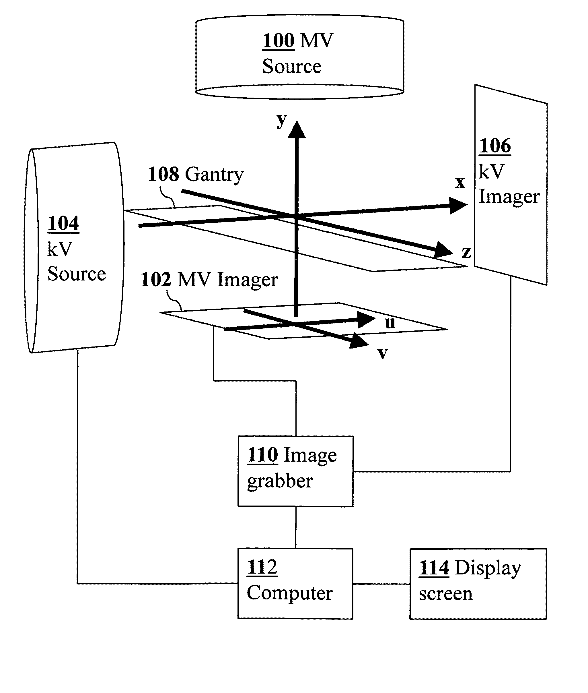
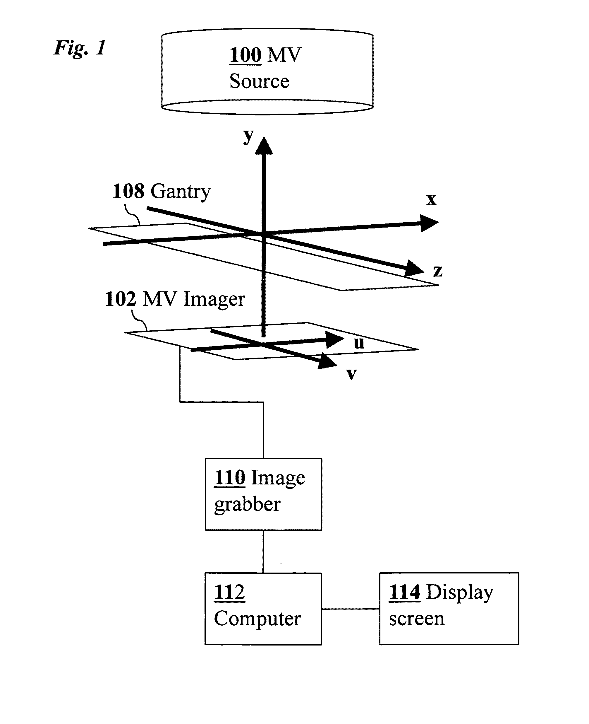
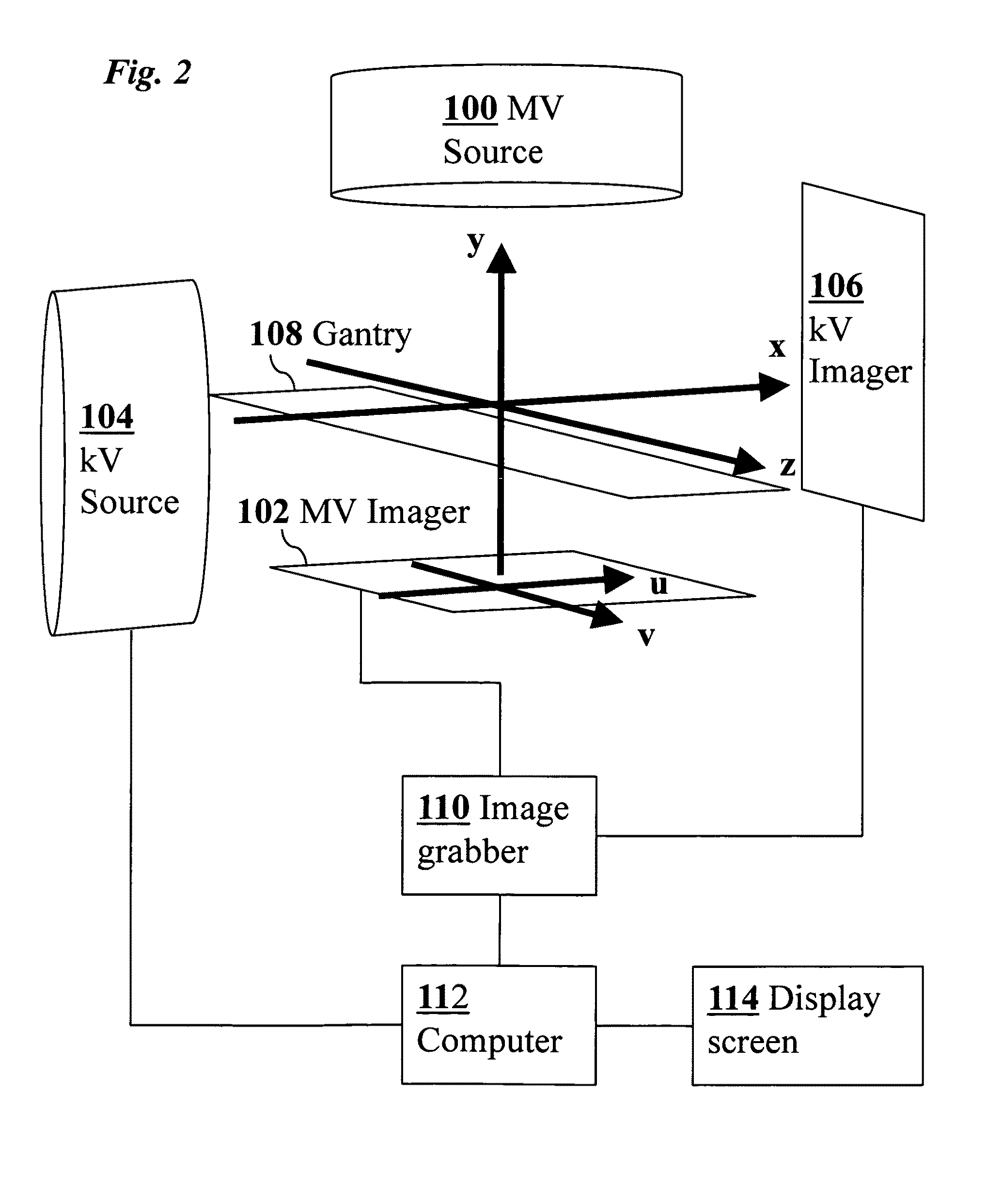
[0030]In a preferred embodiment of the invention, techniques are provided for real-time position monitoring of anatomical position (e.g., prostate) through novel use of cine-megavoltage (MV) imaging, optionally combined with as-needed kilovoltage (kV) imaging. The techniques are especially valuable in the context of modern arc radiotherapy.

[0031]A commercially available radiotherapy system (e.g., Trilogy™, Varian Medical System, Palo Alto, Calif.) may be used to practice one embodiment of the invention. Successive MV treatment beam images were acquired during an arc radiotherapy delivery. Using the present and prior acquired images together with their known angular separation, a computational method was used to determine 3D fiducial locations as a function of time. The fiducial position information was updated at the rate of MV imaging frame rate. Each frame can be used in conjunction with the previous images to reconstruct the 3D fiducial position. From phantom studies the geometri...


PUM


Abstract

Real-time 3D tracking of anatomical positions during radiation therapy uses acquired image data from an MV treatment beam as it is rotated around the patient during arc radiotherapy treatment. The acquired image data and associated angular positions are computationally combined during the arc radiotherapy treatment to estimate in real time 3D positions of anatomical features of the patient, e.g., combining present image data and prior image data at earlier times. Supplementary image data from a kV imaging system may be acquired on an as-needed basis if MV position estimates indicate movement exceeding a predetermined threshold, and the supplementary kV image data combined with the acquired MV image data to improve an accuracy of the estimated 3D positions.

Description

CROSS-REFERENCE TO RELATED APPLICATIONS[0001]This application claims priority from U.S. Provisional Patent Application 61 / 268,883 filed Jun. 16, 2009, which is incorporated herein by reference.FIELD OF THE INVENTION[0002]This invention relates generally to methods and systems for medical imaging. More specifically, it relates to improved techniques for determining and tracking anatomical positions as a function of time.BACKGROUND OF THE INVENTION[0003]A goal of x-ray radiotherapy systems is to precisely deliver MV radiation beam to desired locations while minimizing exposure to undesired locations. Movement of internal organs (e.g., prostate) during treatment poses a challenge to this goal. A necessary step in combating the adverse effects of intrafraction prostate motion is real-time monitoring of the target position. Several methods for obtaining the data in real time have been proposed. Prostate tracking using electromagnetic transponders has recently been developed. However, due...

Claims


Application Information

Patent Timeline
Patent Type & Authority Applications(United States)
IPC IPC(8): G06K9/00
CPCA61N5/1042A61N5/1047A61N5/1067A61N2005/1054A61N2005/1061G06T2207/10112G06T2207/30081G06T7/55G06T7/246
Inventor LIU, WUWIERSMA, RODNEY D.RAIZ, NADEEMWANG, JINGXING, LEI
Owner THE BOARD OF TRUSTEES OF THE LELAND STANFORD JUNIOR UNIV
Why Eureka
Try Eureka