Eureka AIR delivers breakthrough ideas for toughest innovation challenges, trusted by R&D personnel around the world.

Synovial villi for use with tissue engineering

a technology of tissue engineering and villi, which is applied in the field of tissue engineering methods and tools, can solve the problems of further reducing the likelihood of a synvovial hernia or leakage of synovial fluid, and achieves the effects of promoting cartilage, ligament healing, and repairing or preventing further damage to injured or diseased tissu

Active Publication Date: 2011-01-06
JOHNSON LANNY L
View PDF29 Cites 12 Cited by
  • Summary
  • Abstract
  • Description
  • Claims
  • Application Information

AI Technical Summary

Benefits of technology

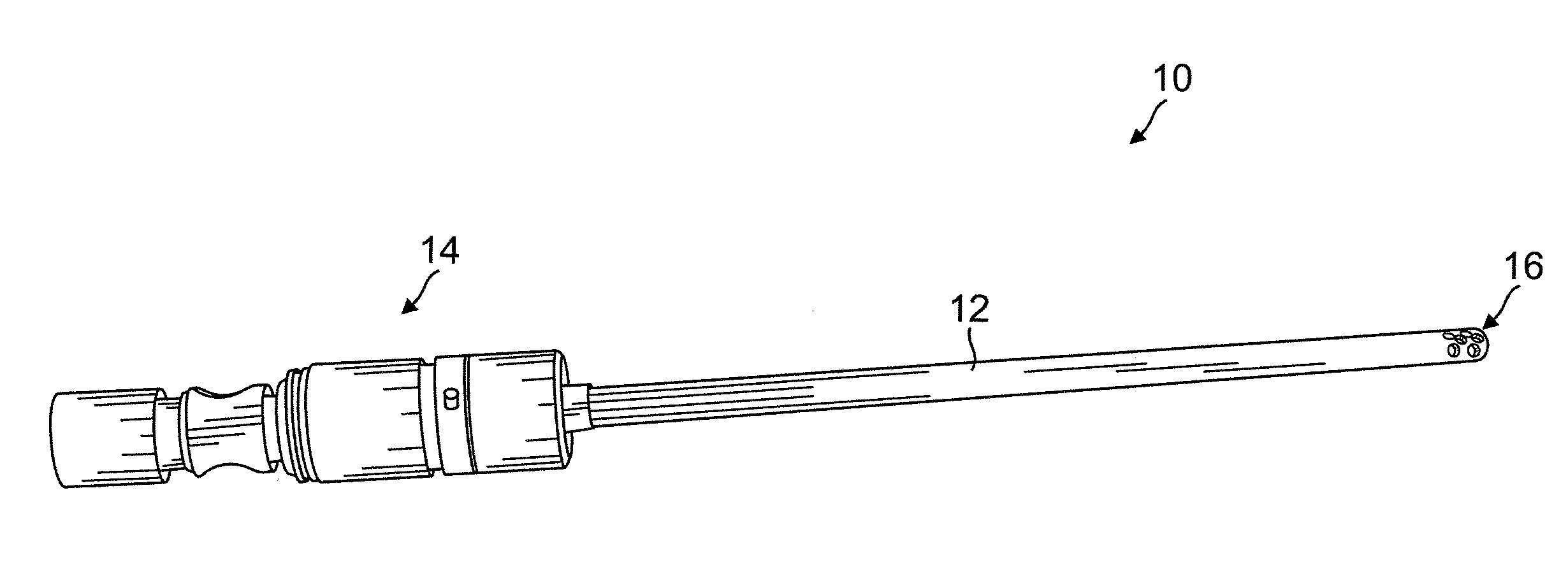
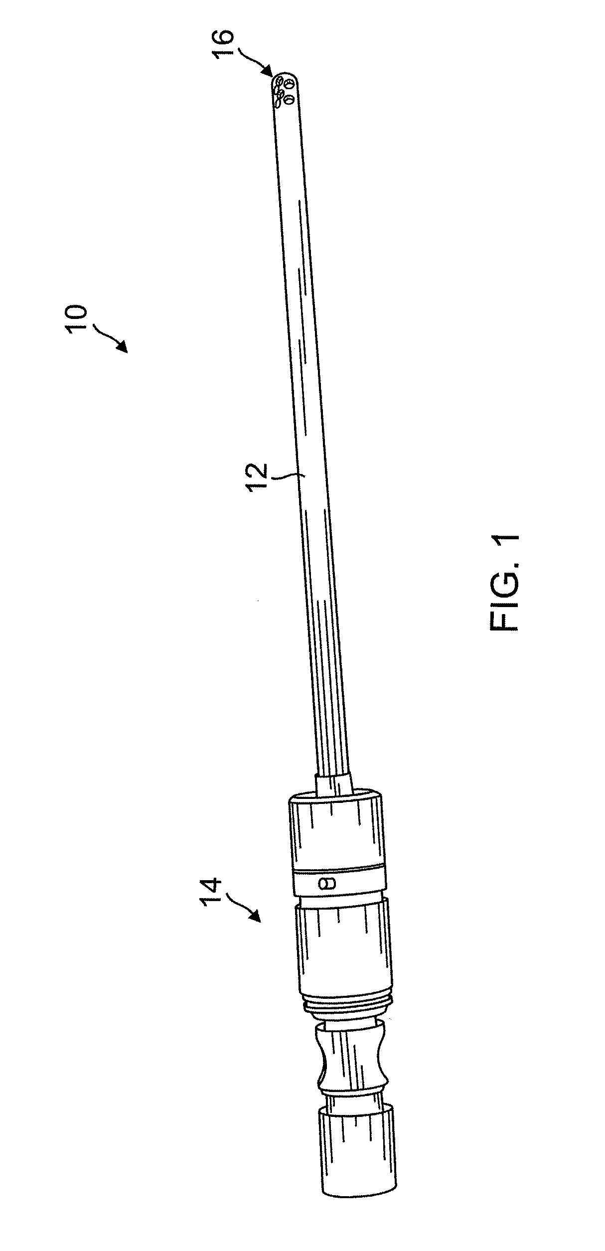
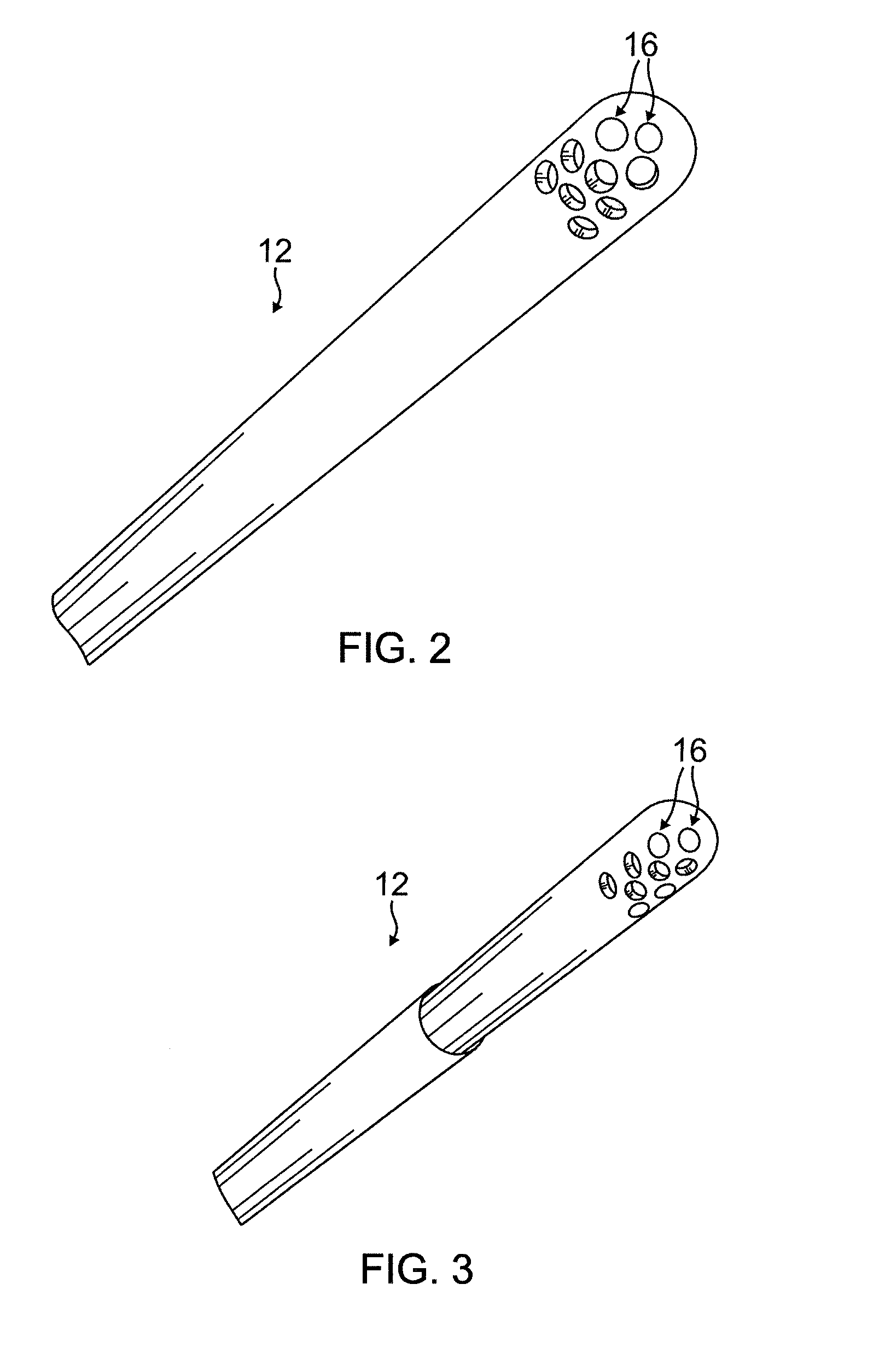
[0010]Harvest is preferably selective to prevent or reduce damage of the base synovial membrane disposed between the synovial villi in the joint. Thus, in addition to targeting the beneficial villi, selective harvest reduces the severity of injury at the harvest site. As such, selectively harvesting synovial villi from the joint prevents compromising the joint capsule integrity, which further reduces the likelihood of a synvovial hernia or leak of synovial fluid post op. In an exemplary embodiment selective harvest is performed using a harvesting tool, which includes a hollow shaft having at one end a plurality of apertures for receiving synovial villi and a cutting means for surgical removal of received villi. Harvest may also include an aspiration means or step where excised or cut portions of synovial villi are collected from an aspirate. In some embodiments the portions of synovial villi are collected in a trap resembling a syringe body. In another embodiment harvesting synovial villi is performed using a harvesting tool including a syringe body engaged to a needle having a bulbous tip. Synovial fluid may also be collected for later culturing or co-administration.
[0014]In some instances the amount of beneficial components, such as beneficial cells or growth factors, found in the synovial villi can be increased by pretreating the patient with one or more compositions that stimulate or increase their production or localization at the synovial Compositions such as IGF-1 and anthocyanins or anthocyanidins and optionally dextrose may increase local concentration of beneficial components.
[0016]The tissue engineering system can be used to treat a variety of damaged tissues such as a sternoclavicular meniscus, an acromioclavicular meniscus, a tempromandibular meniscus, and a radioulnar meniscus. Alternatively, the system may treat a joint labrum, a shoulder labrum or a hip labrum. Treatment methods may be used alone or combined with other procedures such as a microfracture surgery, arthroscopic abrasion surgery or arthroplasty surgery to enhance treatment.

Problems solved by technology

As such, selectively harvesting synovial villi from the joint prevents compromising the joint capsule integrity, which further reduces the likelihood of a synvovial hernia or leak of synovial fluid post op.

Method used


Image

  • Synovial villi for use with tissue engineering
  • Synovial villi for use with tissue engineering
  • Synovial villi for use with tissue engineering

Examples


example 1

Harvesting Synovial Villi from the Knee

[0096]As a pilot study synovial villi are removed from synovium from total knee patient. The synovium is surgically removed and placed in a Ringer's lactate bath. Synovial villi are removed from the synovium using arthroscopic equipment. The collected material is placed in the meniscal and articular cartilage explants for later use.

example 2

Storage of Cells Obtained from Harvested Synovial Villi

[0097]Samples harvested or collected from synovial villi may be frozen for later use or analysis. Fresh freezing medium is prepared by mixing 80% FCS and 20% DMSO. Cryovials are pre-chilled to −70 C in a Nalgene freezing box and cultured cells at a concentration of between 5×10⁵ and 1×10⁶ cells / mL are harvested with trypsin-EDTA. The cell / media material is centrifuged for 5′ at 3500 rpm. The majority of the media is removed by aspiration and the cells are chilled on ice for 1-2′. The final cell pellet is re-suspended in ice-cold freezing medium. The cell solution (0.5 mL per cryovial) is transferred into the pre-chilled cryovials in the freezing box. The freezing box containing the cryovials is placed in a −70 C freezer. Twenty four (24) hours after the cryovials are placed in the freezer, the frozen cells are transferred to liquid nitrogen for long term storage.

example 3

Meniscus Replacement

[0098]After determining that the patient is a suitable candidate for meniscus replacement, the replacement meniscus is prepared. Synovial villi are identified in a joint of the patient and are harvested. They synovial villi are morcellized and placed into a biologically compatible replacement meniscus. The replacement meniscus is made from type 1 collagen. The replacement (containing the cells) is then placed into an appropriate tissue culture medium and maintained under aseptic conditions.

[0099]The patient is prepared for surgery using typical procedures. After the anesthetic is administered and knee examined, a tourniquet is placed on the upper thigh and the thigh is secured to the table in a padded limb holder. The knee and lower leg are cleansed and draped and a diagnostic arthroscopy is performed. The instruments are inserted through three to four 1 cm incisions around the knee. One incision is for sterile saline inflow, used to improve visualization within ...


PUM

PropertyMeasurementUnit
Fractionaaaaaaaaaa
Meniscusaaaaaaaaaa
Biocompatibilityaaaaaaaaaa

Abstract

Synovial villi derived explants and related surgical tools used to repair or prevent damage to injured or diseased tissue, including connective tissue. The tools including a harvesting tool for selectively harvesting synovial villi from the joint of patient and a delivery tool having a bulbous end for delivering the synovial villi derived explants while reducing damage to surrounding tissue.

Description

CROSS REFERENCE TO RELATED APPLICATIONS[0001]This application is a continuation-in-part of U.S. patent application Ser. No. 11 / 210,077 filed on Aug. 23, 2005, which claims benefit of priority to U.S. provisional patent application Ser. Nos. 60 / 622,446 filed on Oct. 27, 2004 and 60 / 607,676 filed on Sep. 7, 2004; the contents of which are herein incorporated by reference in their entiretyTECHNICAL FIELD[0002]The invention relates to methods and tools for the treatment of damaged or diseased tissue by harvesting synovial villi from a patient to form an explant and delivering the explant to the damaged tissue, optionally together with an implantable device.BACKGROUND OF THE INVENTION[0003]There are various types of cartilage, e.g., hyaline cartilage and fibrocartilage. Hyaline cartilage is found at the articular surfaces of bones, e.g., in the joints, and is responsible for providing the smooth gliding motion characteristic of moveable joints. Articular cartilage is firmly attached to t...

Claims


Application Information

Patent Timeline
IPC IPC(8): A61K35/18A61K35/12A61P19/02C12N5/071A61K35/32
CPCA61B10/025A61B17/00491A61B17/1635A61B17/32002A61B2017/00561A61L27/3645A61B2017/320028A61B2017/3225A61K35/32A61L27/3612A61B2017/00969B33Y80/00A61P19/02
Inventor JOHNSON, LANNY L.
Owner JOHNSON LANNY L
Why Eureka
Eureka Blog
Learn More