
ECG Rhythm Advisory Method

a technology of ecg and advisory method, applied in the field of cardiac resuscitation, can solve the problems of unstable vt, life-threatening condition, uncomfortable dizziness, etc., and achieve the effect of accurate analysis of ecg waveform

Active Publication Date: 2011-02-10
ZOLL MEDICAL CORPORATION
View PDF99 Cites 40 Cited by
  • Summary
  • Abstract
  • Description
  • Claims
  • Application Information

AI Technical Summary

Benefits of technology

[0027]These and other implementations may have one or more of the following advantages. The method uses frequency-domain analysis methods for ECG processing / advisory during chest compressions. This method allows ECG analysis without interruption of chest compression and thus significantly reduces the interruption time during chest compressions, leading to an increase in the success rate of resuscitation.
[0028]Some implementations allow for the more complete specification of the ECG waveform spectrum for different cardiac states in a mathematically tractable form that provides improved receiver-operator characteristics (ROC) of the detection algorithm, while reducing the performance burden on the processor.

Problems solved by technology

If there is no pulse associated with this VT rhythm, then the VT is considered to be unstable and a life threatening condition.
Although SVT is rarely life-threatening, the symptoms which include a feeling of a racing heart, fluttering or pounding in the chest or extra heartbeats (palpitations), or dizziness can be uncomfortable.
VF is usually an immediate life threat.
One limitation associated with many AEDs is that current automated ECG rhythm analysis methods cannot function with extra noise due to CPR chest compressions.
Long interruptions of chest compressions have been shown to result in higher failure rate of resuscitation.
For example, the median frequency of the ECG spectrum for almost all patients in ventricular fibrillation decreases initially then increases again after several minutes, making it difficult to use median frequency to predict how long a patient has been in cardiac arrest.
Using amplitudes of the frequency spectrum of the ECG can be limited because the amplitudes are dependent on both the cardiac electrical output as well as position of the ECG lead electrodes on the patient.
One problem with these conventional methods is that CPR changes the assumption behind the methods so that VF and other dangerous rhythms cannot be typically detected during chest compressions.
However, there is usually a significant part of the artifact left in the estimated ECG signal.
Moreover, the adaptive-filter algorithm sometimes has a high computational complexity.
(2002) further suggested that the source of the noise in the signal during chest compressions is the electrode motion and related to the electrode's electrical properties, which makes the relation between the noise and the compression depth more complicated.
Thus, the artifact cannot be sufficiently attenuated for satisfactory results with the conventional advisory algorithm for fibrillation detection.

Method used


Image

  • ECG Rhythm Advisory Method
  • ECG Rhythm Advisory Method
  • ECG Rhythm Advisory Method

Examples


Embodiment Construction

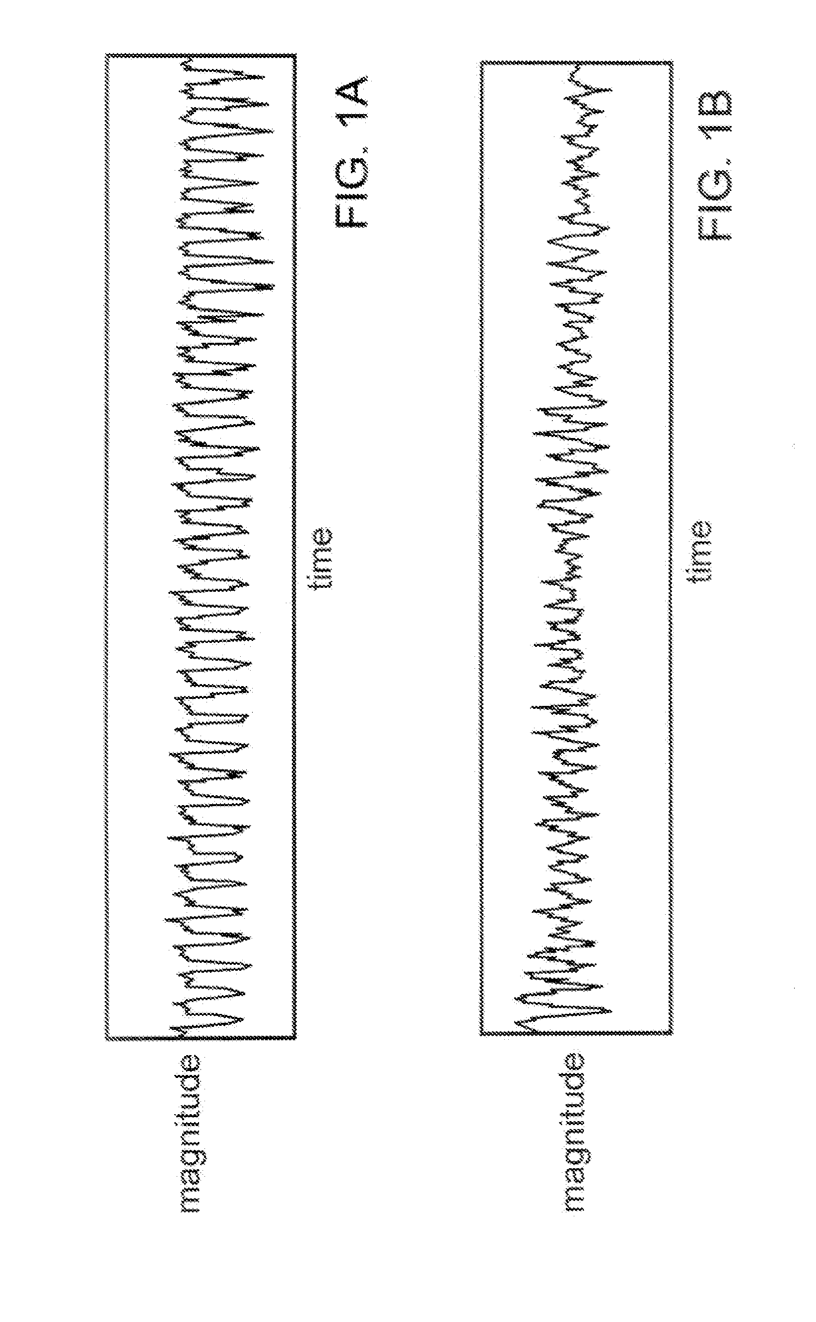
a ventricular tachycardia (VT) rhythm.

[0031]FIG. 1B is a magnitude versus time plot of a ventricular fibrillation (VF) rhythm.

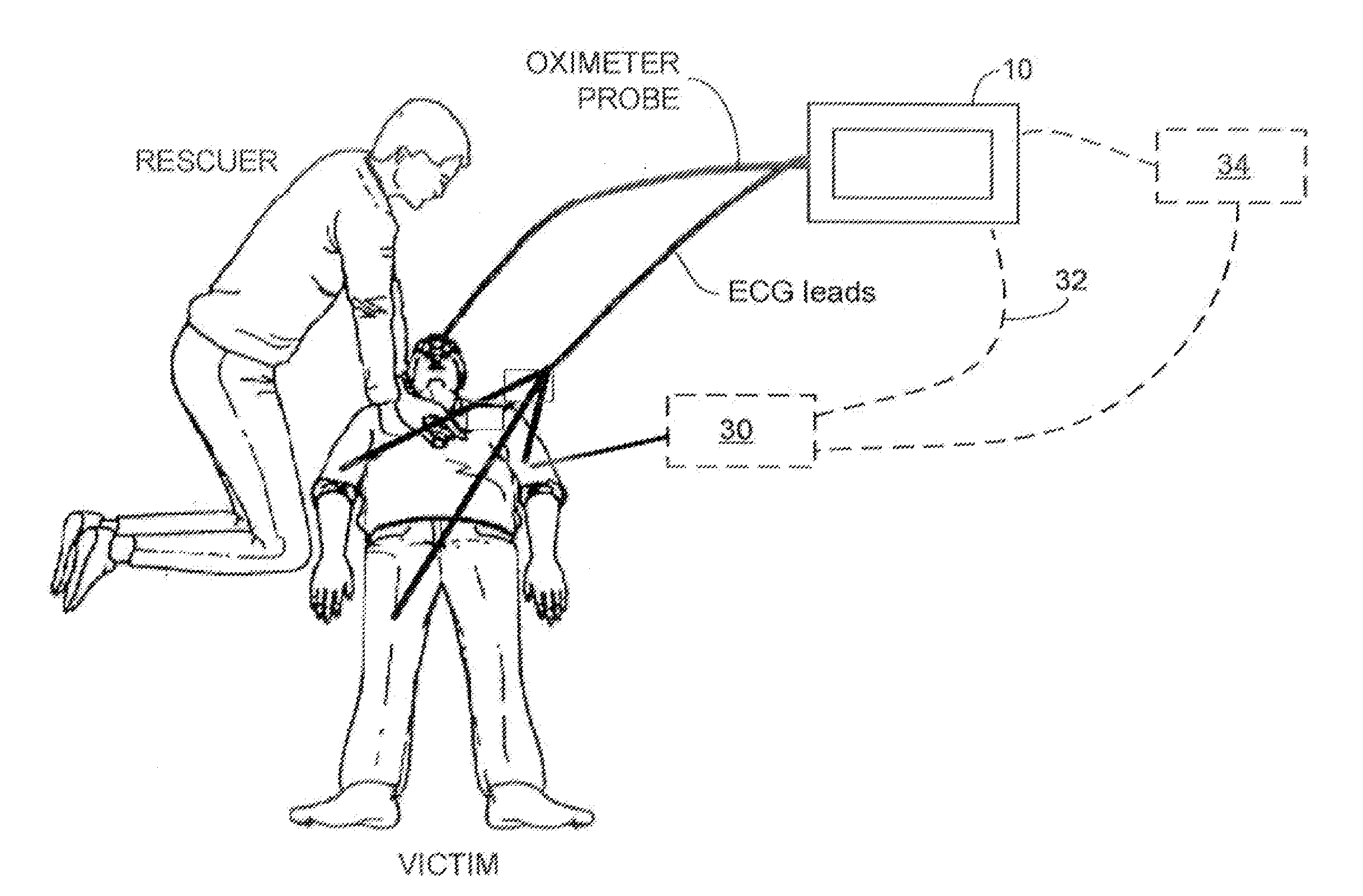
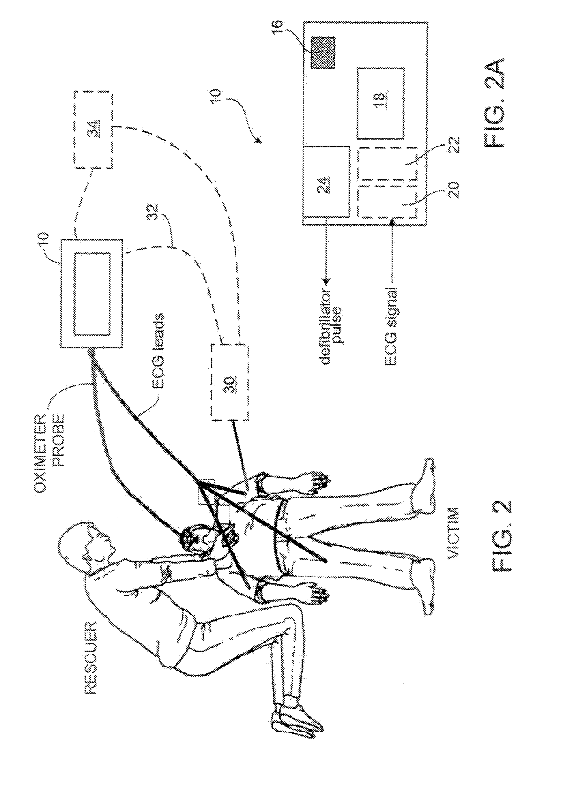
[0032]FIG. 2 is a diagram of one implementation including an automatic electronic defibrillator (AED) and a multiple lead electrocardiograph (ECG) device.

[0033]FIG. 2A is a diagram of the AED of FIG. 2.

[0034]FIG. 3A is an example of a frequency spectrum plot with the energy concentrated within a small frequency range.

[0035]FIG. 3B is an example of a frequency spectrum plot with the energy distributed over a relatively larger frequency range.

[0036]FIG. 4 is an example of an ECG spectrum as a function of time. The magnitude (or energy) of the spectrum is encoded by the grayscale. A darker color corresponds to a higher magnitude.

[0037]FIG. 5 is an EFV score of the signal in FIG. 4 as a function of time.

[0038]FIG. 6 is a flow chart of a process for detecting VF in a patient during chest compressions.

[0039]FIGS. 7A and 7B are examples of an ECG spectrum at two poi...


PUM


Abstract

A method of automatically determining which type of treatment is most appropriate for a cardiac arrest victim, the method comprising transforming one or more time domain electrocardiogram (ECG) signals into a frequency domain representation comprising a plurality of discrete frequency bands, combining the discrete frequency bands into a plurality of analysis bands, wherein there are fewer analysis bands than discrete frequency bands, determining the content of the analysis bands, and determining the type of treatment based on the content of the analysis bands.

Description

CROSS-REFERENCE TO RELATED APPLICATIONS[0001]This application is a continuation application of and claims priority to U.S. application Ser. No. 11 / 430,579, filed on May 9, 2006, which is a continuation application of U.S. application Ser. No. 10 / 844,253, filed on May 12, 2004.TECHNICAL FIELD[0002]This invention relates to the field of cardiac resuscitation, and in particular to devices for assisting rescuers in performing cardio-pulmonary resuscitation (CPR).BACKGROUND[0003]The heart relies on an organized sequence of electrical impulses in order to beat effectively. Any deviation from this normal sequence is known as “arrhythmia.” A class of devices includes signal processing software that analyzes electrocardiography (ECG) signals acquired from the victim to determine when a cardiac arrhythmia such as ventricular fibrillation (VF) or shockable ventricular tachycardia (VT) exists. These devices include automated external defibrillators (AEDs), ECG rhythm classifiers, or ventricular...

Claims


Application Information

Patent Timeline
IPC IPC(8): A61B5/0402A61B5/361A61B5/363A61N1/365A61N1/39
CPCA61B5/0464A61B5/7257A61B5/726A61H31/005A61H2201/5015A61H2230/04A61N1/39A61B5/7207A61B5/04014A61B5/046A61B5/0402A61B5/4839A61N1/3925A61N1/3904A61N1/39044A61B5/363A61B5/361A61B5/347
Inventor TAN, QINGFREEMAN, GARY A.GEHEB, FREDERICKBREWER, JAMES E.
Owner ZOLL MEDICAL CORPORATION
Learn More