Ultrasound garment

Inactive Publication Date: 2011-03-31
VOLUSONICS MEDICAL IMAGING
View PDF4 Cites 55 Cited by
  • Summary
  • Abstract
  • Description
  • Claims
  • Application Information

AI Technical Summary

Benefits of technology

[0097]In some embodiments of the invention, the ultrasound processing unit includes a software module configured to utilize data from at least one of: the at least one sub-array, the at least one array, the plurality of

Problems solved by technology

However, ultrasound imaging suffers from some disadvantages, chief of which are low image quality compared to other imaging modalities, for example Computed Tomography (CT) and Magnetic Resonance Imaging (MRI); and limited volume coverage.
Conventional phased array transducers have a limited field of view due to limitatio

Method used


Image

  • Ultrasound garment
  • Ultrasound garment
  • Ultrasound garment

Examples


Example

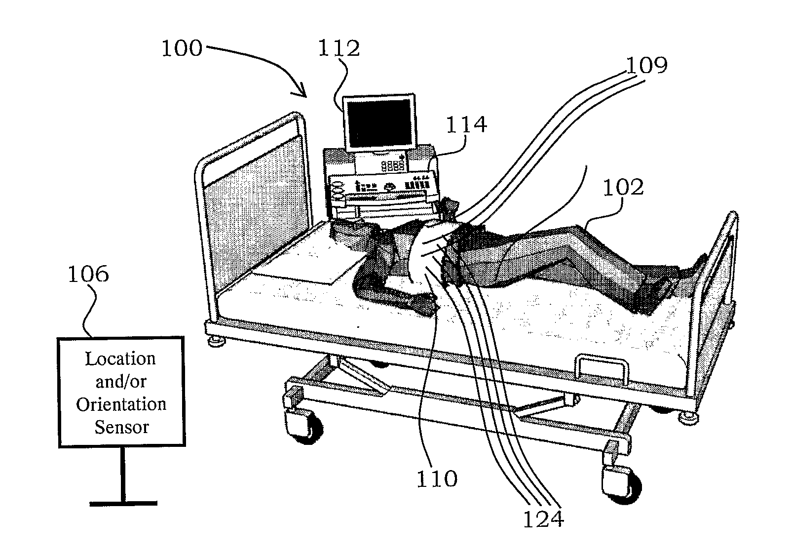
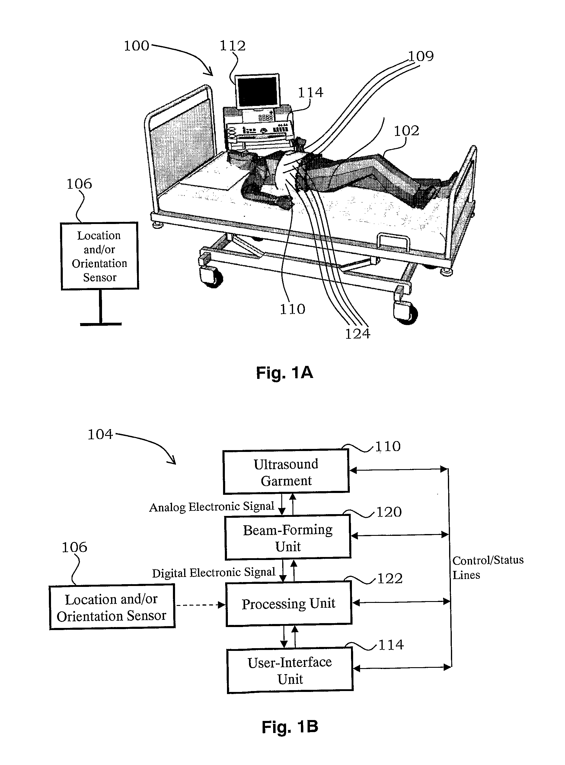
[0130]The present invention, in some embodiments thereof, relates to fixing the position of an ultrasound transducer with respect to a subject and, more particularly, but not exclusively, to fixing the position of an ultrasound transducer within a garment affixed to the subject.

[0131]Before explaining at least one embodiment of the invention in detail, it is to be understood that the invention is not necessarily limited in its application to the details of construction and the arrangement of the components and / or methods set forth in the following description and / or illustrated in the drawings and / or the examples. The invention is capable of other embodiments or of being practiced or carried out in various ways.

[0132]Referring now to the drawings:

Ultrasonic Imaging System

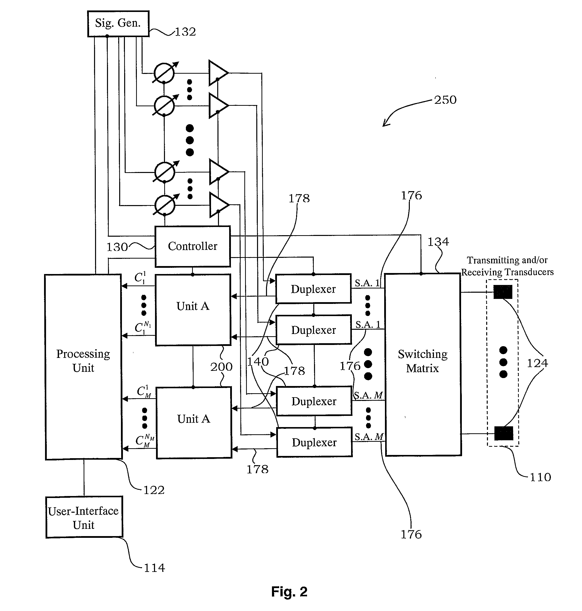
[0133]In FIGS. 2-6, triangles stand for amplifiers or attenuators and crossed-out circles stand for phase shifters and / or true time-delay components. ‘S.A.’ stands for ‘Sub-Array’, ‘Sig. Gen.’ stands for ‘Signal Gen...


PUM


Abstract

Disclosed is an ultrasound assembly. The ultrasound assembly includes a garment configured to be affixed to a portion of a living body, and at least one ultrasound transducer having a fixed position on the garment and configured to provide at least one of: produce and receive, ultrasound signals that pass through the living body. The ultrasound assembly further includes an ultrasound processing unit operatively associated with the at least one ultrasound transducer and configured to process the ultrasound signals following passage through the living body, and an ultrasound operator-interface unit operatively associated with the ultrasound processing unit and configured to provide information with respect to the ultrasound signals following passage through the living body.

Description

RELATED APPLICATION[0001]This application claims priority from U.S. Patent Application Ser. No. 61 / 056,069 filed 27 May 2008, the content of which is hereby incorporated by reference as if fully set forth herein.FIELD AND BACKGROUND OF THE INVENTION[0002]The present invention, in some embodiments thereof, relates to fixing the position of an ultrasound transducer with respect to a subject and, more particularly, but not exclusively, to fixing the position of an ultrasound transducer within a garment affixed to the subject.[0003]Ultrasound is a well matured medical imaging modality. It provides two-dimensional (2D) and / or three-dimensional (3D) anatomic information as well as a plurality of physiological and functional parameters at relatively high refresh rates, reaching an order of 100 frames per second for 2D imaging.[0004]The imaging platforms are portable and reasonably priced. However, ultrasound imaging suffers from some disadvantages, chief of which are low image quality comp...

Claims


Application Information

Patent Timeline
IPC IPC(8): A61B8/14
CPCA61B5/0095A61B8/483A61B5/6805A61B5/6831A61B8/06A61B8/08A61B8/13A61B8/4281A61B8/485A61B8/488A61B8/543A61B8/145A61B8/4416A61B8/4494A61B5/745A61B5/6804
Inventor ZWIRN, GIL
Owner VOLUSONICS MEDICAL IMAGING
Why Eureka
Try Eureka