
Ultrasound-enhanced stenosis therapy

a technology of stenosis and ultrasound, applied in the field of medical devices and methods, can solve the problems of limited long-term effectiveness of angioplasty and stent implantation, far from solving the restless problem, and the method and device for delivering anti-stenotic therapeutic agents to blood vessel wall tissue are as yet not fully satisfactory

Inactive Publication Date: 2011-04-07
CARDIOPROLIFIC INC
View PDF42 Cites 8 Cited by
  • Summary
  • Abstract
  • Description
  • Claims
  • Application Information

AI Technical Summary

Benefits of technology

[0015]Embodiments of the method for treating stenosis or inhibiting restenosis may further include repositioning or moving the ultrasound drug delivery catheter during ultrasound energy activation and a therapeutic agent delivery to further enhance drug delivery.
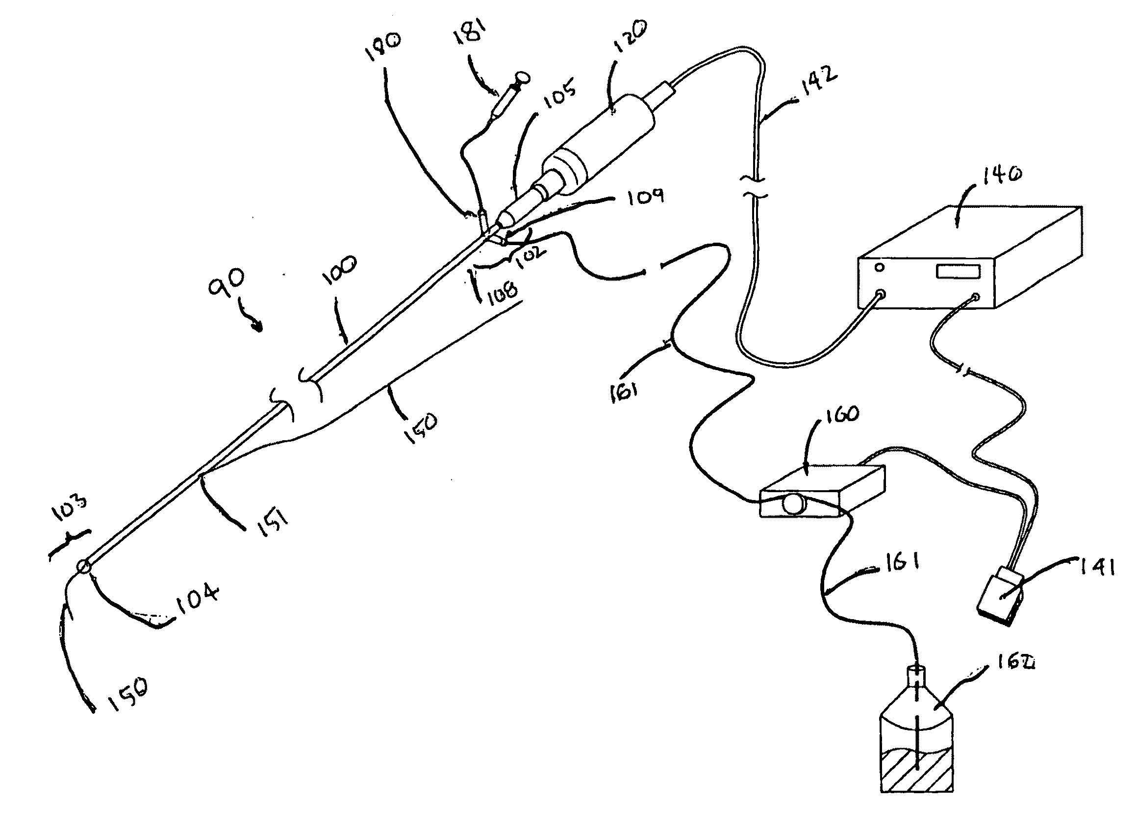
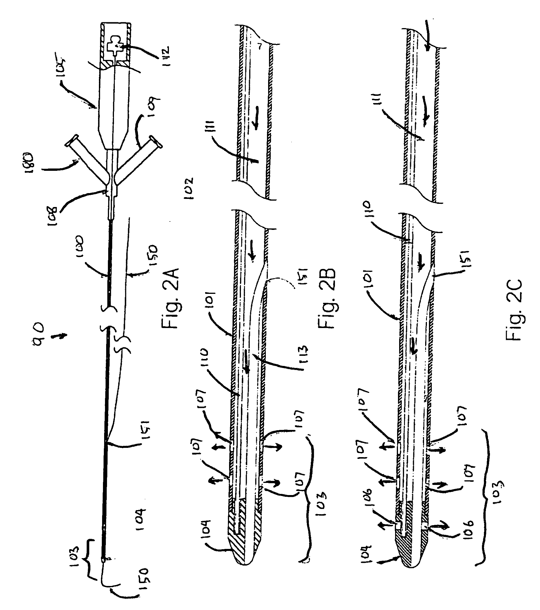
[0021]In another aspect, the present invention provides a method for treating stenosis and inhibiting restenosis in an artery by dilating the artery, delivering a therapeutic agent to the artery, and at the same time enhancing absorption of the therapeutic agent using ultrasound energy. In this aspect, the method may include advancing a distal portion of a combined dilation, ultrasound, drug delivery catheter to an area of stenosis or restenosis in an artery; expanding an arterial dilator of the catheter to dilate the artery at the area of stenosis or restenosis; delivering a stenosis inhibiting therapeutic agent into the artery through the catheter; and activating the catheter to emit ultrasound energy while delivering the therapeutic agent.
[0027]Embodiments of the method of antistenotic treatment include positioning a distal end of a combined ultrasound drug delivery catheter proximate the treatment site in a blood vessel. This positioning of the catheter proximate the site may be accomplished without ablating or removing any plaque material that may be present. Embodiments of the method further include delivering a fluid formulation including a therapeutic agent to the site from the ultrasound drug delivery catheter; and emitting ultrasound energy from the ultrasound catheter while delivering the therapeutic agent. In some embodiments of the method, a dilatormay also be positioned at the treatment site and dilated, such dilation increasing the efficiency and consistency of ultrasound delivery to areas of the internal vessel surface at the treatment site. While, as noted above, some embodiments of the method do not include direct physical or energy delivery attack on plaque, other embodiments may include ablating, removing, or compressing plaque material at the treatment site.
[0029]As noted above, some embodiments may include repeated applications, or multiple applications at the same site, or at another portion of a larger treatment site. Thus, for example, embodiments of the method may include repositioning the ultrasound drug delivery catheter; and repeating the step of emitting ultrasonic energy. Positioning the ultrasound drug delivery catheter at the target site may include positioning the catheter nearby the target site, or it may include contacting the vessel wall at the site. In some embodiments, the contacting may be optimized by dilation of the treatment site, so as to optimize and make uniform a therapeutically effective contact between the ultrasound catheter and the target tissue.

Problems solved by technology

Atherosclerosis and its consequences, including arterial stenosis and hypertension, represent a major health problem both in the U.S. and throughout the world.
Angioplasty and stent implantation, however, often are of limited long term effectiveness due to restenosis.
While these approaches have met with some success, the restenosis problem is far from solved, as drug eluting stents and balloons have had mixed results in clinical studies.
While holding considerable promise, the methods and devices for delivering antistenotic therapeutic agents to blood vessel wall tissue are as yet not fully satisfactory.
Absorption of the therapeutic agent into the blood vessel wall, for example, represents a significant challenge.

Method used


Image

  • Ultrasound-enhanced stenosis therapy
  • Ultrasound-enhanced stenosis therapy
  • Ultrasound-enhanced stenosis therapy

Examples


Embodiment Construction

[0058]The present application provides new methods to improve the treatment of vascular stenosis and re-stenosis using ultrasound technology to enhance delivery of therapeutic agents directly to a targeted therapeutic site, such as a stenotic site on an arterial wall. These methods may be understood as forms of anti-stenosis treatment, which may include treatment of a stenotic site to reduce plaque and to increase the patency of the afflicted vessel, or it may also include treatment of a site previously treated or contemporaneously treated to inhibit or prevent restenosis. Aspects of the invention, including the types of therapeutic agents whose efficacy may be enhanced by the provided technology will be described first in general terms, and then, further below, will be described in the context of FIGS. 1-8.

[0059]The methods described herein employ endovascular sonophoresis and induce vasodilatation, a process that creates micro-indentations in a vessel wall during ultrasound energy...


PUM


Abstract

Apparatus and methods are disclosed for vascular stenosis and vulnerable plaque therapies, including enhancing permeability of the vessel wall using ultrasound energy and delivering a therapeutic agent into the arterial wall. Ultrasound energy may also be delivered at frequencies less than 10 MHz and power less than 20 watts using transcutaneous techniques and endovascular methods. Therapeutic agents may be delivered to the treatment area via intravenous and endovascular methods. In some embodiments, ultrasound energy and therapeutic agent application may be combined with angioplasty techniques and with blood flow protection devices.

Description

CROSS-REFERENCES TO RELATED APPLICATIONS[0001]This application claims priority to U.S. Provisional Application No. 61 / 278,353, of Wallace, filed on Oct. 6, 2009, and is also a continuation-in-part of co-pending application Ser. No. 12 / 807,129, filed Aug. 27, 2010, which is in turn a continuation-in-part of co-pending application Ser. No. 12 / 661,853, filed Mar. 25, 2010.TECHNICAL FIELD OF THE INVENTION[0002]The present invention is related to medical devices and methods. More specifically, the invention is related to ultrasound-enhanced delivery of therapeutic agents to treat vascular stenosis and prevent restenosis following a treatment.BACKGROUND[0003]Atherosclerosis and its consequences, including arterial stenosis and hypertension, represent a major health problem both in the U.S. and throughout the world. A common treatment for arterial stenosis involves balloon angioplasty, more specifically percutaneous transluminal balloon angioplasty (PTA), a procedure in which a balloon cat...

Claims


Application Information

Patent Timeline
IPC IPC(8): A61N7/00
CPCA61B17/22012A61B17/22004
Inventor WALLACE, MICHAEL P.
Owner CARDIOPROLIFIC INC
Learn More