Imaging Mass Spectrometry for Improved Prostrate Cancer Diagnostics

Inactive Publication Date: 2011-06-09
EASTERN VIRGINIA MEDICAL SCHOOL
View PDF7 Cites 6 Cited by
  • Summary
  • Abstract
  • Description
  • Claims
  • Application Information

AI Technical Summary

Benefits of technology

[0006]The invention is directed to detecting and determining prostate health by proteomic profiling. The invention provides biomarkers that have been profiled and characterized from clinical samples such as prostatic tissue. These biomarkers may be used to develop proteomic profiling systems for detection and diagnosis of prostate disease. Compared to a negative diagnosis (e.g., normal or disease-free), the markers are, variously, more frequently detected

Problems solved by technology

However, PSA lacks specificity as a screening tool for prostate cancer, and there is really no lower limit of PSA that entirely excludes cancer (3).
Thus, clinical decision making in PCa places a significant burden upon biopsy results.
Ultrasound guided needle biopsy is the standard for diagnosis however, a negative result does not exclude the presence of cancer.
In practice, false negative results engender a need for repeat biopsies which can delay diagnosis and treatment or unnecessarily subject cancer-free men to repeat biopsies and their attendant anxiety and risk (4, 5).
The heterogeneity of prostate cancer is also a significant problem as the death rates from prostate cancer are relatively low as compared to those from the other major cancers such as lung, pancreas, and colon.
However, a major limitation of this grading system, and a result of aggressive screening procedures, is that a majority of newly diagnosed prostate cancer cases are Gleason grade 6 or 7 tumors.

Method used


Image

  • Imaging Mass Spectrometry for Improved Prostrate Cancer Diagnostics
  • Imaging Mass Spectrometry for Improved Prostrate Cancer Diagnostics
  • Imaging Mass Spectrometry for Improved Prostrate Cancer Diagnostics

Examples


example 1

Identification of Prostate Cancer Tumor-Specific Biomarkers

A. Materials and Methods

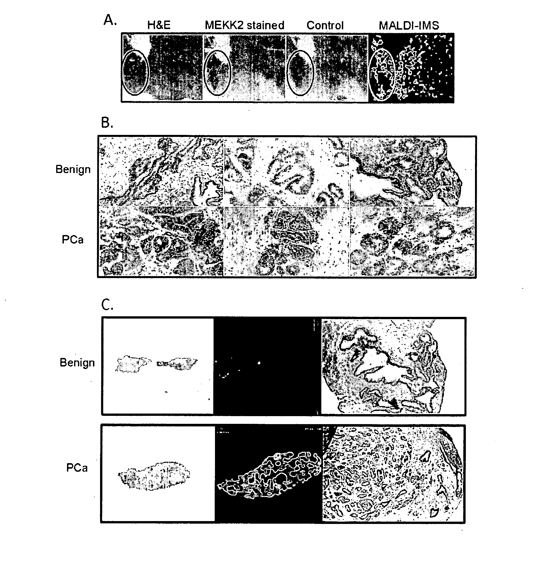
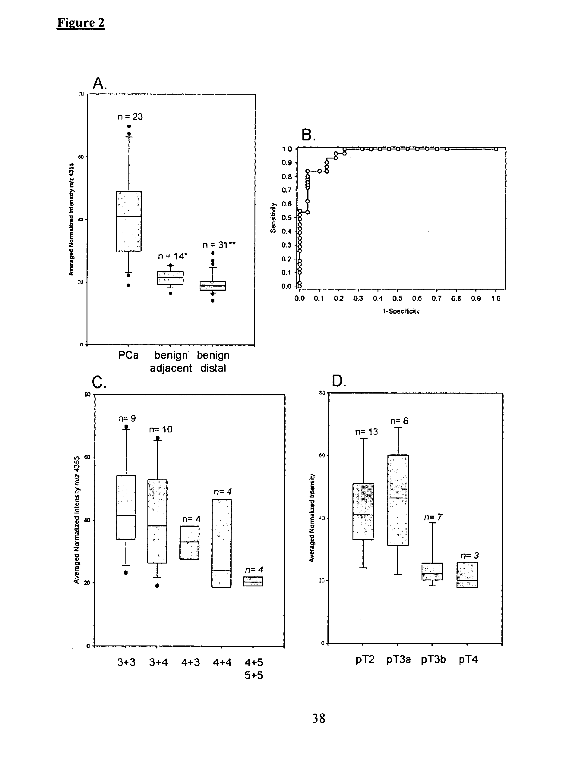
[0061]Patients and Tissue Samples. Patients were consented prior to undergoing radical prostatectomy at Sentara Norfolk General Hospital. Study protocols were approved by the institutional review board at EVMS. The age range of the patients was 46-82 with a mean age of 58.8 years. A total of 75 patients (21 for the discovery set and 54 for the validation set) were recruited for this study. Two cored specimens were harvested from each prostate immediately after removal of the gland. Each core is divided longitudinally to create mirrored cores; one was fixed and paraffin embedded and the other is embedded in OCT (optimal cutting temperature compound, Sakura Finetek USA) and frozen at 80° C. The frozen blocks yielded 41 sections (10 for the discovery set and 31 for the validation set) of benign tissue harvested from prostate tissue distal from the tumor site and 34 sections (11 for the discovery set and ...

example 2

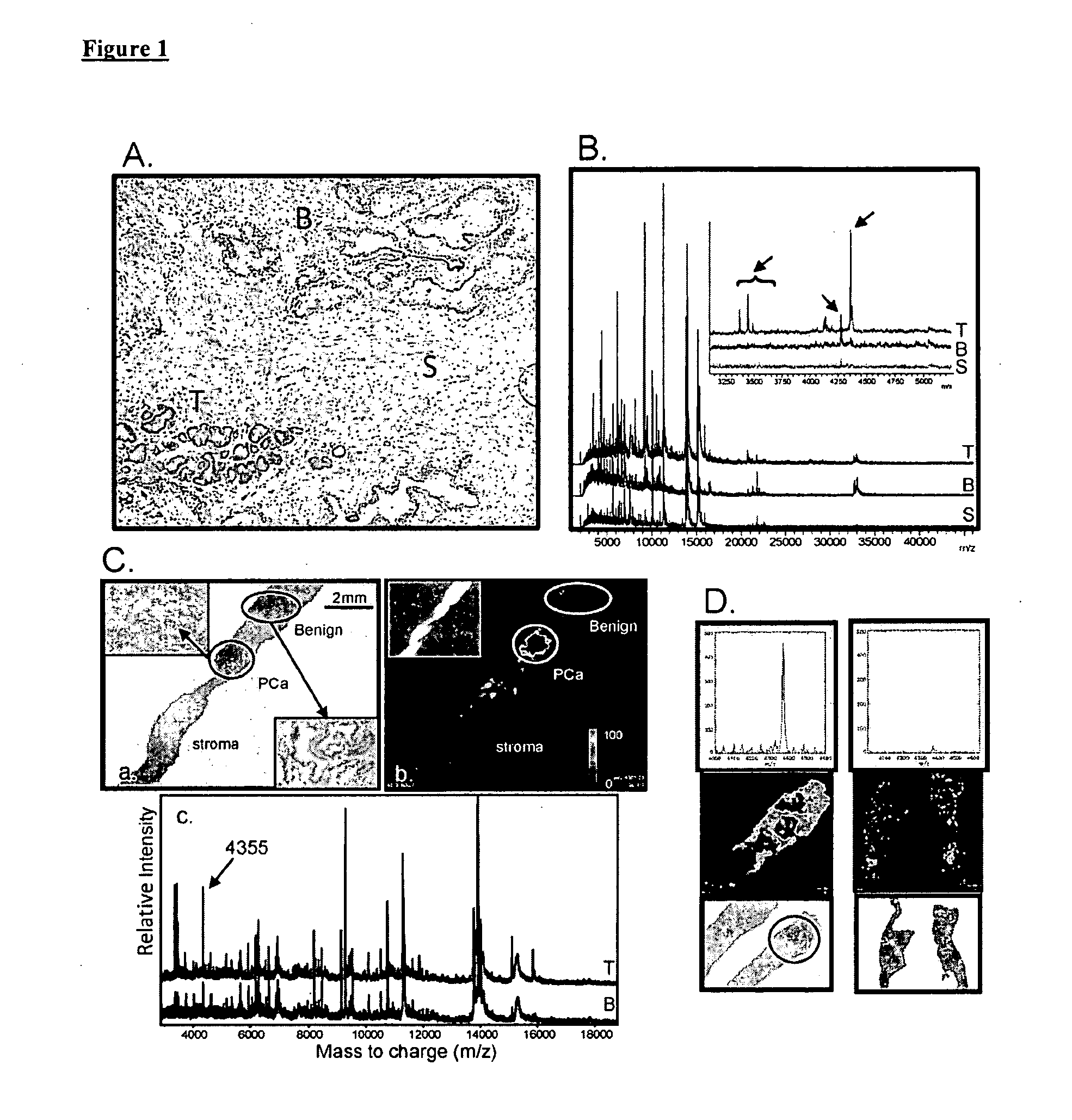
Differential Protein Expression Using MALDI-IMS for the Detection of Metastatic Disease

[0083]Frozen prostate sections of similar stage disease were processed in which the case / control design is the discovery of metastatic disease after surgery in the case group. This study concentrated on differential expression patterns of the tumor tissue regions between case and control. From the examination of eight pairs of case / control samples, a list of top discriminating spectral peaks was generated and is presented in Table 2. Several of the markers were plotted for image generation and the images were subsequently compared to the mirrored histologically stained and pathology-read sections. FIG. 10 shows the ability to detect cancer in tumors associated with distal metastatic disease using expression of select markers.

example 3

Utilization of UMFix Treated Tissue for MALDI-IMS

[0084]The utility of the inventive biomarkers is evident at the biopsy stage. Since frozen sections are not suitable for biopsy-driven diagnostics, a pathology-friendly fixation method known as UMFix is incorporated that is acceptable to clinical histology but also able to preserve protein. The UMFix approach was developed at the University of Miami and is a commercially viable system for tissue preparation in place and is available to pathologists. The UMFix process preserves both IMS detail and information in a fixed tissue for mirrored histology. FIG. 11 is an image of the profile within the region of the 4360 m / z peak utilizing UMFix preserved tissues.

[0085]While the invention has been illustrated and described in the figures and foregoing description, the same is to be considered as illustrative and not restrictive in character, it being understood that only the preferred embodiments have been shown and described and that all cha...


PUM


Abstract

The invention provides biomarkers that can discriminate between prostate cancer and normal tissue as well as identification of associated metastatic disease. One biomarker was identified as a peptide fragment of MEKK2. Methods of diagnosing prostate cancer, including metastatic cancer, by detecting the differential expression of one or more biomarkers are also provided.

Description

RELATED APPLICATION[0001]This application claims the benefit under 35 U.S.C. §119(e) of U.S. Provisional Application No. 61 / 036,837 filed on Mar. 14, 2008, which is hereby incorporated by reference herein in its entirety.STATEMENT REGARDING FEDERALLY SPONSORED RESEARCH[0002]Work described herein was performed with government support under Grant 2 U0 CA085067 awarded by the NIH / NCI Early Detection Research Network. The U.S. Government has certain rights in the invention.BACKGROUND OF THE INVENTION[0003]Prostate cancer (PCa) is one of the most common malignancies in the US (1). It is clinically heterogeneous, with a highly variable natural history (2). The discovery and widespread utilization of serum prostate specific antigen (PSA) monitoring for early detection has greatly changed the way prostate cancer is diagnosed and treated. However, PSA lacks specificity as a screening tool for prostate cancer, and there is really no lower limit of PSA that entirely excludes cancer (3). Thus, ...

Claims


Application Information

Patent Timeline
IPC IPC(8): C12Q1/00
CPCG01N33/57434G01N2800/56G01N2333/9121
Inventor SEMMES, O. JOHNCAZARES, LISA H.DRAKE, RICHARD R.MENDRINOS, SAVVASLANCE, RAYMOND S.
Owner EASTERN VIRGINIA MEDICAL SCHOOL
Why Eureka
Try Eureka