Method and apparatus for cancer screening

a technology of apparatus and cancer, applied in the direction of fluid pressure measurement, liquid/fluent solid measurement, peptide, etc., can solve the problems of long urine preparation time, low detection limit of oncopterin reported in the study, and resistance to blood giving for diagnostic tests, so as to improve the stability and reproducibility of urine pteridine detection, enhance detection, and reduce laser scatter

Active Publication Date: 2011-06-23
UNIVERSITY OF MISSOURI
View PDF4 Cites 7 Cited by
  • Summary
  • Abstract
  • Description
  • Claims
  • Application Information

AI Technical Summary

Benefits of technology

[0016]The present invention is further directed to a capillary electrophoresis lased induced fluorescence (CE-LIF) apparatus. The CE-LIF employs a combination of lenses, filters, and irises to enhance stability and reproducibility of the urine pteridine detection. The lenses, filters, and irises enhance detection by substantially reducing laser scatter from the excitation source and from the capillary, thereby producing a stable, low noise baseline that enables low limits of detection. The CE-LIF apparatus employs a combination of a confocal lens having greater than 10× magnification placed in the range of 45 to 90 degrees from the laser path to collect and focus fluorescence emission from the sample. The CE-LIF apparatus employs at least one physical iris for removing scattered light either from the filtered non-polarized laser or from any fluorescence generated by the device. A device structured with the high magnification confocal lens and iris yields a very low noise baseline, enabling low limits of detection of pteridines in urine and other samples.
[0017]In some embodiments, the inventive apparatus includes 1) a non-polarized excitation source emitting non-polarized laser at a non-traditional solid state laser wavelength. In some embodiments, the inventive apparatus includes 2) a first filtration means for filtering the non-polarized laser. In some embodiments, the inventive apparatus includes 3) a focal lens that focuses the filtered, non-polarized laser onto a capillary, the pathway by which the intended sample is separated and exposed to the filtered, non-polarized laser via a detection window on the capillary. In some embodiments, the inventive apparatus includes 4) a sample holder with a detection window that facilitates the mounting of an electrophoresis capillary. In some embodiments, the inventive apparatus includes 5) a capillary electrophoresis device that is capable of separating the specific compounds of interest (pteridines) within the samples and delivering the sample to the detection window of the capillary mounted within the sample holder.
[0018]In some embodiments, the inventive apparatus includes 6) a confocal lens greater than 10× magnification placed in the range of 45 to 90 degrees from the laser path to collect and focus fluorescence emission from the sample. In some embodiments, the inventive apparatus includes 7) a second filtration means for filtering non-pteridine fluorescence generated by any compound other than those in the pteridine family or for filtering background scattering light from the laser. In some embodiments, the inventive apparatus includes 8) a physical iris for removing any scattering either from the filtered non-polarized laser or from any fluorescence generated by the device. The physical iris controls the light beam size and minimizes the scattered light. In some embodiments, the inventive apparatus includes 9) a detection means for detecting the filtered fluorescence emission generated by the pteridines in the samples. In some embodiments, the inventive apparatus includes 10) a current to voltage (I to V) converter to communicate the raw output from the electrons from the compounds of interest undergoing fluorescent decay to the digital input of the device collecting the output. In some embodiments, the inventive apparatus includes 11) a control means for independently controlling the stations of the laser focal lens, capillary mount, collection lens and detector to prevent any mechanical cross-talk from one stage to the others that may adversely affect the resulting data.

Problems solved by technology

They may also be reluctant to give blood for diagnostic tests.
However, the detection limit for oncopterin reported in the study is relatively high at about 1.43 mol / L.
Moreover, the reported urine preparation time is still long (about 2 hr), since the study utilized full hydrolysis vs. oxidation of the pteridines.
Even though the patented CE-LIF technology has shown promising improvements in feasibility for clinical laboratories, the patented CE-LIF technology has limitation on the accuracy and reproducibility because of the complexity of urine samples.

Method used


Image

  • Method and apparatus for cancer screening
  • Method and apparatus for cancer screening
  • Method and apparatus for cancer screening

Examples


example 1

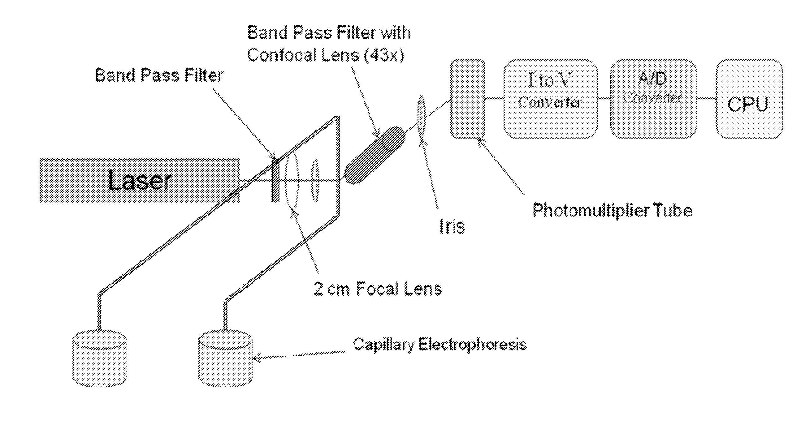
[0084]The following example describes a high performance capillary electrophoresis (HPCE) separation of pteridines in a biological sample with laser-induced fluorescence (LIF) detection method (HPCE-LIF) for quantitative analysis of pteridines in urine samples. HPCE-LIF was used to investigate pteridine level patterns in 38 urine samples from a variety of cancer patients. Some types of cancer were not studied mainly due to the unavailability of urine samples and not the limitation of the technique. In order to assure that the pteridine levels represent the physiological concentration, the amount of pteridines was reported here as a ratio of pteridine to creatinine See Shi H., Ma Y., Ma Y. A Simple and Fast Method to Determine and Quantify Urinary Creatinine, Anal. Chim. Acta., 1995, 312, pp. 79-83.

Materials and Methods.

[0085]Chemicals. Pteridine standard solutions of 6-biopterin, D-(+)-neopterin, pterin, isoxanthopterin, xanthopterin pteridine were purchased from Sigma-Aldrich (St. ...

example 2

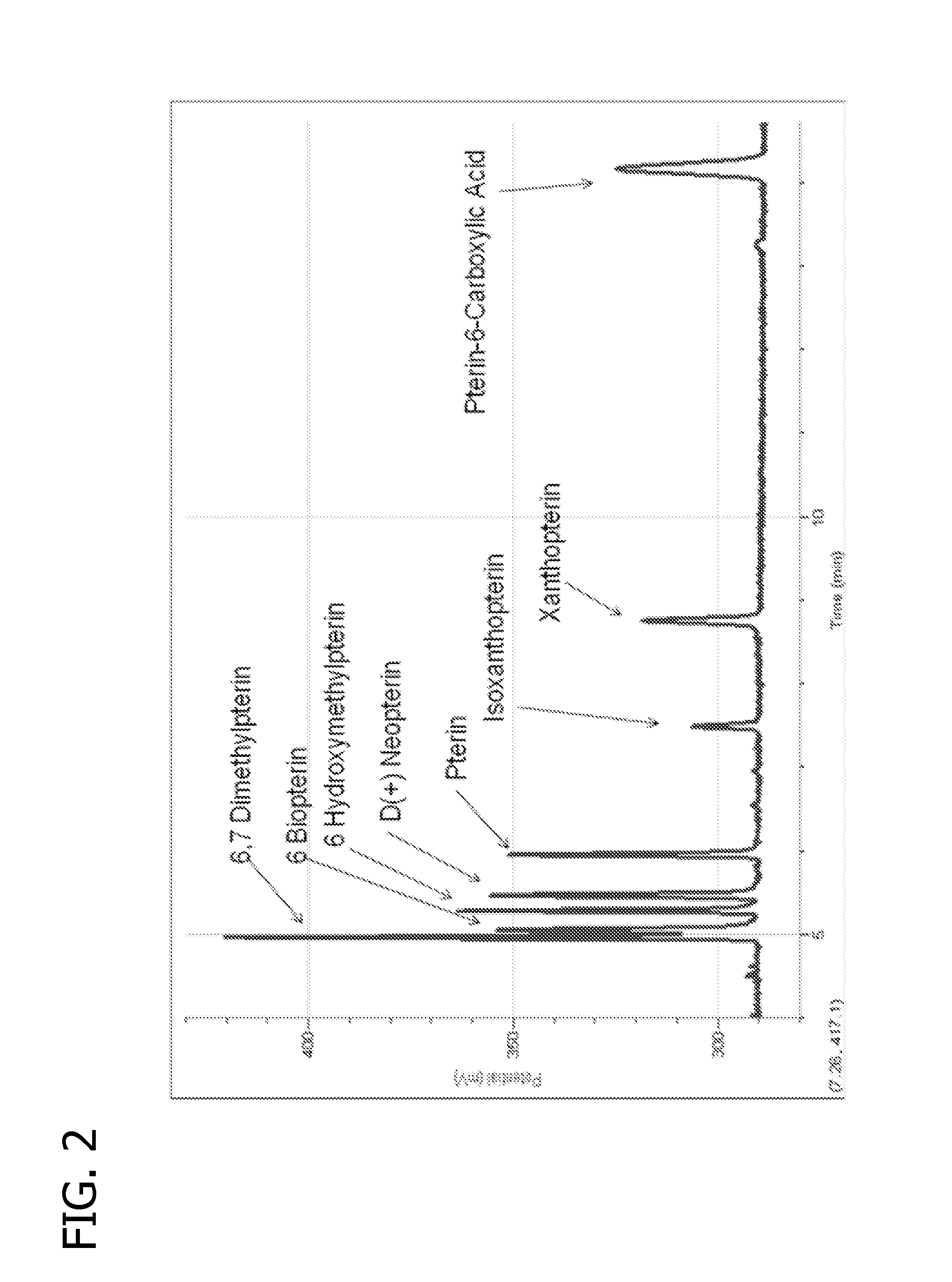
[0102]The foregoing method was also performed to analyze for cancer by detection of oncopterin. Cancer and cancer-free urine samples were analyzed and plotted as shown in FIG. 14. The experimental conditions were as follows: Running buffer: 0.1 M Tris-0.1 M borate-2 mM EDTA, pH 9.63; Capillary: 50 mm i.d.×70 cm (35 cm effective column length); Injection: gravimetric (17.5 cm from the of the sample to the instrument table and the injection time was 10 seconds). Running voltage: 371 V / cm; LIF detection at 325 nm / 445 nm (ex / em); concentration of each pteridine standard: 1.0×10⁻⁹ M. FIG. 14 shows these eight known peaks in the cancer samples and has them labeled 1-8: 1) 6,7-dimethylpterin; 2) 6-biopterin; 3) D-(+)-neopterin; 4) 6-hydroxymethylpterin; 5) Pterin; (6) isoxanthopterin; 7) xanthopterin; (8) carboxypterin. FIG. 14 also shows two peaks not identified with any of (1) through (8).

[0103]Previous work shows that oncopterin is elevated in patients with cancer. Since this experiment...


PUM

PropertyMeasurementUnit
voltageaaaaaaaaaa
poweraaaaaaaaaa
wavelengthaaaaaaaaaa

Abstract

A method and apparatus for detection of pteridine levels in a biological sample using CE-LIF which is useful for early cancer screening involving fully oxidizing pteridine compounds in a sample such as a urine sample, subjecting to CE-LIF to assess compound concentration, and compare to expected levels in for healthy or cancer-bearing patients.

Description

CROSS REFERENCE TO RELATED APPLICATIONS[0001]This application claims priority from U.S. Provisional Application Ser. No. 61 / 284,336, which has a filing date of Dec. 17, 2009.FIELD OF THE INVENTION[0002]The present invention generally relates to an apparatus and method for screening for cancer. In particular, the apparatus and method for screening for cancer is based on the detection of pteridine molecules in biological samples, for example, urine samples.BACKGROUND OF THE INVENTION[0003]Cancer is the second leading cause of death in the United States. To date, approximately 50% of all neoplasms are treated with multi-disciplinary cancer treatment. Neoplastic disorders differ widely in etiology, pathology, and natural disease history. The principle behind detection at earlier stages of disease progression yielding higher cure rates applies to virtually all solid tumors. As a result, cancer screening has been an area of elevated research and clinical interest and has greatly impacted ...

Claims


Application Information

Patent Timeline
Patent Type & Authority Applications(United States)
IPC IPC(8): G01N27/447G01N33/50
CPCG01N21/645G01N33/5308G01N33/52G01N27/44721
Inventor MA, YINFAGIBBONS, STEPHEN
Owner UNIVERSITY OF MISSOURI
Why Eureka
Try Eureka