Diagnosis of Melanoma and Solar Lentigo by Nucleic Acid Analysis

a nucleic acid and melanoma technology, applied in the field of diagnosis of melanoma and solar lentigo by nucleic acid analysis, to achieve the effect of less trauma

Inactive Publication Date: 2011-06-30
DERMTECH INT
View PDF6 Cites 21 Cited by
  • Summary
  • Abstract
  • Description
  • Claims
  • Application Information

AI Technical Summary

Benefits of technology

[0010]In one embodiment, the method involves use of a non-invasive approach for recovering nucleic acids such as DNA or messenger RNA or proteins from the surface of skin via a tape stripping procedure that permits a direct quantitative and qualitative assessment of biomarkers. Although tape-harvested nucleic acid and prote

Problems solved by technology

Although tape-harvested nucleic acid and protein expression products are shown to be comparable in quality and utility to recovering such molecules by

Method used


Image

  • Diagnosis of Melanoma and Solar Lentigo by Nucleic Acid Analysis
  • Diagnosis of Melanoma and Solar Lentigo by Nucleic Acid Analysis
  • Diagnosis of Melanoma and Solar Lentigo by Nucleic Acid Analysis

Examples


example 1

RNA Quantitation and Profiling

[0103]The core hypothesis of this study is that epidermal cells overlying in situ or invasive melanoma, including but not limited to the stratum corneum, stratum lucidum, and stratum granulosum, can be recovered by adhesive means and that the quality and quantity of gene expression in the form of RNA contained within this sample is differently expressed than from a nearby epidermal sample, i.e. that the sampled RNA is diagnostic because of the underlying melanoma. It has been previously shown that changes in gene expression of specific genes are detectable in epidermal hyperplasia overlying cutaneous human melanoma samples obtained from surgical specimens of the epidermis (McCarty et al., 2003).

[0104]The present study is divided into two separate phases, a sample collection and characterization phase (phase 1) and an RNA profiling phase (phase 2). In phase 1 the tape stripped specimens and biopsied sample collections were performed by the principal inve...

example 2

Preliminary Power and Sample Size Studies

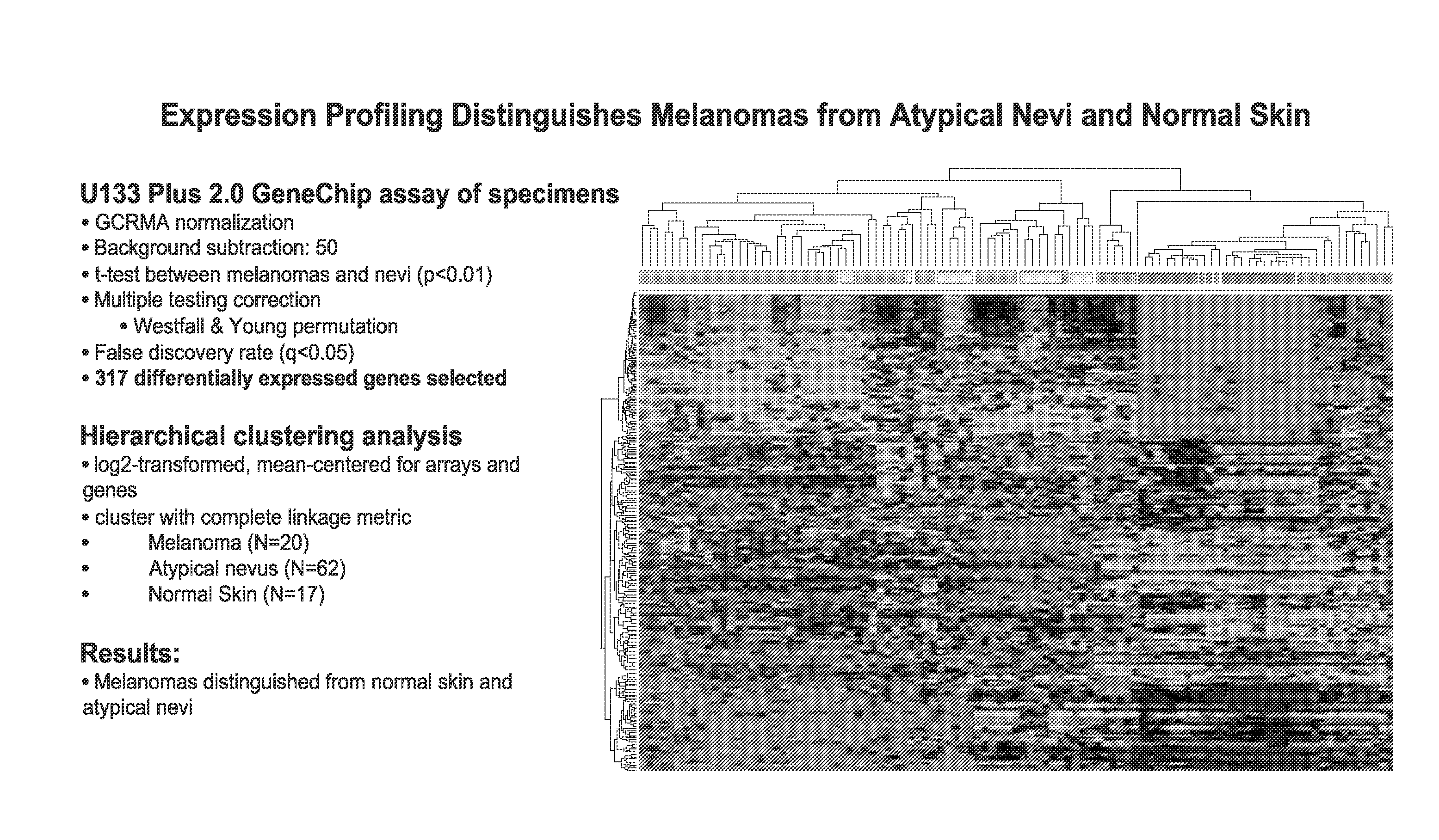
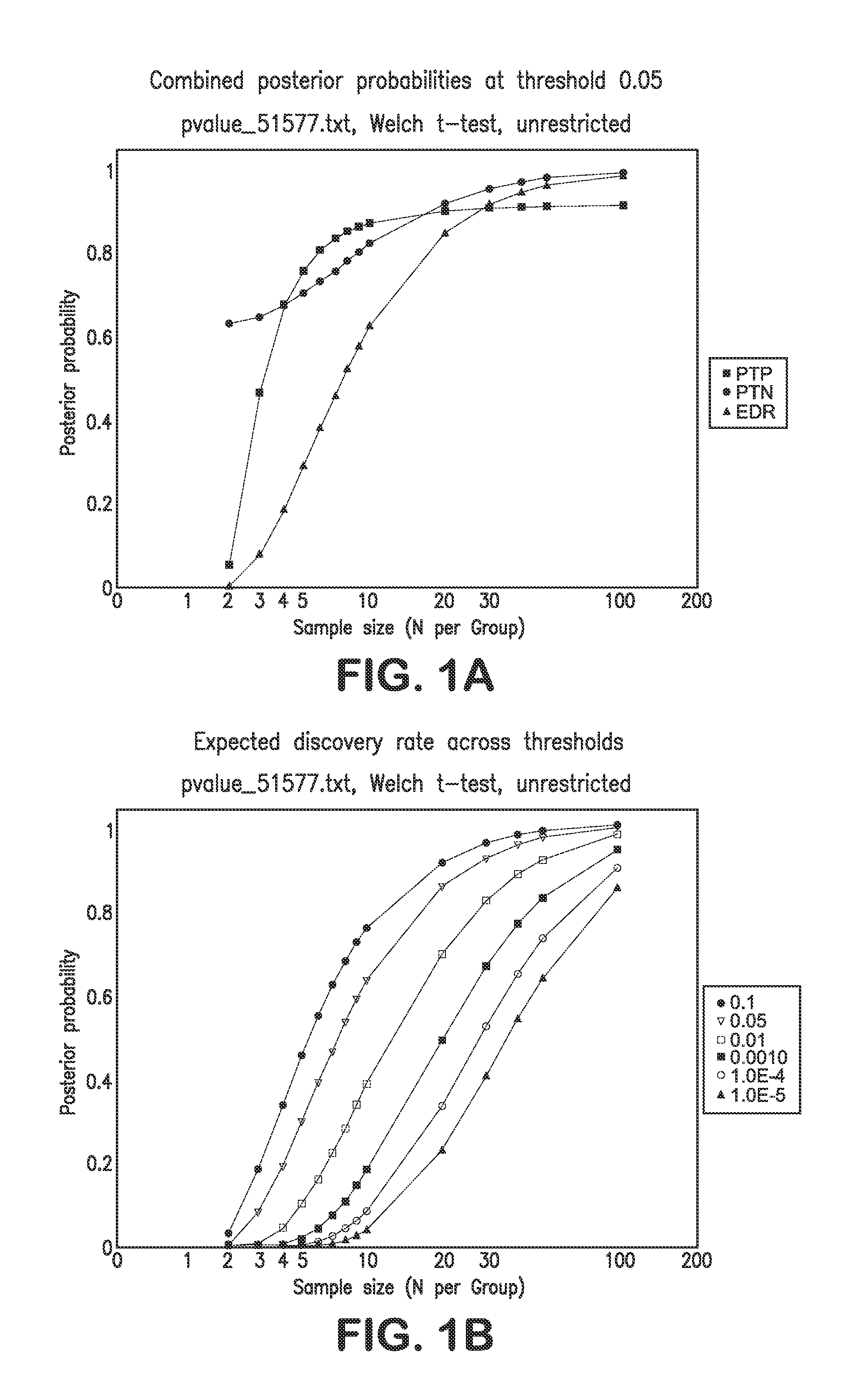
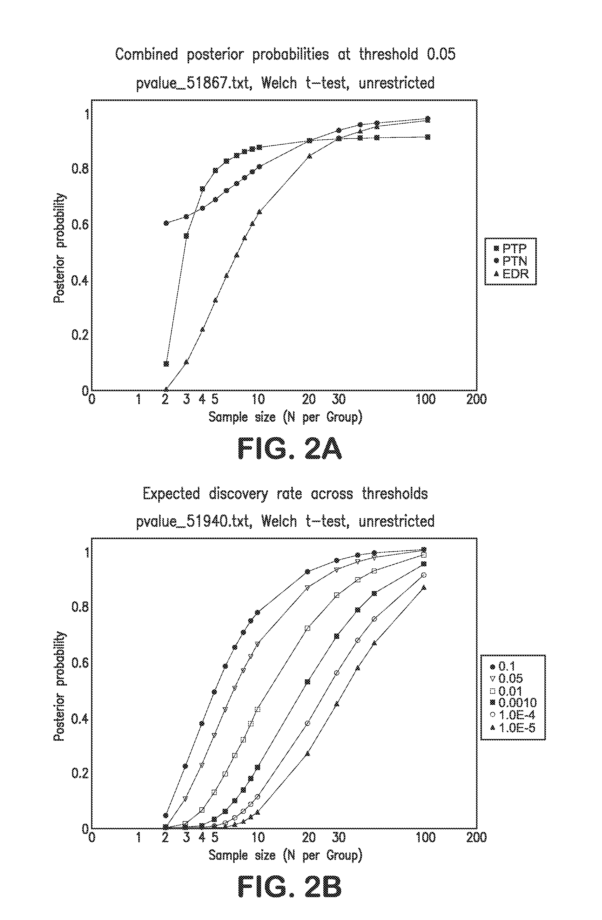
Nevi vs. Primary Melanoma

[0124]The following sample size and power calculations are based exclusively on the large-scale cDNA study data provided in Haqq et al (2005). That data focused on normal skin (n=3 samples), nevi (n=9), primary melanomas (n=6) and metastatic melanomas (n=19). For purposes of the sample size calculations, the focus was on the comparison of nevi to primary melanomas. Power and sample size assessments were calculated based on the bootstrap strategy outlined by Page et al. Using the raw data available from the Haqq et al (2005) study, gene expression differences—based on all 14,772 probes used in their cDNA assay—between nevi and primary melanomas were computed using simple t-tests for each probe / gene. Note that multiple probes can be used interrogate individual genes. In addition, normal skin, nevi, and primary melanoma gene expression differences were also assessed in a three group analysis of variance (ANOVA), with the...

example 3

Tape Stripping to Recover Nucleic Acids from Normal Skin

[0132]The following procedure was used to recover nucleic acids from normal skin (e.g., the mastoid or upper back areas) of a subject.

[0133]Tapes were handled with gloved hands at all times. Locate a particular site that is relatively blemish-free and healthy, unless otherwise specified by the protocol. Preferred normal skin sites are the mastoid process (the bony process behind the ear at the base of the skull) and the upper back, immediately superior to the scapular spine. Shave the site if necessary to remove non-vellus hairs. Cleanse the site with an alcohol wipe (70% isopropyl alcohol). Let the site air dry completely before application of the tape. It is recommended to wait approximately 2 minutes to ensure the site is completely dry before application of the tape.

[0134]Apply the tape to the skin site. If more than one tape is used, apply tapes in sequential order starting from the left side. Use a surgical skin marker an...


PUM

PropertyMeasurementUnit
Currentaaaaaaaaaa
Volumeaaaaaaaaaa
Volumeaaaaaaaaaa

Abstract

The present invention provides methods for diagnosing melanoma and/or solar lentigo in a subject by analyzing nucleic acid molecules obtained from the subject. The present invention also provides methods for distinguishing melanoma from solar lentigo and/or dysplastic nevi and/or normal pigmented skin. The methods include analyzing expression or mutations in epidermal samples, of one or more skin markers. The methods can include the use of a microarray to analyze gene or protein profiles from a sample.

Description

BACKGROUND OF THE INVENTION[0001]1. Field of the Invention[0002]The invention relates generally to methods of characterizing pigmented skin lesions suspected of being melanomas using primarily non-invasive skin sampling.[0003]2. Background Information[0004]Melanoma is a serious form of skin cancer in humans. It arises from the pigment cells (melanocytes), usually in the skin. The incidence of melanoma is increasing at the fastest rate of all cancers in the United States with a lifetime risk of 1 in 68. Although melanoma accounts for only 4% of all dermatologic cancers, it is responsible for 80% of all deaths from skin cancers. It has long been realized that recognition and diagnosis of melanoma, when it is early stage disease, is key to its cure.[0005]Given this, it is imperative that research be carried out not only on therapeutics for melanoma, but also on all aspects of melanoma including prevention and detection. Most of these deaths from melanoma could have been prevented if th...

Claims


Application Information

Patent Timeline
IPC IPC(8): C40B30/04C12Q1/68G01N33/574G16B35/00
CPCC12Q1/6886C12Q2600/112C12Q1/68C12Q2600/178C12Q2600/158G16B35/00G16C20/60C12Q2600/16
Inventor CHANG, SHERMAN H.
Owner DERMTECH INT
Why Eureka
Try Eureka