System and method for volumetric analysis of medical images

Active Publication Date: 2011-06-30
BRAINREADER APS
View PDF8 Cites 26 Cited by
  • Summary
  • Abstract
  • Description
  • Claims
  • Application Information

AI Technical Summary

Benefits of technology

[0002]A plurality of medical imaging techniques exists that enables creation of 2D and / or 3D images of the human (or animal) body for clinical purposes. Techniques such as X-rays (used for instance in mammography and computed tomography (CT)), ultrasound, positron emission tomography (PET), and magnetic resonance imaging (MRI). CT provides good spatial resolution, which is necessary to be able to distinguish two structures an arbitrarily small distance from each other, i.e. to determine whether the structures are separate. MRI provides a resolution comparable with CT but with far better contrast resolution, i.e. the ability to distinguish the differences between two arbitrarily similar but not identical tissues. This ability makes MRI superior in connection with medical images, e.g. medical images of the brain.
[0005]The medical images, also termed clinical images, provided by different medical imaging techniques, can provide a visualisation of the inside of the human body, whereby it is possible to for example detect bone fractures, aneurysms or other internal lesions. Medical imaging also provides the possibility of detecting diseases in advance of normal physical indications, characteristics or symptoms, e.g. screening for breast cancer with mammography. Mental illness is often caused by damages and / or abnormalities in the brain, and medical images of the brain can thereby help diagnosing mental diseases. Likewise, damages and / or abnormalities in the brain can be an indication of a mental illness or initial stages of same. For many diseases an early diagnosis dramatically improves the chance of recovery. Medical imaging is therefore an important tool in screening for, detecting, predicting and diagnosing diseases and initial stages of diseases.
[0027]The individual data for the patient(s) or person(s) used in the system or by the method according to the invention can be any combination of the previously mentioned data types. In some cases it is only necessary to include a few individual data types in the statistical model, thereby simplifying the statistical analysis.
[0050]During the course of a disease the different stages may result in variations of various anatomical structures. E.g. when a disease such as Alzheimers progresses, certain neuro-anatomical regions can change in size and / or shape. In one embodiment of the invention a variation over time of the volume and / or the shape of at least one anatomical structure in at least two medical images acquired at different times can be analysed, evaluated and illustrated. The analysis is preferably combined with individual data of the patient. It is straightforward to compare different images and detect the changes of the same anatomical structure of the same patient over a period of time. But if the time period of the different acquired medical images is running over a substantial period of time, e.g. several years, the change in size and / or shape of the anatomical structure can be caused by a plurality of reasons. By comparing the analysis with individual data of the patient, the size and / shape of the anatomical structure can be corrected for the expected changes that happens in the period of time. The changes happens partly due to the course of time, i.e. the patient is getting older, but for instance changed habits and known diseases in that period may also influence the variation of the anatomical structure. Thereby a much more precise and trustworthy analysis and evaluation of the medical images can be provided.
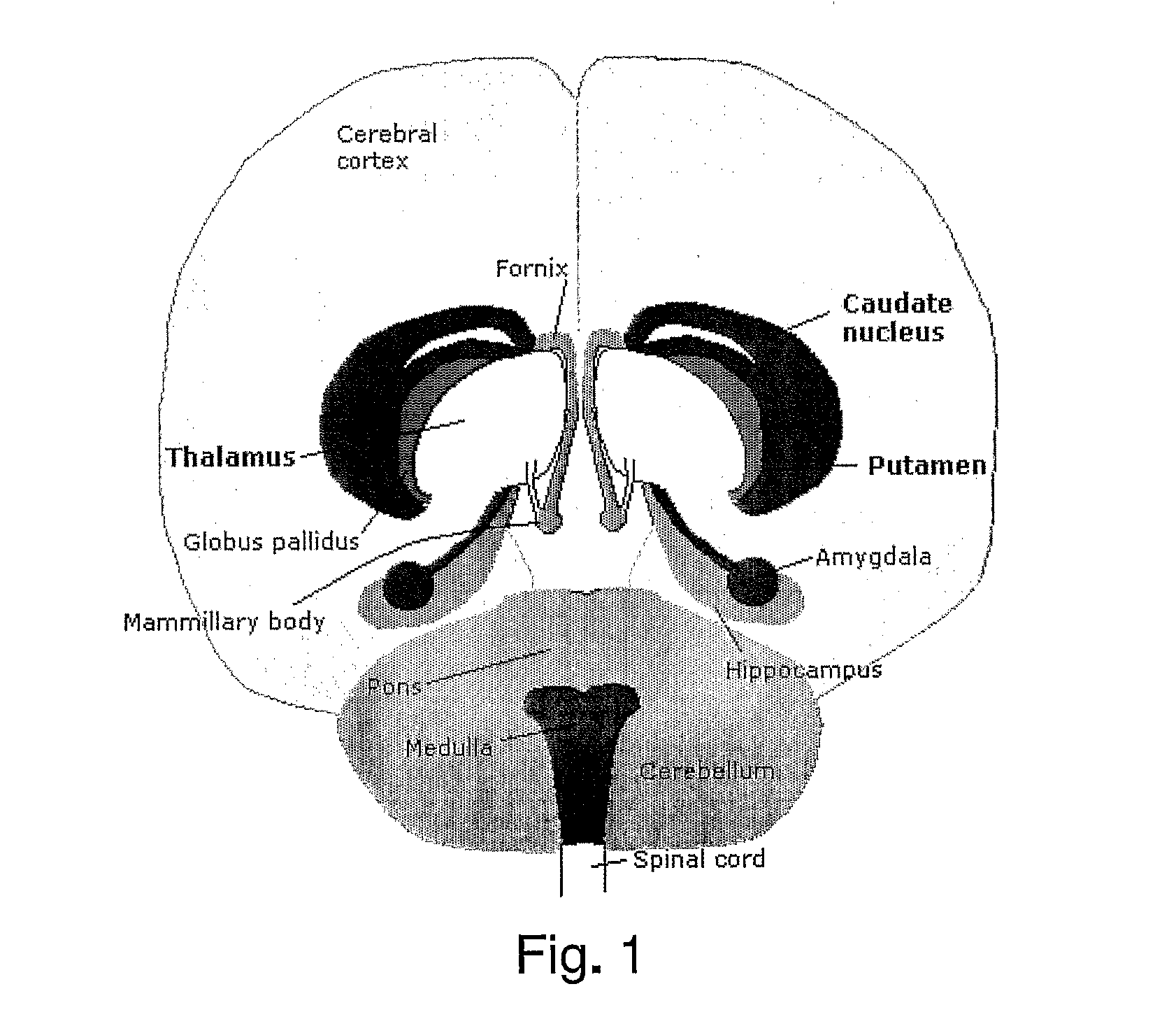
[0059]Diagnosing of diseases and / or initial stages of diseases can be provided by feeding the system with information of the characteristics of the diseases. For example a certain change in size and / or shape of a neuro-anatomical structure like hippocampus might indicate the initial stages of Alzheimer's disease. Access to such information is preferably provided to the system and method according to the invention, e.g. by means of databases, and the segmentation and analysis of a patient's brain provided by the system can thereby provide a diagnosis. The analysis is furthermore combined with the information of individual data of the patient, whereby small deviations from normality in the anatomical structures of the patient can be detected. The result is that diseases can be detected and diagnosed sooner than what is hitherto known. That is the major advantage of the current invention.

Problems solved by technology

For example, comparing the size and shape of the neuro-anatomical structure hippocampus, provided from MRI data of the brain of an eleven-year old left handed Asian female patient, to the average human hippocampus does not make sense.
It will not be possible to conclude on any potential diagnosis of the patient.

Method used


Image

  • System and method for volumetric analysis of medical images
  • System and method for volumetric analysis of medical images
  • System and method for volumetric analysis of medical images

Examples


example

[0092]Below is provided an example of the procedure for brain structure model-based segmentation and statistical characterisation in the form of a confidence interval based on a reference model.[0093]1. A reference model is initially created, wherein the structures of interest are segmented and measured.[0094]2. A formula is calculated using the demographic and / or individual data, such as sex (gender), age, height, weight, genotype, ethnicity, habits, handedness, cognitive measures and the like, of the persons included in the model, and returns a confidence interval of the volume of the structures of interest.[0095]3. Finally, a patient's 3D image is provided as input to the system, with the patient's demographic and / or individual data, and the system returns a 3D image of the structure(s) of interest, the volume(s), and confidence interval.[0096]4. The 3D image of the segmented structure can be coloured reflecting the movement applied to the structure(s) of interest to make it rese...


PUM


Abstract

The invention relates to a system and a method for analysis and evaluation of at least one anatomical structure in a medical image, said medical image representing at least a part of a patient or person, said system comprising image analysing means for calculating the volume and / or providing the shape of the at least one anatomical structure, and statistical analysing means for providing a confidence interval of the volume and / or the shape of the analysed anatomical structure(s) based on individual data of the patient or person, thereby quantifying the normality of said anatomical structure(s). By the present system and method a detailed and trustworthy confidence interval can be provided in connection with analysis and evaluation of a medical image of a patient, thereby illustrating the expected normality of the patient. The invention can be applied to images acquired from magnetic resonance imaging (MRI). One example of use is for diagnosing diseases and / or initial stages of diseases.

Description

[0001]The invention relates to a system and a method for analysis of medical images, such as magnetic resonance images, for automatic characterisation of the volume and / or shape of at least one anatomical structure, whereby diseases, or initial stages of diseases, can be diagnosed. Moreover, the invention relates to a computer-readable medium for implementing such a system on a computer network.[0002]A plurality of medical imaging techniques exists that enables creation of 2D and / or 3D images of the human (or animal) body for clinical purposes. Techniques such as X-rays (used for instance in mammography and computed tomography (CT)), ultrasound, positron emission tomography (PET), and magnetic resonance imaging (MRI). CT provides good spatial resolution, which is necessary to be able to distinguish two structures an arbitrarily small distance from each other, i.e. to determine whether the structures are separate. MRI provides a resolution comparable with CT but with far better contr...

Claims


Application Information

Patent Timeline
IPC IPC(8): A61B5/00G06K9/00
CPCG06T7/0012G06T7/401G06T7/602G06T2207/30016G06T2207/10088G06T2207/20076G06T2207/20128G06T2207/10016G06T7/41G06T7/62
Inventor MADSEN, JAMILA AHDIDAN
Owner BRAINREADER APS
Why Eureka
Try Eureka