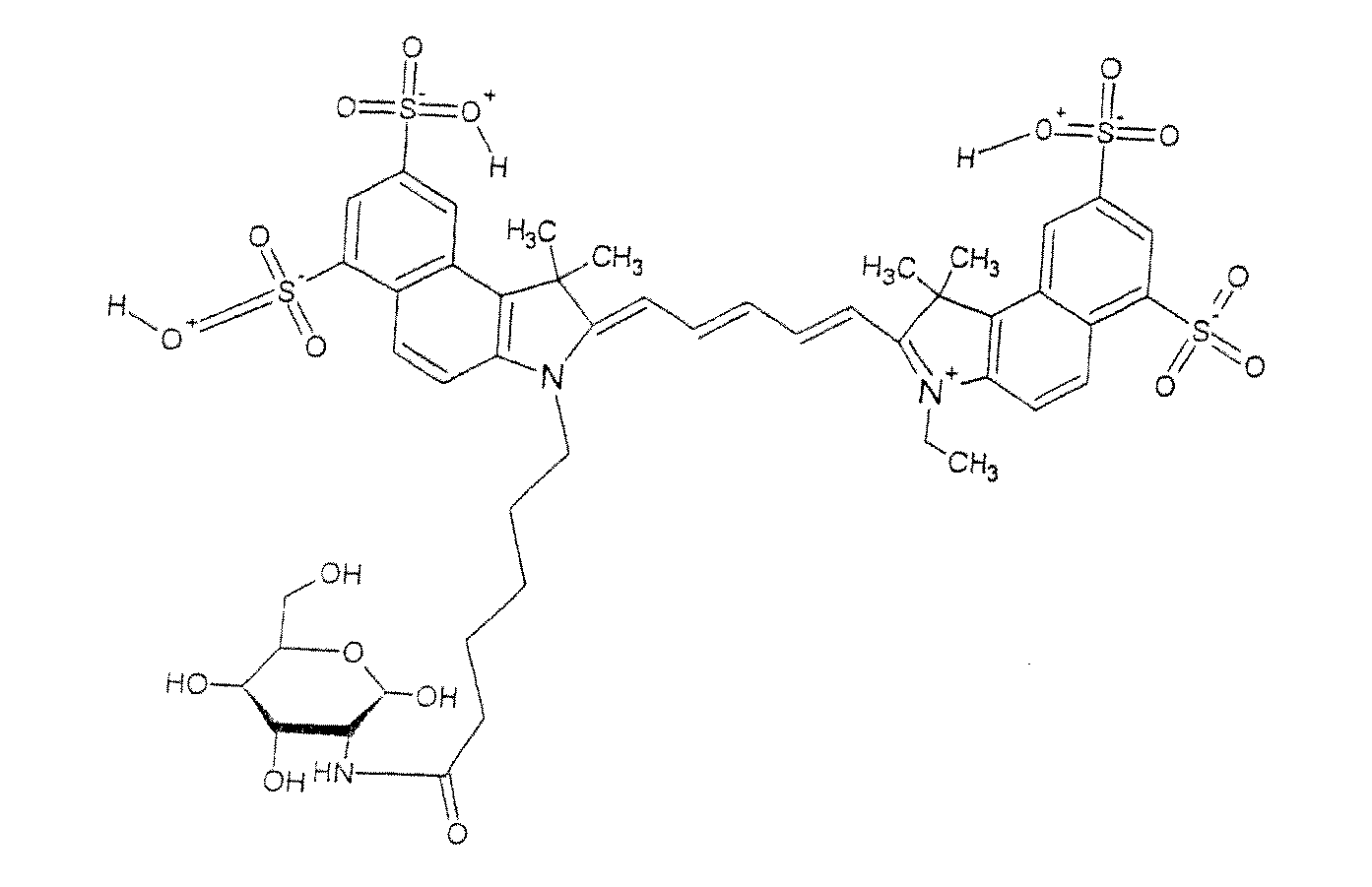
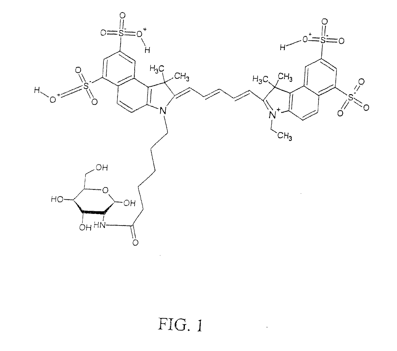
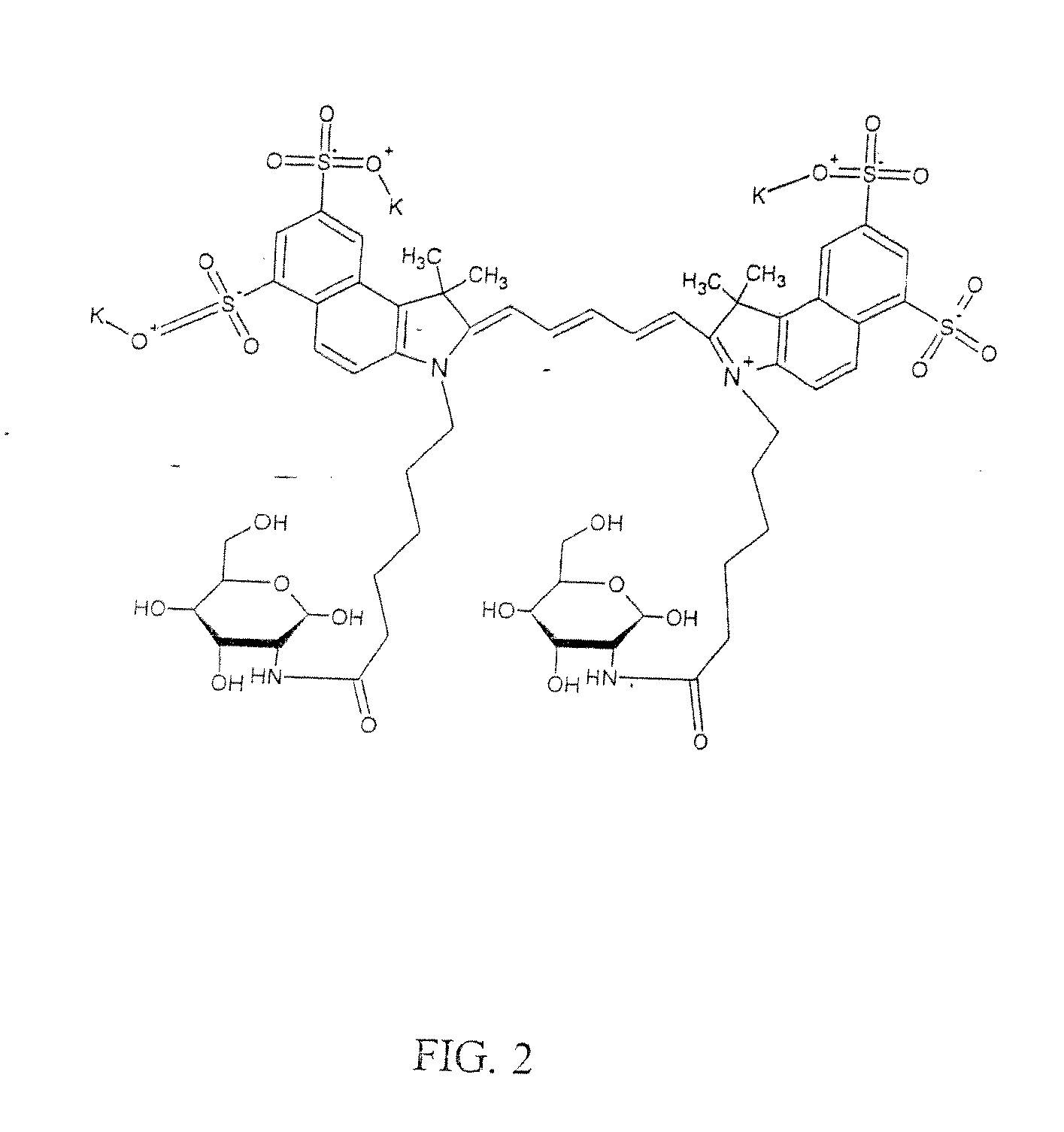
Optical imaging probes

a technology of optical imaging and probes, applied in the field of optical imaging probes, can solve the problems of limited nature, inability to obtain specific molecular information using these modalities, and significant limitations of these imaging approaches

Inactive Publication Date: 2011-07-14
VISEN MEDICAL INC
View PDF29 Cites 32 Cited by
  • Summary
  • Abstract
  • Description
  • Claims
  • Application Information

AI Technical Summary

Benefits of technology

[0018]L is another metabolically recognizable molecule or helper ligand to improve substrate binding and / or delivery.
[0020]The metabolically recognizable molecules can be chemically linked to F, and can total 1-30 per entire optical imaging probe. In one embodiment, M is 2-30. In preferred embodiments, M is 2 or 3. The metabolically recognizable molecule itself may itself be polyvalent, i.e., have more than one repeating structural unit. After derivatization with a single reporter molecule, many metabolites remain metabolically active, but usually at lower rates compared to the underivatized metabolite. A key aspect of this present invention therefore relates to strategies to improve on metabolite or substrate activity in order to optimize imaging of metabolic alterations. While this can be achieved by: 1) optimizing linker systems, 2) rational design and ligand / target molecular modeling and 3) chemically modifying the substrate for optimized in vivo performance, degrees of polyvalency (including bivalency) can result in superior optical metabolite imaging probes with greater “activity” and affinity for imaging metabolic processes. Polyvalency is therefore often important to improve the “activity” and metabolic rates of derivatized NIRF imaging agents, and thus enhancing imaging of metabolic activity.
[0021]A “fluorochrome” includes, but is not limited to, a fluorochrome, a fluorophore, a fluorochrome quencher molecule, or any organic or inorganic dye. Preferred fluorochromes are red and near infrared fluorochromes (NIRFs) with absorption and emission maximum between 600 and 1200 nm. Preferred NIRFs have an extinction coefficient of at least 50,000 M−1 cm−1 in aqueous medium. Preferred NIRFs also have (1) high quantum yield (i.e., quantum yield greater than 5% in aqueous medium), (2) narrow excitation / emission spectrum, spectrally separated absorption and excitation spectra (i.e., excitation and emission maxima separated by at least 15 nm), (3) high chemical and photostability, (4) nontoxicity, (5) good biocompatibility, biodegradability and excretability, and (6) commercial viability and scalable production for large quantities (i.e., gram and kilogram quantities) required for in vivo and human use. Methods for measuring these parameters are known to one of skill in the art.
[0030]A “helper ligand” is any moiety that can be chemically linked to the imaging probe of the present invention that enhances accumulation, targeting, binding, recognition, metabolic activity of the probe, or enhances the efficacy of the probe in any manner. This includes but is not limited to membrane (or transmembrane) translocation signal sequences, which could be derived from a number of sources including, without limitation, viruses and bacteria. Also included are moieties such as monoclonal antibodies (or antigen-binding antibody fragments, such as single chain antibodies) directed against a target-specific marker, a receptor-binding polypeptide directed to a target-specific receptor, a receptor-binding polysaccharide directed against a target-specific receptor and other molecules that target internalizing receptors including but not limited to nerve growth factor, oxytocin, bombesin, calcitonin, arginine vasopressin, angiotensin II, atrial natriuretic peptide, insulin, glucagons and glucagon-like peptides, prolactin, gonadotropin, and various opioids.
[0031]Derivatization of a fluorochrome may also change the biological properties of the NIRF itself. For instance, mono-, bi-, or polyvalent derivatization of a fluorochrome may improve the pharmacokinetics, toxicity, solubility, and fluorescence properties of the fluorochrome molecule itself, thereby making it a more suitable in vivo imaging agent, that could be used in any number of different applications which may or may not include imaging metabolic or physiologic activity.

Problems solved by technology

Specific molecular information using these modalities often cannot be obtained, or is of limited nature.
Although nuclear imaging of radioactively labeled metabolites has demonstrated some clinical utility, there remain significant limitations with these imaging approaches.
Specifically, the short half-life of many radionuclides, including 18F, 11C, 17O, and 99mTc, severely limits the time available for synthesis and subsequent imaging, and therefore any facilities using these technologies require skilled radiochemists on staff to synthesize the imaging agents immediately prior to use.
In addition, the clinical hardware systems required to detect positron and gamma emitting radionuclides are relatively expensive and therefore, require a significant upfront capital investment.
Because of these limitations, few clinical centers have the necessary expertise, resources, and money to operate a nuclear imaging center effectively.
Another significant disadvantage to nuclear imaging is that patients are exposed to radioactivity.
Because strict clinical guidelines govern the amount of radiation a patient can receive over a given timeframe, the number of imaging procedures a patient can receive per year is limited.
Therefore, nuclear imaging is limited for routine monitoring of a patient's disease state or response to therapy over time.
While receptor targeted fluorochromes such as those described by Becker et al. and enzyme activatable probes such as those described by Weissleder et al. are able to image some forms of molecular activity, these probes are not optical metabolite imaging probes.
However, because these fluorescent agents do not absorb or emit light in the red or near infrared range, their in vivo use is very limited, i.e., for cancer detection in deep tissues.

Method used


Image

  • Optical imaging probes
  • Optical imaging probes
  • Optical imaging probes

Examples


Embodiment Construction

[0050]In one embodiment, the imaging agent (i.e., optical imaging probe) accumulates in diseased tissue at a different rate than in normal tissue. For example, the rate of accumulation of the agent can be at least 5%, 10%, 20%, 30%, 50%, 75%, or 90% faster in diseased tissue compared to normal tissue. Alternatively, the rate of accumulation of the agent can be at least 5%, 10%, 20%, 30%, 50%, 75%, or 90% slower in diseased tissue compared to normal tissue

[0051]In another embodiment, the imaging agent is metabolized in diseased tissue at a different rate than in normal tissue. For example, metabolism of the imaging agent can occur at a rate that is at least 5%, 10%, 20%, 30%, 50%, 75%, or 90% faster in diseased tissue compared to normal tissue. Alternatively, metabolism of the imaging agent can occur at a rate that is at least 5%, 10%, 20%, 30%, 50%, 75%, or 90% slower in diseased tissue compared to normal tissue.

[0052]In another embodiment, the imaging agent becomes trapped in cells...


PUM

PropertyMeasurementUnit
optical imagingaaaaaaaaaa
timeaaaaaaaaaa
wavelengthaaaaaaaaaa

Abstract

This invention relates to optical imaging probes and the use of such probes for diagnosing and monitoring disease, and disease treatment. The optical imaging probes of the current invention can be used to identify and characterize normal and diseased tissues with regards to altered metabolic activity.

Description

RELATED APPLICATIONS[0001]This application is a continuation of U.S. Ser. No. 10 / 938,744, filed Sep. 10, 2004, which in turn is a continuation of International Application No. PCT / US03 / 07579, which designated the United States and was filed on Mar. 11, 2003, published in English, which claims the benefit of U.S. Provisional Application No. 60 / 363,409, filed on Mar. 11, 2002. The entire teachings of, and each of the above applications are incorporated herein by reference in their entirety.BACKGROUND OF THE INVENTION[0002]This invention relates to optical imaging probes and the use of such probes for diagnosing and monitoring disease, and for disease treatment. The optical imaging probes of the current invention can be used to identify and characterize normal and diseased tissues with regards to altered metabolic or physiologic activity.[0003]With the sequencing of the human genome, there is an enormous effort underway to understand the precise molecular basis of different disease sta...

Claims


Application Information

Patent Timeline
Patent Type & Authority Applications(United States)
IPC IPC(8): A61K49/10A61K49/00A61K49/04C07H5/06A61P25/28A61P9/10A61P3/10A61P25/16A61B10/00G01N33/533G01N33/58
CPCA61K49/0017A61K49/0032G01N33/582A61K49/0052G01N33/533A61K49/0041A61P25/16A61P25/28A61P9/10A61P3/10
Inventor POSS, KIRTLAND G.MADDEN, KAREN N.JONES, ELLAKOSSODO, SYLVIE
Owner VISEN MEDICAL INC
Why Eureka
Try Eureka