Non-invasive ocular monitoring

a non-invasive, ocular monitoring technology, applied in the field of ocular diagnosis, can solve the problems of complex methods, requiring clinical setting, and affecting the normal function of the bab, and achieve the effect of accurately measuring the concentration level of analytes

Active Publication Date: 2011-07-28
CHROMOLOGIC
View PDF19 Cites 18 Cited by
  • Summary
  • Abstract
  • Description
  • Claims
  • Application Information

AI Technical Summary

Benefits of technology

One illustrative embodiment of the disclosure provides a device for accurately measuring an analyte concentration level in the aqueous humour by spectroscopic analysis of the reflected / scattered light using a broadband continuous wavelength visible light source (200 nm to 900 nm). Herein, a radiation source illuminates an anterior region of the eye of the subject with a substantially broad spectrum at an angle substantially tangent to the eye. The reflected and scattered radiations are collected and analyzed to obtain a characteristic spectrum of the detected scattered and reflected radiations. The characteristic spectrum is a combination of the light absorbed and scattered by the analyte based on its refractive index, size of the analyte, natural optical absorption, as well as its optical activity. This characteristic spectrum is then compared with a reference characteristic spectrum to compute the analyte concentration. Illumination at a tangential angle may avoid the total internal reflection characteristics of the eye and enable light collection normal to the corneal surface. Analysis with a broad illumination spectrum enables the determination of the optical properties of the analyte over a large response region.
In one embodiment of the present invention, the analyte concentration is measured by a non-invasive method using optical reflection spectroscopy. Here, measurements are rapid since the method is passive and the analyte is not excited optically with a coherent or monochromatic source. This method is reliable for diagnosis and does not require any baseline measurement. In addition, a broad illumination spectrum primarily in the visible light range is used and hence the device is cost effective, and easy to operate.
The device may further include a modulator configured for modulating the incident light to reduce extraneous effects.

Problems solved by technology

During duress, which may include traumatic injury, radiation exposure or diseases, such as cancer, the normal function of the BAB is adversely affected and results in a range of typically locally blocked analytes crossing into the anterior chamber of the eye.
On the one hand, some of the methods typically used for accurately determining the hydration status, such as Total Body Water (“TBW”) measurements and invasive measurements of plasma osmolality, are complex and require a clinical setting.
However, the techniques available are invasive, inconvenient and often painful, which may lead to a high degree of inadequate treatment.
On the other hand, some of the more rapid and non-invasive techniques, such as bioimpedence measurements made through the skin using weak electrical currents, salivary osmolality measurements (for hydration), and monitoring physical signs of dehydration or diabetes, require baseline characterizations and may not be very specific.
Additionally, non-invasive measurement modalities that require electromagnetic / optic probing made through the skin are easily corrupted by skin temperature, perspiration and body position of the subject, therefore limiting their application.

Method used


Image

  • Non-invasive ocular monitoring
  • Non-invasive ocular monitoring
  • Non-invasive ocular monitoring

Examples


Embodiment Construction

The detailed description set forth below in connection with the drawings is intended as a description of embodiments of a non-invasive method and device for determining health condition of a subject through measurements of concentrations of analytes in the eye in accordance with the present invention and is not intended to represent the only forms in which the invention may be constructed, or utilized. It is to be understood that the same or equivalent functions and structures may be accomplished by different embodiments that are also intended to be encompassed within the spirit and scope of the invention. As denoted elsewhere herein, like element numbers indicate like elements or features.

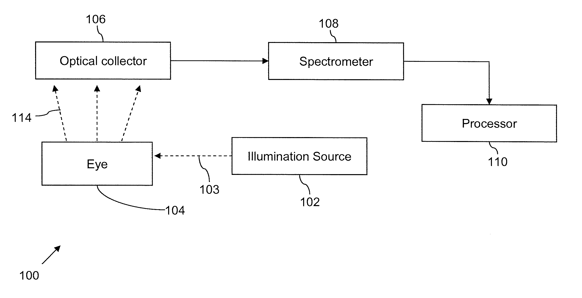
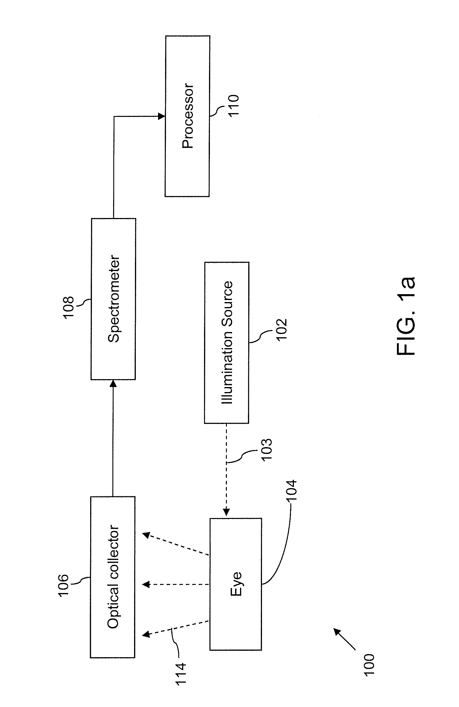
An embodiment of the present invention provides a device and a method for non-invasively measuring concentration of an analyte in the human eye.

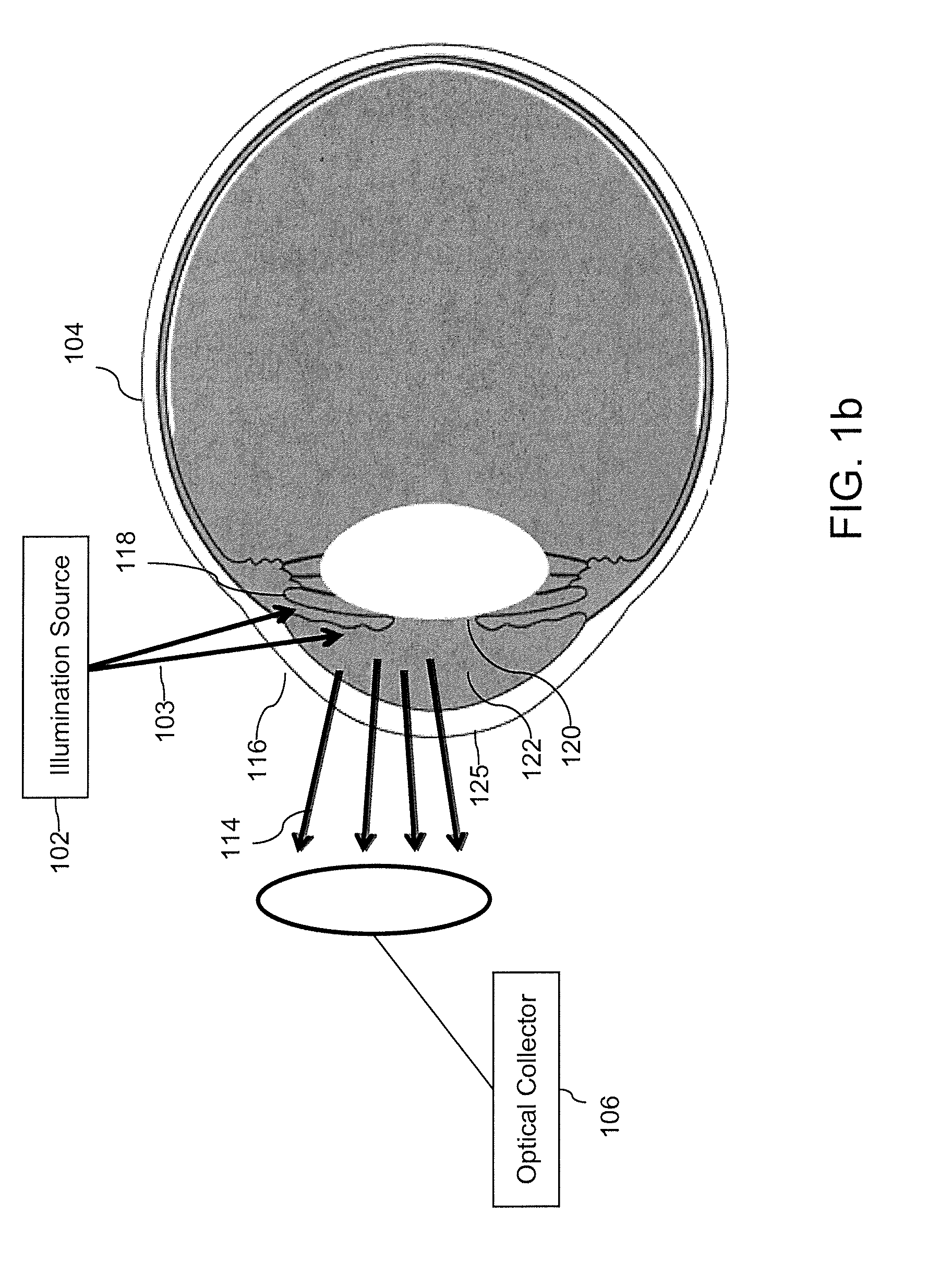
More specifically, an embodiment of the present invention provides a device for accurately measuring an analyte concentration level in the aqueous humor i...


PUM


Abstract

A device for measuring an analyte concentration level in a subject. The device includes: a light source configured for illuminating at least a portion of an anterior region of an eye of the subject with incident light having a substantially broad illumination spectrum at an angle substantially tangential to the surface of the eye; an optical collector configured for detecting reflected light from the at least a portion of the anterior region of the eye; an analyzer configured for analyzing the detected reflected light; and a processor configured to determine the analyte concentration level in the subject based on the analyzed reflected light.

Description

FIELD OF THE INVENTIONThe disclosure relates generally to ocular diagnosis, and, more particularly, to a non-invasive method and device for determining health condition of a subject through measurements of concentrations of analytes in the eye.BACKGROUNDThe Blood-Aqueous Barrier (“BAB”) is a metabolic or cellular structure in the anterior chamber of the eye (“ACE”) that restricts the passage of various chemical substances and microscopic objects, such as bacteria, between the bloodstream and the ACE tissue itself, while still allowing the passage of substances essential to metabolic function, such as oxygen, sodium and potassium salts and glucose. During duress, which may include traumatic injury, radiation exposure or diseases, such as cancer, the normal function of the BAB is adversely affected and results in a range of typically locally blocked analytes crossing into the anterior chamber of the eye. As such, detection of such analytes in the human eye can be a reliable indicator ...

Claims


Application Information

Patent Timeline
Patent Type & Authority Applications(United States)
IPC IPC(8): A61B5/1455
CPCA61B5/14532A61B5/4869A61B5/1455A61B5/14546
Inventor MENON, NARESH
Owner CHROMOLOGIC
Why Eureka
Try Eureka