Cardiac and or respiratory gated image acquisition system and method for virtual anatomy enriched real time 2d imaging in interventional radiofrequency ablation or pace maker replacement procecure

a gated image and virtual anatomy technology, applied in the field of cardiac and or respiratory gated image acquisition system and virtual anatomy, can solve the problems of affecting the electrical conduction leading to an irregular heart beat, radiofrequency ablation (rfa) for the treatment, and a very challenging ep procedure, so as to achieve the effect of difficult and inaccurate registration of two modalities

Active Publication Date: 2011-08-18
KONINKLIJKE PHILIPS ELECTRONICS NV
View PDF7 Cites 127 Cited by
  • Summary
  • Abstract
  • Description
  • Claims
  • Application Information

AI Technical Summary

Benefits of technology

[0012]In view of the above-described facts, it is an object of the present invention to enhance the accuracy of the image-guided interventional procedure by reducing inaccuracies which may arise when registering and fusing images which have been acquired by distinct, independently operated imaging modalities. As most contrast agents are quite expensive and not always well supported by the patients, it is a further object of the present invention to visualize the cardiac vessel structures and / or cardiac chambers anatomy of a patient without needing to inject a contrast agent.
[0013]To solve these problems, a first exemplary embodiment of the present invention is directed to an image acquisition method for tracking navigation motions of an interventional instrument while being navigated through a patient's cardiovascular system and / or cardiac chambers anatomy by intraoperatively acquiring and recording a sequence of 2D live images from the same projection angle and object distance, said images showing the interventional instrument during different stages of such a navigation motion. According to the present invention, said method comprises the steps of recording the particular phases of the patient's cardiac and / or respiratory cycles for each track during the acquisition and recording of the 2D fluorograms and selecting a set of those images which correspond to a predefinable specific phase of the patient's cardiac and / or respiratory cycles by means of cardiac and / or respiratory gating. While executing said selection step and further navigating the interventional instrument, a virtual 2D reconstruction of the patient's cardiovascular system and / or cardiac chambers anatomy is generated by fusing the 2D-gated positions of the interventional instrument which have been selected until the time of said generation step, wherein said 2D reconstruction is dynamically enriched with each newly acquired and selected 2D fluorogram, thus yielding a superimposed 2D view of the interventional instrument's different navigation motion stages. Finally, a dynamically updated version of the two-dimensionally reconstructed virtual anatomy is displayed on a monitor screen or display by overlaying and fusing newly selected 2D-gated positions of the interventional instrument to the current version of the reconstructed anatomy. In this way, it can be avoided to perform a preoperative CT, MR or 3DRA scan for acquiring voxel data needed for reconstructing a 3D model of the patient's cardiovascular system and / or cardiac anatomy which, as known from the prior art, is to be coregistered with intraoperatively acquired fluorograms showing the current position of the interventional instrument while being navigated during an image-guided intervention, thus allowing to avoid registration inaccuracies which are to be expected when using distinct imaging modalities for preoperatively acquiring the voxel data of the reconstructed 3D model and the image data of the intraoperatively acquired fluorograms.

Problems solved by technology

Congenital problems or diseased tissue in the heart can affect the electrical conduction leading to an irregular heart beat.
Aside therefrom, radiofrequency ablation (RFA) for the treatment of atrial fibrillation (AF) is a very challenging EP procedure.
Atrial fibrillation is a cardiac arrhythmia associated with significant morbidity and mortality which arises when the two upper chambers of the heart, the left atrium (LA) and the right atrium (RA), do not beat efficiently such that, as a consequence thereof, blood is not pumped completely out of them and may thus pool and clot.
If a part of the clot leaves the heart, it may cause a stroke or a pulmonary embolism.
The procedures to locate and ablate the appropriate areas of tissue are extremely lengthy.
Electrophysiology diagnosis and treatment does not require the injection of contrast agent into the coronary arteries to produce detailed images and therefore requires somewhat lower imaging capability.
On the other hand, such a registration does not take into account a patient's respiratory and cardiac motions, which may lead to the fact that the obtained registration accuracy is not precise enough for some applications.

Method used


Image

  • Cardiac and or respiratory gated image acquisition system and method for virtual anatomy enriched real time 2d imaging in interventional radiofrequency ablation or pace maker replacement procecure
  • Cardiac and or respiratory gated image acquisition system and method for virtual anatomy enriched real time 2d imaging in interventional radiofrequency ablation or pace maker replacement procecure
  • Cardiac and or respiratory gated image acquisition system and method for virtual anatomy enriched real time 2d imaging in interventional radiofrequency ablation or pace maker replacement procecure

Examples


Embodiment Construction

[0030]In the following, the proposed image acquisition device and method according to the present invention will be explained in more detail with respect to special refinements and referring to the accompanying drawings.

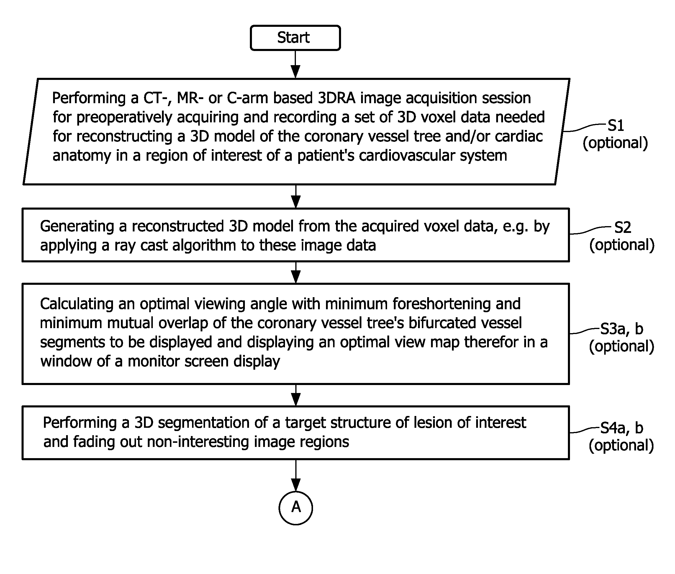
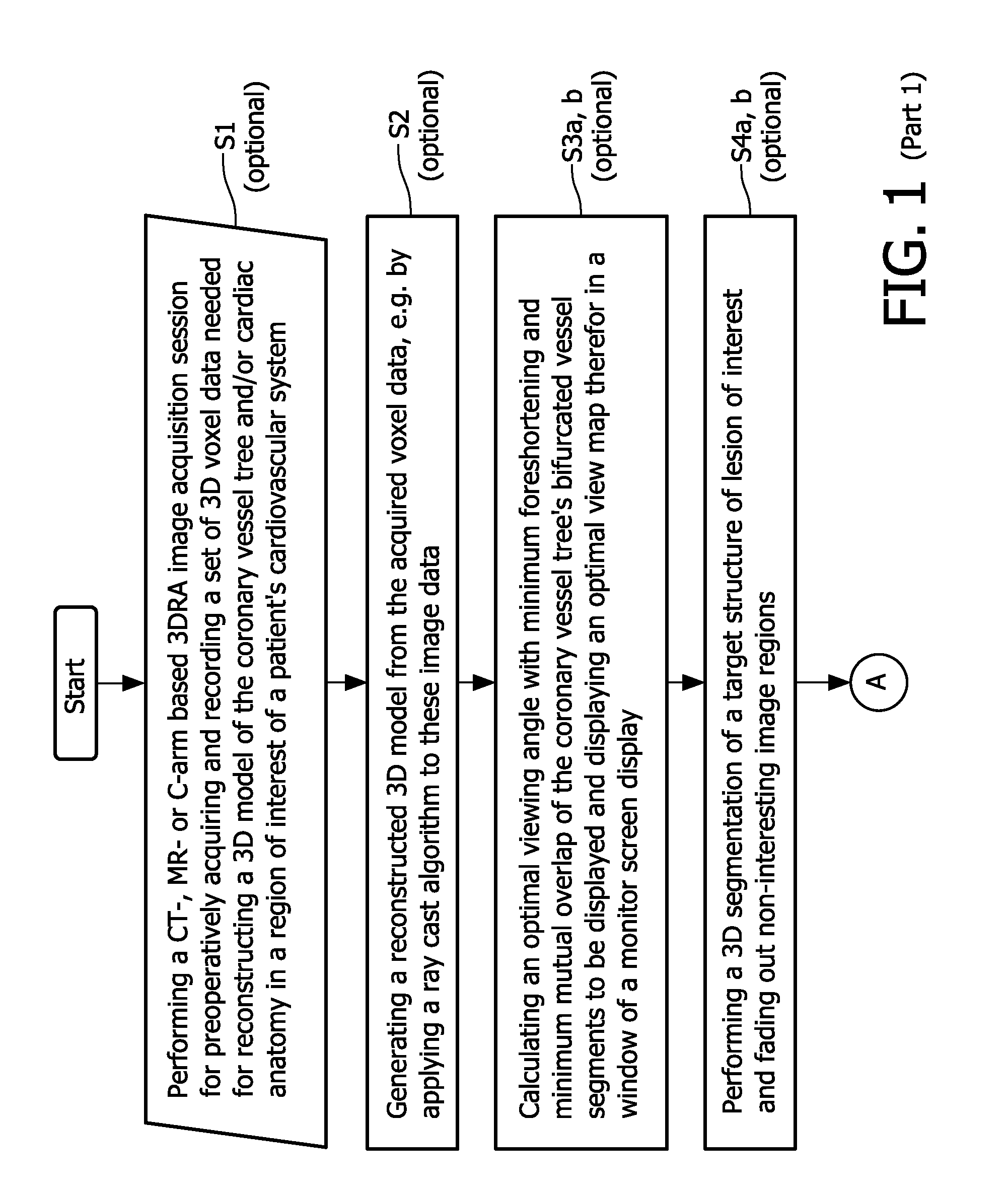
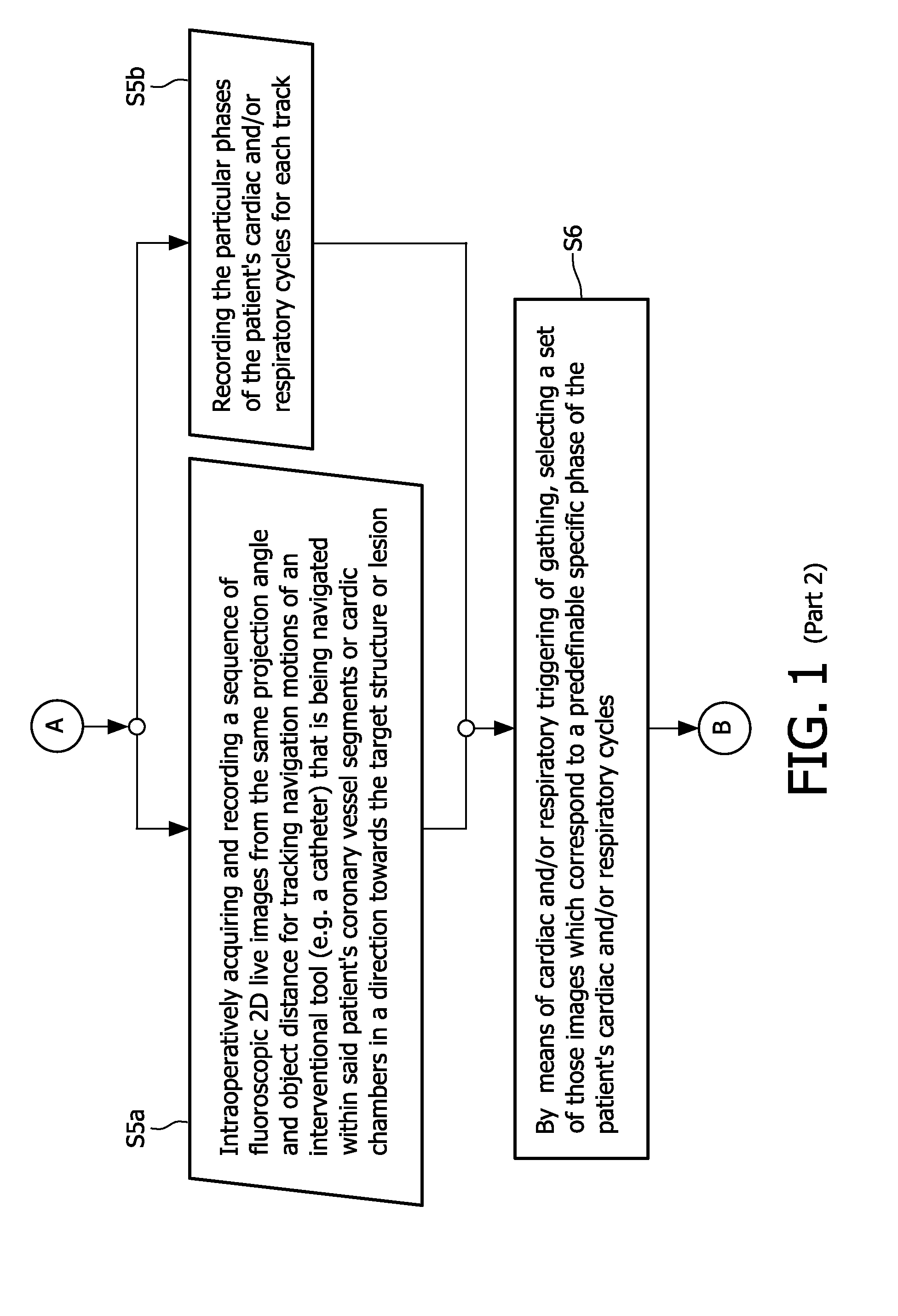
[0031]The flowchart depicted in FIG. 1 illustrates the proposed image acquisition method according to the above-described first exemplary embodiment of the present invention. As already mentioned described above, the proposed method begins with the optional step of performing a CT-, MR-, C-arm based 3DRA or any other modality type (ultrasound, scintigraphy, etc.) based image acquisition session for preoperatively acquiring and recording (S1) a set of 3D voxel data needed for reconstructing a 3D model of the coronary vessel tree and / or cardiac anatomy in a region of interest of a patient's cardiovascular system and, after this preoperative image data acquisition step, optionally generating (S2) a three-dimensionally reconstructed model or 3D map of a patient's cardiov...


PUM


Abstract

The present invention refers to the field of cardiac electrophysiology (EP) and, more specifically, to image-guided radio frequency ablation and pacemaker placement procedures. For those procedures, it is proposed to display the overlaid 2D navigation motions of an interventional tool intraoperatively obtained from the same projection angle for tracking navigation motions of an interventional tool during an image-guided intervention procedure while being navigated through a patient's bifurcated coronary vessel or cardiac chambers anatomy in order to guide e.g. a cardiovascular catheter to a target structure or lesion in a cardiac vessel segment of the patient's coronary venous tree or to a region of interest within the myocard. In such a way, a dynamically enriched 2D reconstruction of the patient's anatomy is obtained while moving the interventional instrument. By applying a cardiac and / or respiratory gating technique, it can be provided that the 2D live images are acquired during the same phases of the patient's cardiac and / or respiratory cycles. Compared to prior-art solutions which are based on a registration and fusion of image data independently acquired by two distinct imaging modalities, the accuracy of the two-dimensionally reconstructed anatomy is significantly enhanced.

Description

[0001]The present invention refers to the field of cardiac electrophysiology (EP) and, more specifically, to image-guided radiofrequency ablation and pacemaker placement procedures. For those procedures, it is proposed to display the overlaid 2D navigation motions of an interventional tool intraoperatively obtained from the same projection angle during an intervention procedure while being navigated through a patient's bifurcated coronary vessel or cardiac chambers anatomy in order to guide e.g. a cardiovascular catheter to a target structure or lesion in a cardiac vessel segment of the patient's coronary venous tree or to a region of interest within the myocard. In such a way, a dynamically enriched 2D reconstruction of the patient's anatomy is obtained while moving the interventional instrument. By applying a cardiac and / or respiratory gating technique, it can be provided that the 2D live images are acquired during the same phases of the patient's cardiac and / or respiratory cycles...

Claims


Application Information

Patent Timeline
Patent Type & Authority Applications(United States)
IPC IPC(8): A61B5/05G06K9/00A61B5/352
CPCA61B5/0456A61B2019/5289A61B5/7289A61B6/032A61B6/12A61B6/503A61B6/504A61B6/5235A61B6/5247A61B6/527A61B6/5288A61B8/0891A61B8/5276A61B19/5225A61B5/1135A61B2090/364A61B90/37A61B5/352
Inventor GOGIN, NICOLAS PIERRE BRUNOPICARD, CECILE ANNE MARIEVILLAIN, NICHOLAS FRANCOIS
Owner KONINKLIJKE PHILIPS ELECTRONICS NV
Why Eureka
Try Eureka