
X-Ray and Gamma Ray Detector Readout Sytem

a detector and readout technology, applied in the field of detectors and readout electronics, can solve the problems of unfavorable radiation detection, unfavorable radiation detection, and limited diagnostic accuracy of the technique for detecting cancer, and achieve the effects of high efficiency, uniform spatial resolution, and relative insensitivity to magnetic fields

Inactive Publication Date: 2011-10-13
NOVA R&D
View PDF5 Cites 15 Cited by
  • Summary
  • Abstract
  • Description
  • Claims
  • Application Information

AI Technical Summary

Benefits of technology

[0021]The compact geometry and low mass of the APD arrays allow for double-ended readout of the LSO crystals, to make depth of interaction (DOI) measurements, with the added engineering advantage of identical readout electronics for both sides of the crystal array. DOI measurement is critical to achieving a uniform spatial resolution in combination with high efficiency in an affordable instrument, with a ring diameter of about 20 cm. Another advantage of APDs is their relative insensitivity to magnetic fields, possibly enabling co-imaging with PET and NMR techniques in the future.
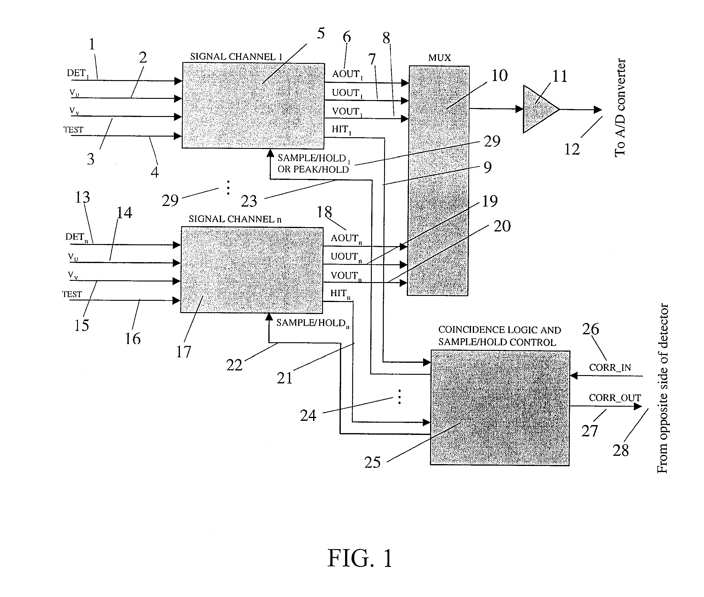
[0022]A complete, highly integrated, low power readout electronics chain optimized for high resolution APD / LSO PET imaging is to date not available, although encouraging results have been reported for individual circuit blocks such as the preamplifier and discriminator. A high resolution PET scanner with DOI for breast cancer imaging will involve 5,000-20,000 channels, making power dissipation a very critical parameter. Position sensitive readout schemes (charge division) can be used to reduce the number of electronics channels, but bring in additional uncertainty in the position measurement and increase the electronics channel hit rate, requiring lower dead time. Since the avalanche gain in an APD is relatively low (compared to a typical PMT (photomultiplier tube)), sophisticated low noise electronics must be placed in close proximity to the APDs, further complicating the power dissipation issue.
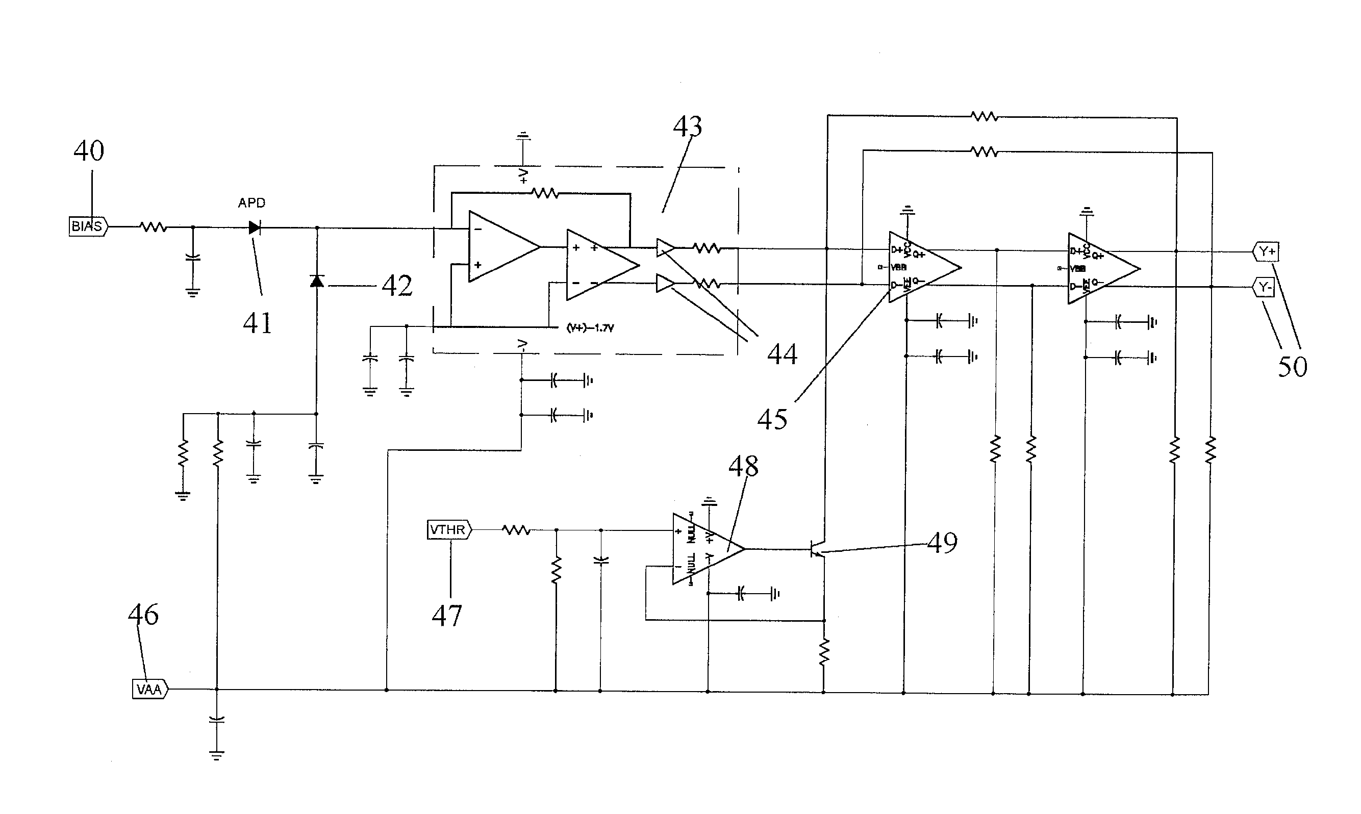
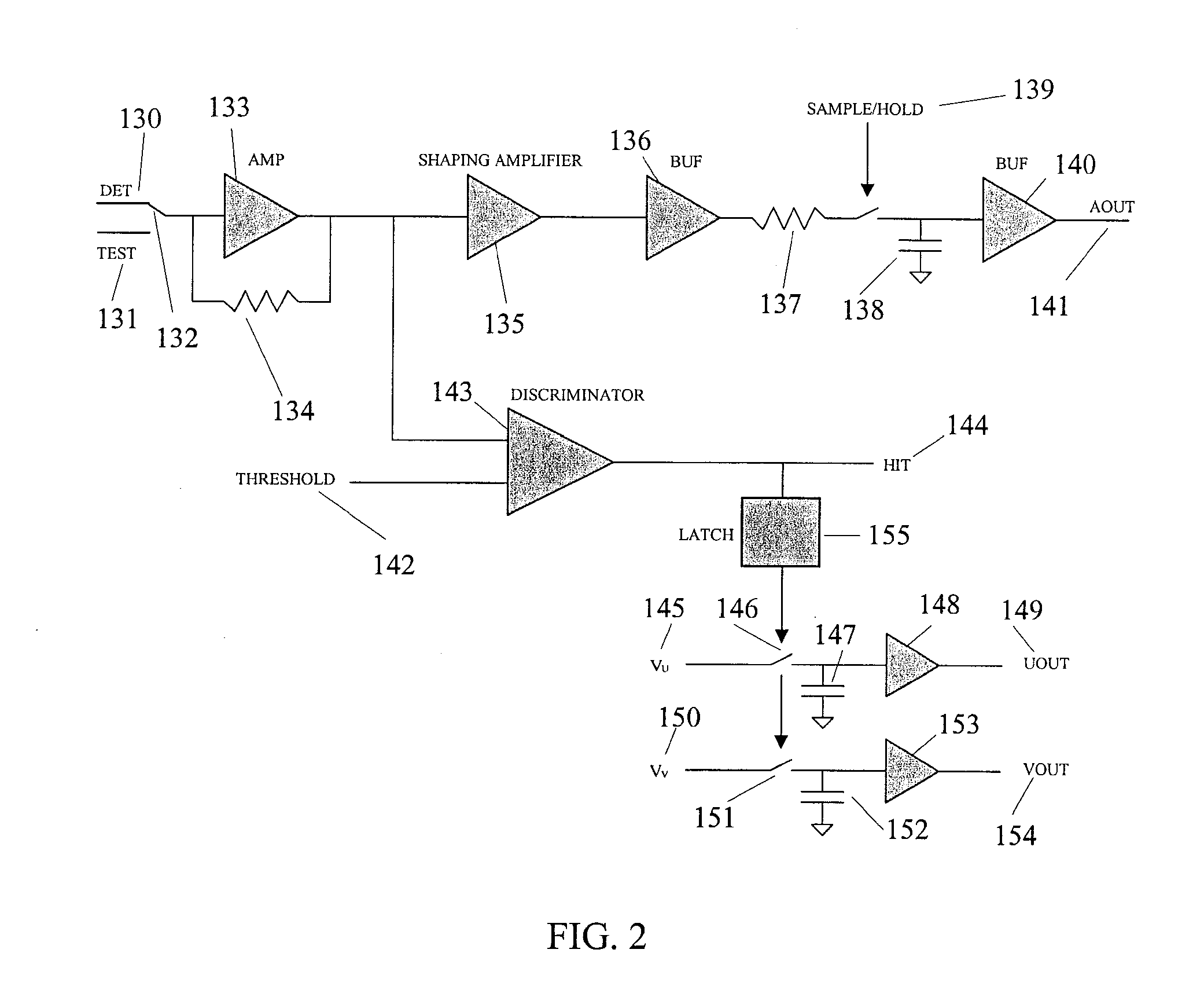
[0023]For minimum system power dissipation, a leading edge level crossing discriminator is used, carefully designed to minimize time walk. For pulses with amplitude greater than about 50% of the photopeak, the time walk can be controlled within 1-2 ns. However, the leading edge of the LSO scintillation light must be observed with the maximum possible bandwidth. A high speed transimpedance preamplifier is used, preserving the bandwidth of the APD. In contrast to the case of a charge sensitive preamplifier, no fast shaper or complex discriminator is required in the timing path, offering considerable power savings. Furthermore, pole / zero cancellation is irrelevant for a transimpedance preamplifier, an important advantage for high rate operation. The pulse amplitude measurement proceeds by using a low power slow shaper and peak stretcher followed by an A / D converter.

Problems solved by technology

However, this technique has a limited diagnostic accuracy for detecting cancer, and image interpretation is subject to considerable inter- and intra-observer variability.
In summary, mammography is a useful screening tool for detecting cancer but is limited by a large number of false positive tests resulting in unnecessary biopsies and, more importantly, a considerable number of false negative tests resulting in missed diagnosis of cancer, which results in unnecessary deaths.
The somewhat lower than desired sensitivity is due to relatively poor accuracy for detecting tumors of less than 1 cm in size.
The detection of malignant breast tumors with PET is limited by the spatial resolution and sensitivity of whole body PET systems.
Whole body PET is also an expensive technology, which is generally only available in the larger medical facilities in the U.S. Therefore, a dedicated compact higher resolution PET system that improves the sensitivity, specificity, and availability of PET imaging for breast cancer detection, which can also be used for many other applications is discussed below.
The approach of placing APD arrays on both front and back sides of the LSO crystals is also an innovative concept that poses design challenges in ensuring that the amount of absorber material in the photons' path is kept to a minimum.

Method used


Image

  • X-Ray and Gamma Ray Detector Readout Sytem
  • X-Ray and Gamma Ray Detector Readout Sytem
  • X-Ray and Gamma Ray Detector Readout Sytem

Examples


Embodiment Construction

[0052]There has been considerable interest in recent years in developing dedicated high resolution positron emission tomography (PET) systems for applications in breast cancer imaging and small-animal imaging. The goal in these systems is to achieve much higher spatial resolution and sensitivity for specific tasks than is possible with whole-body PET scanners designed for general purpose use. A second goal is to produce relatively inexpensive, compact and easy to use systems that make PET more accessible. Generally, these dedicated systems use small scintillator elements read out by position-sensitive or multi-channel PMTs. In most systems, some form of signal multiplexing is used to reduce the number of channels to a manageable number. Since the predominant mode of interaction at 511 keV in all scintillators currently used for PET is Compton scatter, multiplexing can lead to significant loss of position information. Furthermore, depth-of-interaction (DOI) blurring or radial elongat...


PUM


Abstract

A readout electronics scheme is under development for high resolution, compact PET (positron emission tomography) imagers based on LSO (lutetium ortho-oxysilicate, Lu2SiO5) scintillator and avalanche photodiode (APD) arrays. The key is to obtain sufficient timing and energy resolution at a low power level, less than about 30 mW per channel, including all required functions. To this end, a simple leading edge level crossing discriminator is used, in combination with a transimpedance preamplifier. The APD used has a gain of order 1,000, and an output noise current of several pA / √Hz, allowing bipolar technology to be used instead of CMOS, for increased speed and power efficiency. A prototype of the preamplifier and discriminator has been constructed, achieving timing resolution of 1.5 ns FWHM, 2.7 ns full width at one tenth maximum, relative to an LSO / PMT detector, and an energy resolution of 13.6% FWHM at 511 keV, while operating at a power level of 22 mW per channel. Work is in progress towards integration of this preamplifier and discriminator with appropriate coincidence logic and amplitude measurement circuits in an ASIC suitable for a high resolution compact PET instrument. The detector system and / or ASIC can also be used for many other applications for medical to industrial imaging.

Description

PRIORITY CLAIM[0001]This application is a continuation of U.S. application Ser. No. 10 / 291,251 filed Nov. 8, 2002 now issued U.S. Pat. No. 7,818,047 which claims the priority to U.S. Provisional Patent Application No. 60 / 331,161 filed Nov. 9, 2001, both of which are incorporated herein by reference in their entirety.GOVERNMENT RIGHTS NOTICE[0002]There invention was made with U.S. Government support under Contract Numbers DE-FG03-00ER83058 and DE-FG03-00ER83058 / A002, both awarded by Department of Energy. The U.S. Government has certain rights in the invention.FIELD OF INVENTION[0003]These detectors and readout electronics have been developed for high resolution Positron Emission Tomography (PET) for application to medical imaging. PET is an important new modality for imaging metabolism of organic radiopharmaceuticals and radiotracers. The detector system described here can also be used for non-tomographic medical imaging of positron emitting compounds. It can also be used for other g...

Claims


Application Information

Patent Timeline
Patent Type & Authority Applications(United States)
IPC IPC(8): H03L5/00G01T1/29
CPCA61B6/037G01T1/2985
Inventor TUMER, TUMAY O.CLAJUS, MARTINVISSER, GERARD
Owner NOVA R&D
Learn More