

Method of engrafting cells from solid tissues
a solid tissue and cell technology, applied in the field of tissue engrafting, can solve the problems of poor cell survival, muted effects, and the failure to treat the disease of most solid organs as successfully, and achieve the effect of improving the survival rate of cells
- Summary
- Abstract
- Description
- Claims
- Application Information
AI Technical Summary
Benefits of technology
Problems solved by technology
Method used
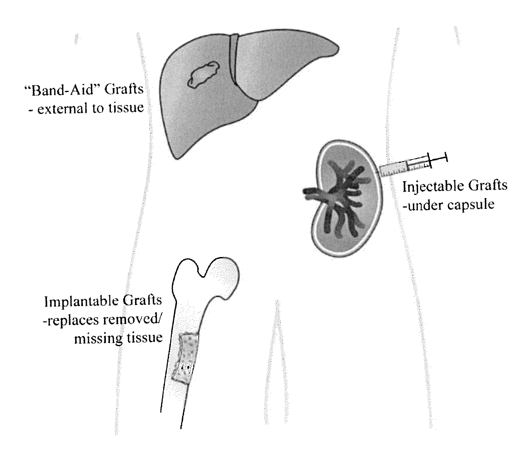
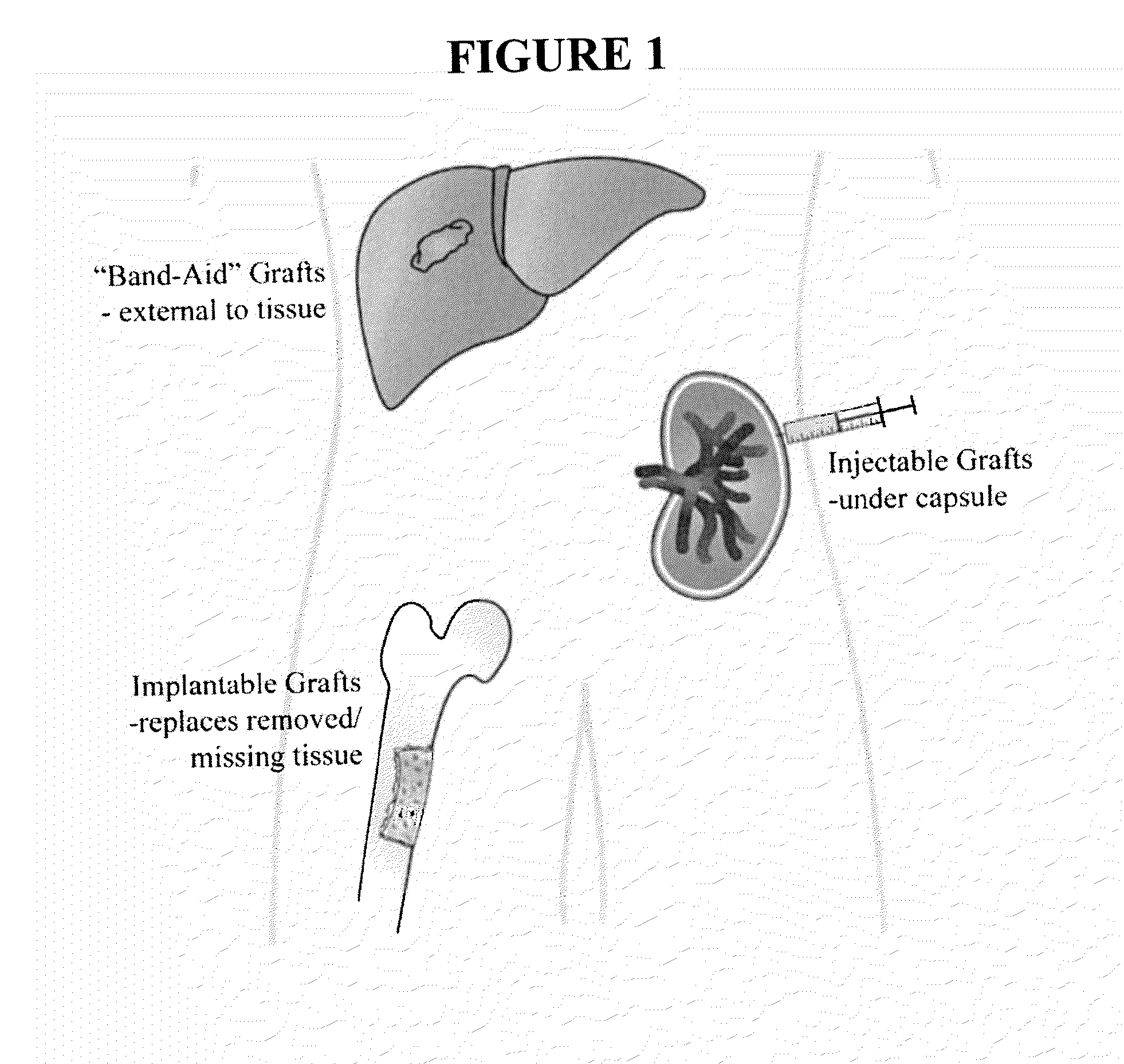
Image

Examples
example 1
[0097]Mouse hepatic progenitor cells were isolated from a host C57 / BL6 mouse (4-5 weeks) according to reported protocols. For the “grafting” studies, a GFP reporter was introduced into the hepatic progenitor cells. The cells were then mixed with hyaluronan (HA) hydrogels and the HA crosslinked with Poly (Ethylene Glycol)-Diacrylate (PEG-DA) prior to introduction into a subject mouse. For introduction / transplantation, mice were anesthetized with ketamine (90-120 mg / kg) and xylazine (10 mg / kg), and their abdomens were opened. The cells, with or without HA, were then slowly injected into the front liver lobe. The incision site was closed and animals were given 0.1 mg / kg buprenorphine every 12 hrs for 48 hrs. After 48 hrs, animals were euthanized, and tissue was removed, fixed, and sectioned for histology.
[0098]To determine cell localization within the murine models, “control” hepatic progenitor cells were infected for 4 hrs at 37° C. with a luciferase-expressing adenoviral vector at 50...
example 2
[0101]Human hepatic progenitor cells were isolated from fetal liver tissue (16-20 weeks) according to reported protocols. A luciferase-expressing adenoviral vector was introduced into the hepatic progenitor cells. The cells were then mixed with thiol-modified carboxymethyl HA (CMHA-S) and in the presence of the crosslinker Poly (Ethylene Glycol)-Diacrylate (PEG-DA) prior to introduction into a subject mouse. More specifically, the hydrogel was constructed by dissolving HA dry reagents in KM to give a 2.0% solution (weight / volume) and the crosslinker was dissolved in KM to give a 4.0% weight / volume solution. Samples were then allowed to incubate in a 37° C. water bath to completely dissolve. Collagen III and laminin were prepared at a concentration of 1.0 mg / ml and blended with crosslinker / hydrogels in a 1:4 ratio.
[0102]For introduction / transplantation, mice were anesthetized with ketamine (90-120 mg / kg) and xylazine (10 mg / kg), and their abdomens were opened. The cells, with or with...
example 3
[0108]Human pancreatic progenitor cells are isolated from pancreatic tissue. A luciferase-expressing adenoviral vector is introduced into the progenitor cells. The cells are then mixed with thiol-modified carboxymethyl HA (CMHA-S) and in the presence of the crosslinker Poly (Ethylene Glycol)-Diacrylate (PEG-DA) as described in Example 2.
[0109]For introduction / transplantation, mice are anesthetized with ketamine (90-120 mg / kg) and xylazine (10 mg / kg), and their abdomens are opened. The cells, with or without HA, are then slowly injected into the pancreas. The incision site is closed and animals are given 0.1 mg / kg buprenorphine every 12 hrs for 48 hrs. After 48 hrs, animals are euthanized, and tissue is removed, fixed, and sectioned for histology.
[0110]To determine cell localization within the murine models, “control” progenitor cells are infected for 4 hrs at 37° C. with a luciferase-expressing adenoviral vector at 50 POI. Survival surgery is performed as described above, and cells ...
PUM
| Property | Measurement | Unit |
|---|---|---|
| shear moduli | aaaaa | aaaaa |
| sizes | aaaaa | aaaaa |
| thick | aaaaa | aaaaa |
Abstract
Description
Claims
Application Information