Cathepsin l proteolytically processes histone h3 during mouse embryonic stem cell differentiation
a cathepsin and embryonic stem cell technology, applied in biochemistry apparatus and processes, peptide/protein ingredients, drug compositions, etc., can solve the problems of endogenous proteolysis that has not been well documented in mammalian cells, and is specific, regulated, and regulated. to achieve the effect of reducing expression and high expression
- Summary
- Abstract
- Description
- Claims
- Application Information
AI Technical Summary
Benefits of technology
Problems solved by technology
Method used
Image
Examples
example 1
Cell Culture and Differentiation
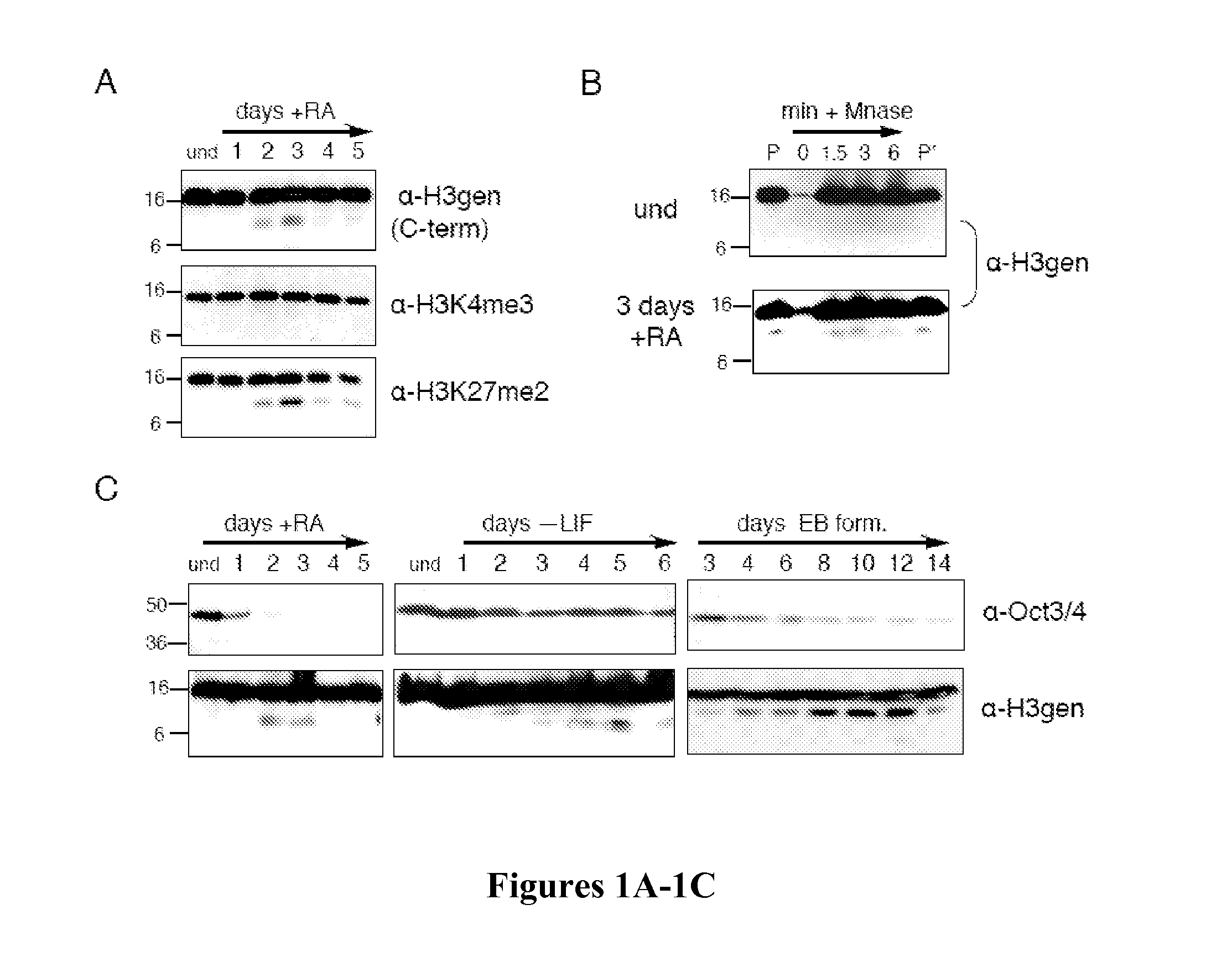
[0141]ES cells (LF2 line) were cultured as previously described (Bernstein et al., “Mouse Polycomb Proteins Bind Differentially to Methylated Histone H3 and RNA and are Enriched in Facultative Heterochromatin,”Mol Cell Biol 26:2560-2569 (2006), which is hereby incorporated by reference in its entirety). Cells were grown on gelatin-coated plates without feeder cells and maintained in an undifferentiated state through culture in KODMEM (Invitrogen 1082-9018)), 2 mM L-glutamine (Sigma G7513), 15% ES grade fetal bovine serum (Gibco 10439-024), 10−4 mM 2-mercaptoethanol, and leukemia inhibitory factor (LIF). To differentiate, cells were plated sparsely at ˜1*104 cells / cm2, allowed to adhere overnight and then fed with differentiation media containing DMEM, 10% FBS, 10−4 mM 2-mercaptoethanol and, for retinoic acid (RA) differentiation, 100 nM all-trans-RA (Sigma R2625). Embryoid body formation was accomplished by splitting partially trypsinized cells onto n...
example 2
Cell Extract Preparation
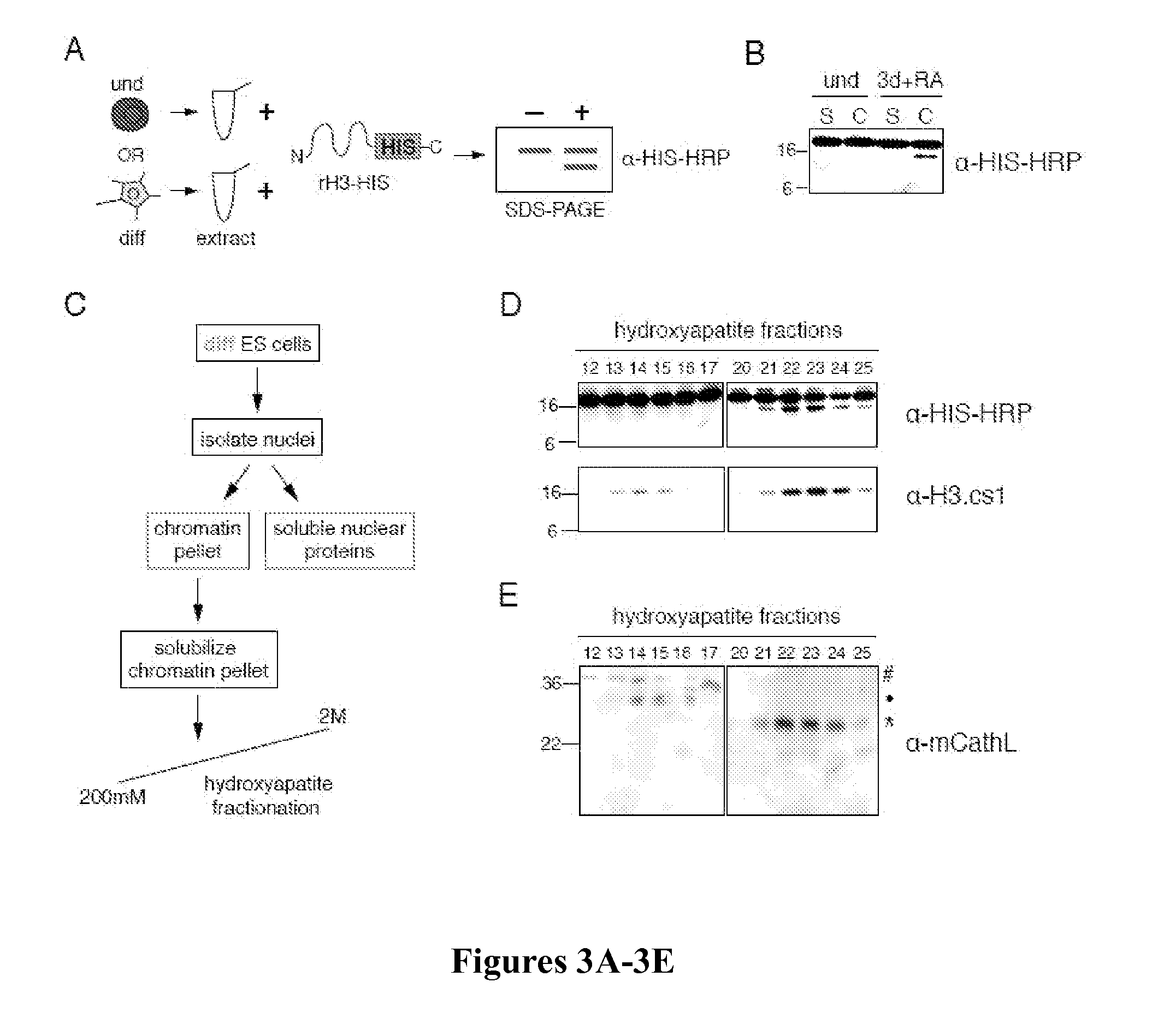
[0142]Whole cell extracts were prepared by resuspending fresh or flash frozen cell pellets in SDS-Laemmli sample buffer and boiling immediately. Chromatin extracts were prepared by sonicating chromatin pellets in buffer (10 mM HEPES, 10 mM KCl, 1.5 mM MgCl2, 0.34M sucrose, 10% glycerol, 5 mM β-mercaptoethanol) after isolation by various methods (high salt extraction as described by Dignam et al., “Accurate Transcription Initiation by RNA Polymerase II in a Soluble Extract From Isolated Mammalian Nuclei,”Nucleic Acids Res 11:1475-1489 (1983), which is hereby incorporated by reference in its entirety, or chromatin fractionation as described by Mendez et al., “Chromatin Association of Human Origin Recognition Complex, cdc6, and Minichromosome Maintenance Proteins During the Cell Cycle: Assembly of Prereplication Complexes in Late Mitosis,”Mol Cell Biol 20:8602-8612 (2000), which is hereby incorporated by reference in its entirety). Briefly, cells were swelled in...
example 3
Histone Purification, Edman Degradation and MS-MS Mapping of H3Cleavage Sites
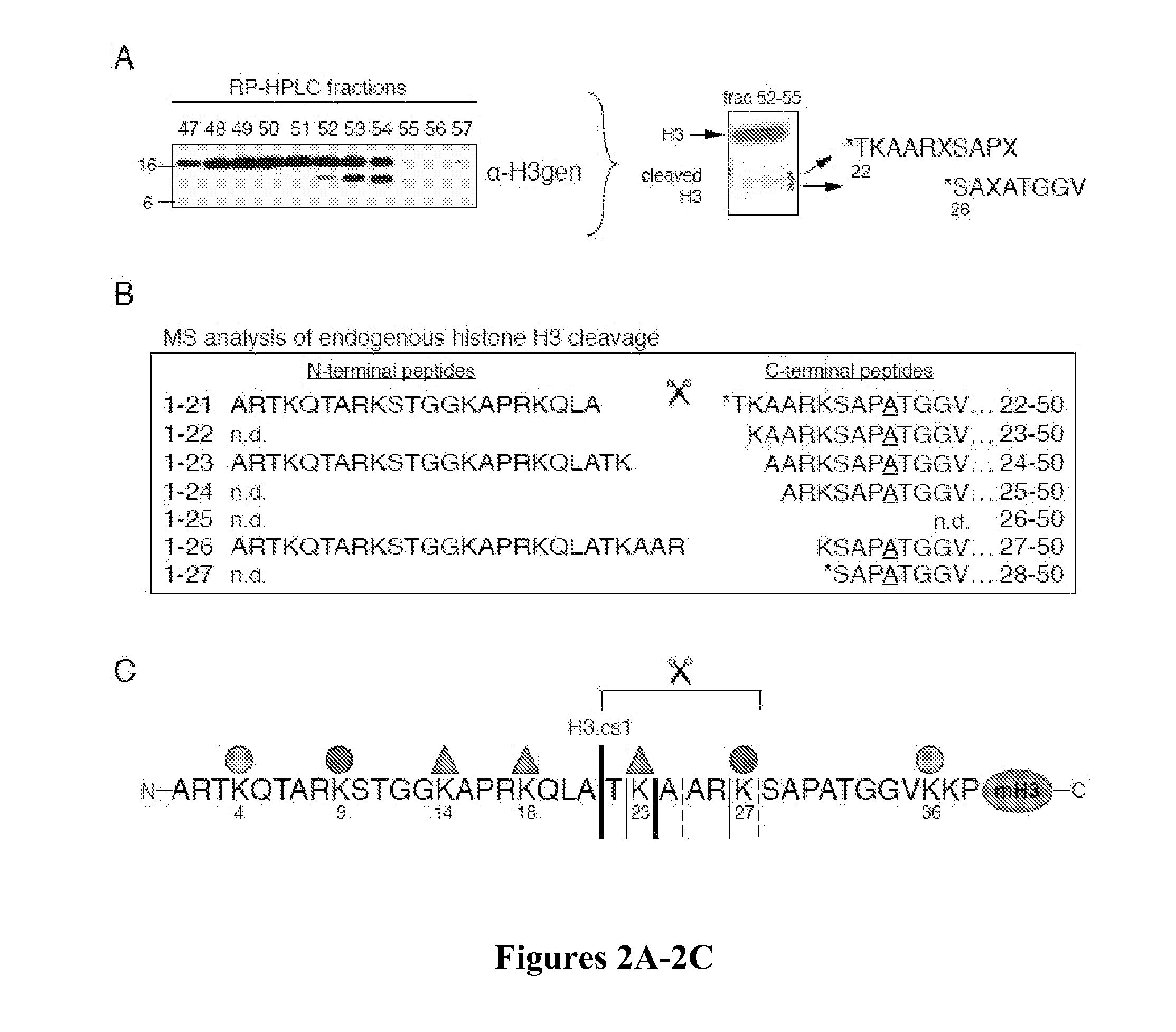
[0143]Histones were acid extracted from nuclei and purified by RP-HPLC using a C8 column as described (Shechter et al., “Extraction, Purification and Analysis of Histones,”Nat Protoc 2:1445-1457 (2007), which is hereby incorporated by reference in its entirety). RP-HPLC fractions containing the H3 sub-band were pooled and repurified by RP-HPLC using a C18 column. Equal amounts of fractions 52-55 were pooled, separated by SDS-PAGE, and blotted onto 0.2 μm pore size membrane (Millipore Cat. No. ISEQ00010). H3 sub-bands were excised and subjected to Edman degradation as described previously (Strahl et al., “Methylation of Histone H3 at Lysine 4 is Highly Conserved and Correlates With Transcriptionally Active Nuclei in Tetrahymena,”Proc Natl Acad Sci USA 96:14967-14972 (1999), which is hereby incorporated by reference in its entirety). Fraction 54 was digested with endoproteinase GluC (Roche Diagnostics, Indian...
PUM
 Abstract
Description
Claims
Application Information
 - R&D
- Intellectual Property
- Life Sciences
- Materials
- Tech Scout
- Unparalleled Data Quality
- Higher Quality Content
- 60% Fewer Hallucinations
Browse by: Latest US Patents, China's latest patents, Technical Efficacy Thesaurus, Application Domain, Technology Topic, Popular Technical Reports.
© 2025 PatSnap. All rights reserved.Legal|Privacy policy|Modern Slavery Act Transparency Statement|Sitemap|About US| Contact US: help@patsnap.com


