Method and apparatus for tissue grafting

a tissue grafting and tissue technology, applied in the field of tissue grafting methods and apparatuses, can solve the problems of extensive healing time at the donor site, limited tissue availability for autografting, and large loss of sheet grafts after placement, etc., and achieve the effect of rapid healing of the donor si

Active Publication Date: 2012-02-16
THE GENERAL HOSPITAL CORP
View PDF8 Cites 113 Cited by
  • Summary
  • Abstract
  • Description
  • Claims
  • Application Information

AI Technical Summary

Benefits of technology

[0020]Exemplary embodiments of the present disclosure provide method and apparatus for obtaining small portions of graft tissue that can be accompanied by rapid healing of the donor site. For example, the exemplary embodiment of the method can be provided for obtaining skin graft tissue by harvesting small portions of the tissue, e.g., micrografts, from a donor site.
[0021]Such micrografts can comprise skin tissue that can include, e.g., epidermal and dermal tissue, and / or tissue obtained from other body organs. The micrografts can have at least one dimension that is relatively small, e.g., less than about 1 mm, or less than about 0.5 mm, or optionally about 0.3 mm or less, or about 0.2 mm. Such exemplary small dimensions of the micrografts can facilitate both healing of the donor site following harvesting and viability of the micrografts by allowing greater diffusional nourishment of the micrograft tissue. The small regions of damage in the donor site caused by a removal of the tissue portions can heal rapidly with little or no formation of visible scars. The micrografts obtained from skin tissue can include, e.g., epidermal and dermal tissue, and can also include stem cells that can be located proximal to the dermal / fatty layer boundary. The micrografts can also be obtained from other types of tissue, e.g., various internal organs or the like.
[0022]A fraction of dermal tissue that is removed from a donor site can be, e.g., less than about 70%, or less than about 50%, although other fractions may be used. The harvested tissue portions can be in the shape of cylinders, elongated strips, or other geometries which can include at least one small dimension. In certain exemplary embodiments, a portion of the tissue at the donor site can be frozen or partially frozen. Such freezing can facilitate cutting, removal and / or viability of the harvested tissue portions.
[0025]A further exemplary embodiment of the apparatus can be provided that includes a plurality of such tubes for simultaneous harvesting of a plurality of micrografts. An enclosure and / or a source of pressure, e.g., a pump or the like, can be provided in communication with the proximal ends of the tubes to facilitate application of pressure and / or suction to the plurality of tubes. A vibrating arrangement can be coupled to the apparatus to facilitate the insertion of the tubes into the donor site.
[0026]The exemplary micrografts can be placed in a biocompatible matrix, e.g., to form a graft or directly into tissue at the recipient site The biocompatible matrix can be formed using collagen, polylactic acid, hyaluronic acid, and / or other substances which can support the harvested micrograft tissue portions and promote their growth. The matrix can optionally include, e.g., nutrients and / or other substances to promote tissue growth. The harvested tissue portions can be bonded to the matrix using techniques such as photochemical tissue bonding to provide structural stability. The matrix can then be applied to the recipient site, which can promote growth and revascularization of the tissue portions to form a continuous sheet of the grafted tissue.

Problems solved by technology

Availability of tissue for autografting can be limited by characteristics of candidate donor sites, including a number and / or total area of tissue grafts, healing behavior of the donor site, similarity of the donor and recipient sites, aesthetic considerations, etc.
Small areas of a sheet graft can be lost after placement because of a buildup of fluid (e.g., a hematoma) can occur under the sheet graft following placement the sheet graft.
Split-thickness grafts may be preferable to full-thickness grafts because removing large amounts of full-thickness skin tissue from the donor site can lead to scarring and extensive healing times at the donor site, as well as an increased risk of infection.
However, skin tissue removed from the donor site for a split-thickness skin autograft can include only a thin epithelial layer, which can lack certain elements of the dermis that improve structural stability and normal appearance in the recipient site.
However, application of conventional full-thickness skin grafts can be limited to relatively small, uncontaminated, well-vascularized wounds, and thus may not be appropriate for as many types of graft procedures as split-thickness grafts.
A meshed skin graft can be used to cover larger areas of open wounds that may be difficult to cover using sheet grafts because of, e.g., a lack of a sufficient area of healthy donor sites.
For example, donor skin can be meshed at a ratio of about 1:1 or 1:2 ratio, whereas larger expansion ratios may lead to a more fragile graft, scarring of the meshed graft as it heals, and / or extended healing times.
However, meshed grafts may be less durable graft than sheet grafts, and a large mesh can lead to permanent scarring after the graft heals.
Large burn sizes can lead to more than one grafting procedure during a hospital stay, and may require long periods of immobilization for healing.
Heterograft skin tissue can also be used for temporary coverage of an excised wound prior to placement of a more permanent autograft, and may be used because of a limited availability and / or high expense of human skin tissue.
In some cases religious, financial, or cultural objections to the use of human cadaver skin may also be factors leading to use of a heterograft.
For example, hair can be more likely to grow from full-thickness grafts than from split-thickness grafts, but such hair growth may be undesirable based on the location of the wound.
Further, certain hair follicles may not be oriented perpendicular to the skin surface, and they can be transected if an incision provided to remove graft tissue is not oriented properly.
Prior to the regeneration, the skin graft tissue may lack normal lubrication of sebum produced by these glands, which can make such grafts more susceptible to injury.
In general, grafting procedures may be limited by the amount of tissue which can be removed from the donor site without causing excessive adverse effects.
Harvesting of graft tissue from the donor site generally can generate undesirable large-scale tissue damage to the donor site.

Method used


Image

  • Method and apparatus for tissue grafting
  • Method and apparatus for tissue grafting
  • Method and apparatus for tissue grafting

Examples


example

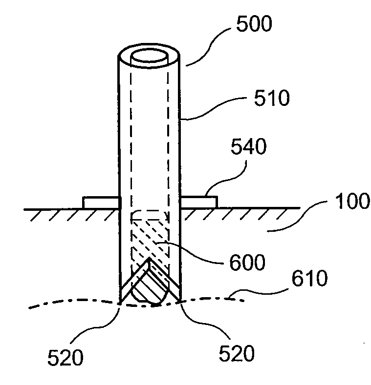
[0089]An image of a distal end of an exemplary apparatus that includes two points is shown in FIG. 8A. This apparatus is similar to the exemplary apparatus 500 illustrated, e.g., in FIG. 5A. A further rotated image of this exemplary apparatus is shown in FIG. 8B. The exemplary apparatus was formed using a tube having an outside diameter of about 1 mm, and an inside diameter of about 0.5 mm. The points or extensions were formed by grinding two opposite sides of the distal end of the tube at an appropriate angle relative to the axis of the tube. The angle used was about 30 degrees, although other angles may also be used. A beveled edge of the tube wall can be seen along the sides of the points or extensions. The shape of these points can facilitate insertion of the apparatus into tissue of a donor site and / or separation of a portion of micrograft tissue from the donor site, as described in more detail herein. For example, such micrografts can be separated and removed from the donor si...


PUM


Abstract

Exemplary embodiments of apparatus and method for harvesting small portions of tissue (“micrografts”) to form grafts can be provided. For example, a hollow tube can be inserted into tissue at a donor site, where a distal end of the hollow tube can have two or more points or extensions to facilitate separation of the micrografts from the surrounding tissue. The exemplary apparatus can be provided that includes a plurality of such tubes for simultaneous harvesting of a plurality of micrografts. The harvested micrografts can have a small dimension, e.g., less than about 1 mm, or less than about 0.3 mm, which can promote healing of the donor site and / or viability of the harvested tissue. The micrografts can be approximately cylindrical or strip-shaped, and can be placed in a biocompatible matrix to form a graft or directly into tissue at the recipient site. Such exemplary micrografts can be obtained from skin or other types of tissue, e.g., various internal organs.

Description

CROSS-REFERENCE TO RELATED APPLICATION[0001]The present application claims priority from U.S. Provisional Patent Application Ser. No. 61 / 041,587 filed Apr. 1, 2008, the disclosure of which is incorporated herein by reference in its entirety.FIELD OF THE INVENTION[0002]The present disclosure relates to exemplary embodiments of method and apparatus for providing tissue grafts using tissue from a donor site.BACKGROUND INFORMATION[0003]An autograft can refer to tissue transplanted from one part of an individual's body (e.g., a “donor site”) to another part (e.g., a “recipient site”). Autografts can be used, for example, to replace missing skin and other tissue and / or to accelerate healing resulting from trauma, wounds, burns, surgery and birth defects. Availability of tissue for autografting can be limited by characteristics of candidate donor sites, including a number and / or total area of tissue grafts, healing behavior of the donor site, similarity of the donor and recipient sites, ae...

Claims


Application Information

Patent Timeline
Patent Type & Authority Applications(United States)
IPC IPC(8): A61B17/322A61B18/20
CPCA61B10/0233A61B17/32053A61B17/322A61B17/32093A61B2017/00969A61B2017/306A61B2017/3225A61B2017/00752A61B17/00A61B17/22A61B17/34A61B5/14503A61B10/02A61B17/1635A61B17/320068
Inventor ANDERSON, RICHARD R.HAMBLIN, MICHAEL R.MANSTEIN, DIETERFARINELLI, WILLIAM A.
Owner THE GENERAL HOSPITAL CORP
Why Eureka
Try Eureka