
Method for treatment of blood tumor using Anti-tim-3 antibody

Active Publication Date: 2012-04-26
KYOWA HAKKO KIRIN CO LTD +1
View PDF1 Cites 27 Cited by
  • Summary
  • Abstract
  • Description
  • Claims
  • Application Information

AI Technical Summary

Benefits of technology

[0053]TIM-3 is known as a gene which controls immune function in a living body.
[0054]In the present invention, the inventors apply the newly found nature of TIM-3, that is, express in blood tumor, to establish an usage of an agent for treatment or diagnosis and further the gene as a target for isolating a tumor cell by using an antitumor substances (preferably anti-TIM-3 antibody) targeting TIM-3 expressing cells.
[0055]Since now, some molecular target drugs have been developed in the field of blood tumor. However, a few of them target a cancer stem cell. For example, AML treating drug, Mylotarg, which targets a CD33 protein is one of them (Bernstein, Leukemia (2000) 14, 474-475). However, as described above, it has limitations such as expression level of CD33, recurrence, age, resistance to chemical therapy.
[0056]In Examples of the present invention, cytotoxicity of the anti-TIM-3 antibody on TIM3 expressing cells or AML primary cells is found. This activity suggests therapeutic effects on various blood diseases by the anti-TIM-3 antibody.

Problems solved by technology

Different from the normal stem cell, studies on cancer stem cells have been delayed for a long time since their true nature could not been found.
Particularly, the characteristics that cancer stem cells express a group of multiple drug resistance genes and are at the stationary phase of cell cycle similar to the normal stem cells could become a therapeutically great problem.
Another problem is that side effects are large since these two treatments affect the entire body.
However, it can be said that that the use of Mylotarg is limited since Mylotarg can be applied only when a patient meets the following four limitations such as the expression rate of CD33 of 80% or more, a case of recurrency, age of 60 or more, and resistance to other chemical therapy.
However, use of mouse antibodies as drugs in vivo is limited due to a large number of reasons.
Further, the Fc region of mouse antibody is not effective for stimulation of human complement or cellular cytotoxicity.

Method used


Image

  • Method for treatment of blood tumor using Anti-tim-3 antibody
  • Method for treatment of blood tumor using Anti-tim-3 antibody
  • Method for treatment of blood tumor using Anti-tim-3 antibody

Examples


example 1

Preparation of Bone Marrow and Peripheral Blood Cells

[0182]Provision of the analytes from patients and healthy volunteers was carried out under the recognition by an ethical committee at the Kyushu University Hospital. The bone marrow cell was collected by a bone-marrow aspiration from a patient of leukemia or myelodysplastic syndrome and a healthy volunteer. The peripheral blood was collected by an intravenous blood collection from a healthy volunteer. The stem cell-mobilized peripheral blood was collected from a patient indicated for autologous peripheral blood stem cell transplantation (a case in which a patient has a relapse of a diffuse large cell type B cell lymphoma after entering into complete remission with chemotherapy and has sensitivity for the chemotherapy). Specifically, after completion of chemotherapy (Cyclophosphamide (CY) massive dose therapy or VP-16 (etoposide) massive dose therapy), peripheral blood stem cells were mobilized by the administration of a G-CSF prep...

example 2

Staining and Analyzing Methods of Bone Marrow Normal Hematopoietic and Tumor Stem Cells

[0184]The cells were stained using 2 μl of an anti-human CD34 antibody (manufactured by Becton Dickinson & Co., (hereinafter referred to as BD), Cat No. 340441), 20 μl of anti-human CD38 antibody (manufactured by CALTAG, Cat No. MHCD3815), 2 μl of anti-human CD90 antibody (manufactured by BD, Cat No. 555595) or 20 μl of anti-TIM-3 antibody (manufactured by R & D Systems, 344823) as the primary antibodies at 4° C. for 40 minutes. Then, PBS was added for wash, and the obtained suspension was centrifuged at 1500 rpm for 5 minutes. After the supernatant was discarded, 100 μl of the staining medium was added thereto. The obtained cells were stained by adding 5 μl of each of anti-human Lineage antibodies (anti-human CD3 (manufactured by BD, Cat No. 555341), CD4 (manufactured by BD, Cat No. 555348), CD8 (manufactured by BD, Cat No. 555636), CD10 (manufactured by BD, Cat No. 555376), CD19 (manufactured by...

example 3

Expression of Human TIM-3 Molecule in Human Blood Tumor

[0185]Expression of human TIM-3 molecule in human blood tumor was examined using the methods of Example 1 and Example 2.

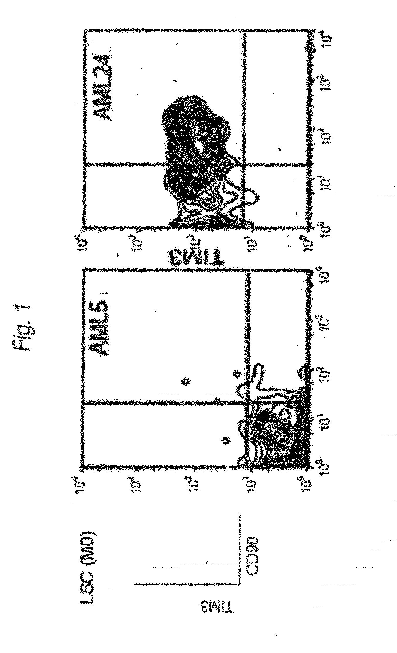
[0186]Expression of human TIM-3 molecule in AML (M0) patient-derived bone marrow Lin(−)CD34(+)CD38(−) cell, Lin(−)CD34(+)CD38(+) cell and Lin(−)CD34(−) cell was examined by analysis using multicolor flow cytometry. The results are shown in FIG. 1 and Table 1. In the patients with AML (M0), expression of TIM-3 was observed in one of the two cases. Accordingly, this example showed usefulness of TIM-3 as a therapeutic target for AML (M0) cells.

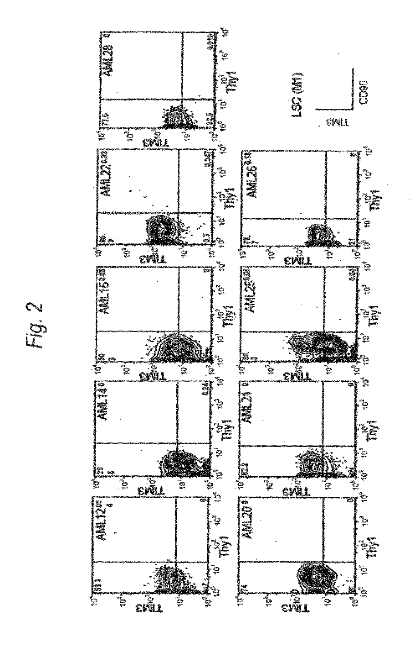
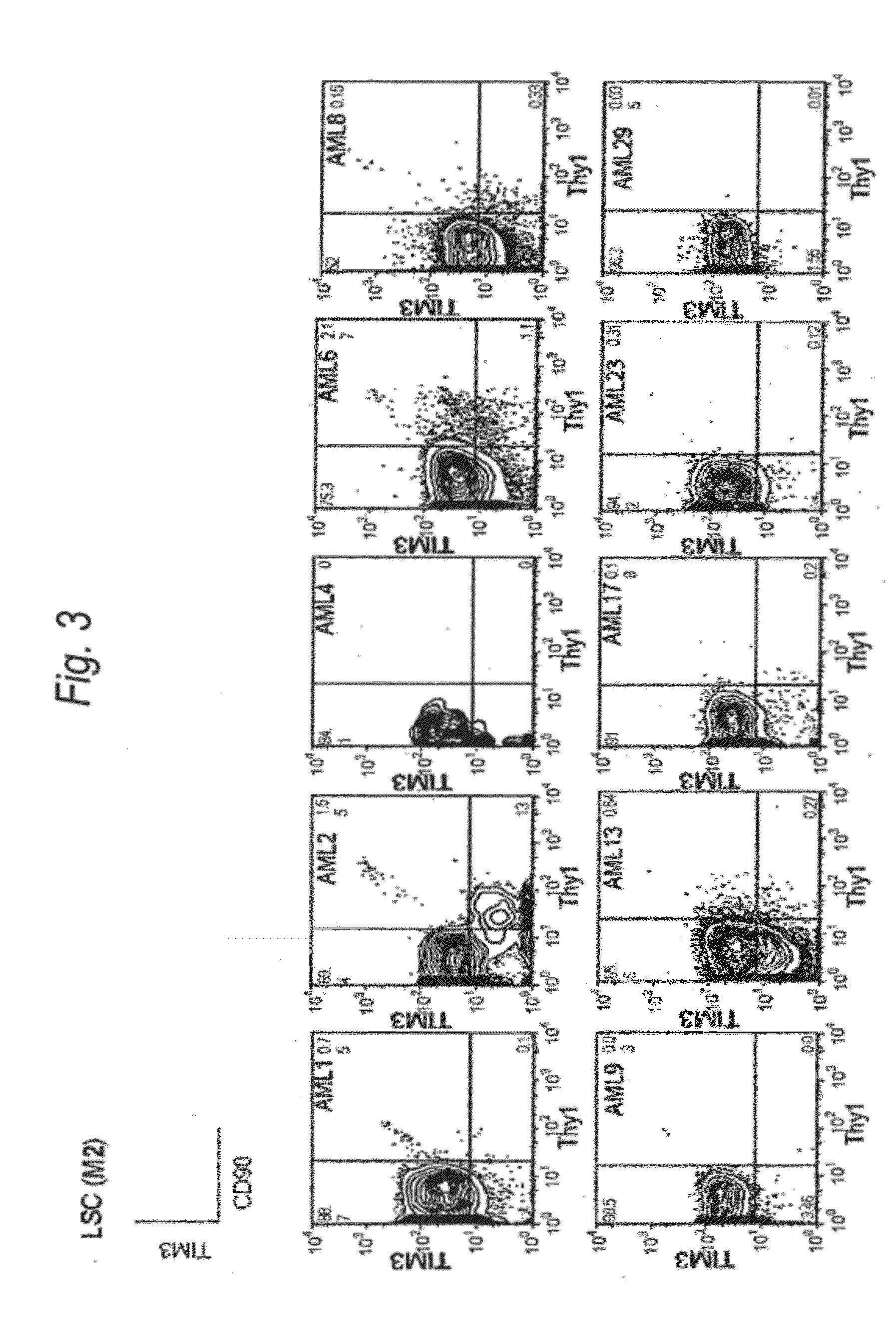
[0187]Expression of human TIM-3 molecule in AML (M1) patient-derived bone marrow Lin(−)CD34(+)CD38(−) cell, Lin(−)CD34(+)CD38(+) cell and Lin(−)CD34(−) cell was examined by analysis using multicolor flow cytometry. The results are shown in FIG. 2 and Table 1. In the patients with AML (M1), expression of TIM-3 was observed in all nine cases. Accordingly, this example showed u...


PUM

PropertyMeasurementUnit
Fractionaaaaaaaaaa
Fractionaaaaaaaaaa
Fractionaaaaaaaaaa

Abstract

Disclosed is a therapeutic method comprising administering a TIM-3 antibody to a subject who is suspected to be suffering from blood tumor and in whom TIM-3 has been expressed in a Lin(−)CD34(+)CD38(−) cell fraction of bone marrow or peripheral blood or a subject who has been received any treatment for blood tumor. Also disclosed is a composition for preventing or treating blood tumor, which comprises a TIM-3 antibody as an active ingredient. Conceived diseases include those diseases which can be treated through the binding or targeting of the TIM-3 antibody to blood tumor cells (AML cells, CML cells, MDS cells, ALL cells, CLL cells, multiple myeloma cells, etc.), helper T cell (e.g., Th1 cells, Th17 cells), and antigen-presenting cells (e.g., dendritic cells, monocytes, macrophages, and cells resembling to the aforementioned cells (hepatic stellate cells, osteoclasts, microglial cells, intraepidermal macrophages, dust cells (alveolar macrophages), etc)), all of which are capable of expressing TIM-3. The diseases for which the therapeutic use is to be examined include blood diseases in which the expression of TIM-3 is observed in bone marrow or peripheral blood, particularly blood tumor.

Description

TECHNICAL FIELD[0001]The present invention relates to an agent for treating or diagnosing myeloid malignancy, especially acute myelogenous leukemia (AML), comprising an antibody to human TIM-3 protein (another name: human HAVCR2) as an active ingredient. In addition, the present invention relates to a method for isolating leukemic stem cells.BACKGROUND ART[0002]Regarding Malignant Tumor:[0003]A malignant tumor (cancer) is the first leading cause of death in Japan and the number of patients is increasing every year, and the development of a drug and a therapeutic method having high efficacy and safety is strongly desired. Examples of the cause of forming a malignant tumor include a mutation of DNA caused by radiation, ultraviolet rays and various carcinogenic substances. Studies on malignant tumors have been focused on identification of these genetic changes molecular biologically. As a result, it is considered that tumorigenic transformation is induced by accumulation of a large num...

Claims


Application Information

Patent Timeline
IPC IPC(8): A61K39/395A61P35/00
CPCA61K2039/505C07K16/2803G01N33/57407C07K2317/734C07K2317/732A61P35/00A61P35/02C07K16/3061C07K2317/73G01N33/57426G01N2333/70503
Inventor TAKAYANAGI, SHINICHIROINAGAKI, YOSHIMASAAKASHI, KOICHIKIKUSHIGE, YOSHIKANE
Owner KYOWA HAKKO KIRIN CO LTD
Learn More