Device for capture, enumeration, and profiling of circulating tumor cells

a tumor cell and profiling technology, applied in the field of diagnostic testing for cancer, can solve the problem of requiring several hours of sample processing

Inactive Publication Date: 2012-04-26
THE JOHN HOPKINS UNIV SCHOOL OF MEDICINE
View PDF0 Cites 24 Cited by
  • Summary
  • Abstract
  • Description
  • Claims
  • Application Information

AI Technical Summary

Benefits of technology

[0018]In one embodiment, the present invention relates to a method of diagnosing pancreatic cancer in a subject. The invention includes collection of a biological sample from a subject suspected of having pancreatic cancer. The sample is passed through a microfluidic d

Problems solved by technology

The microfluidic platform of the prior art has the disadvantages that the sample processing requires several

Method used


Image

  • Device for capture, enumeration, and profiling of circulating tumor cells
  • Device for capture, enumeration, and profiling of circulating tumor cells
  • Device for capture, enumeration, and profiling of circulating tumor cells

Examples


example 1

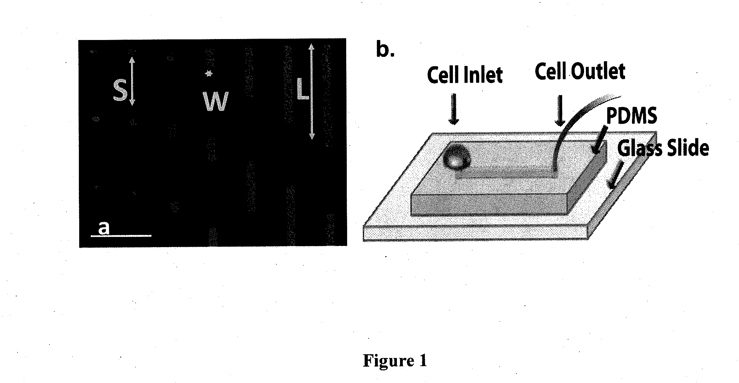
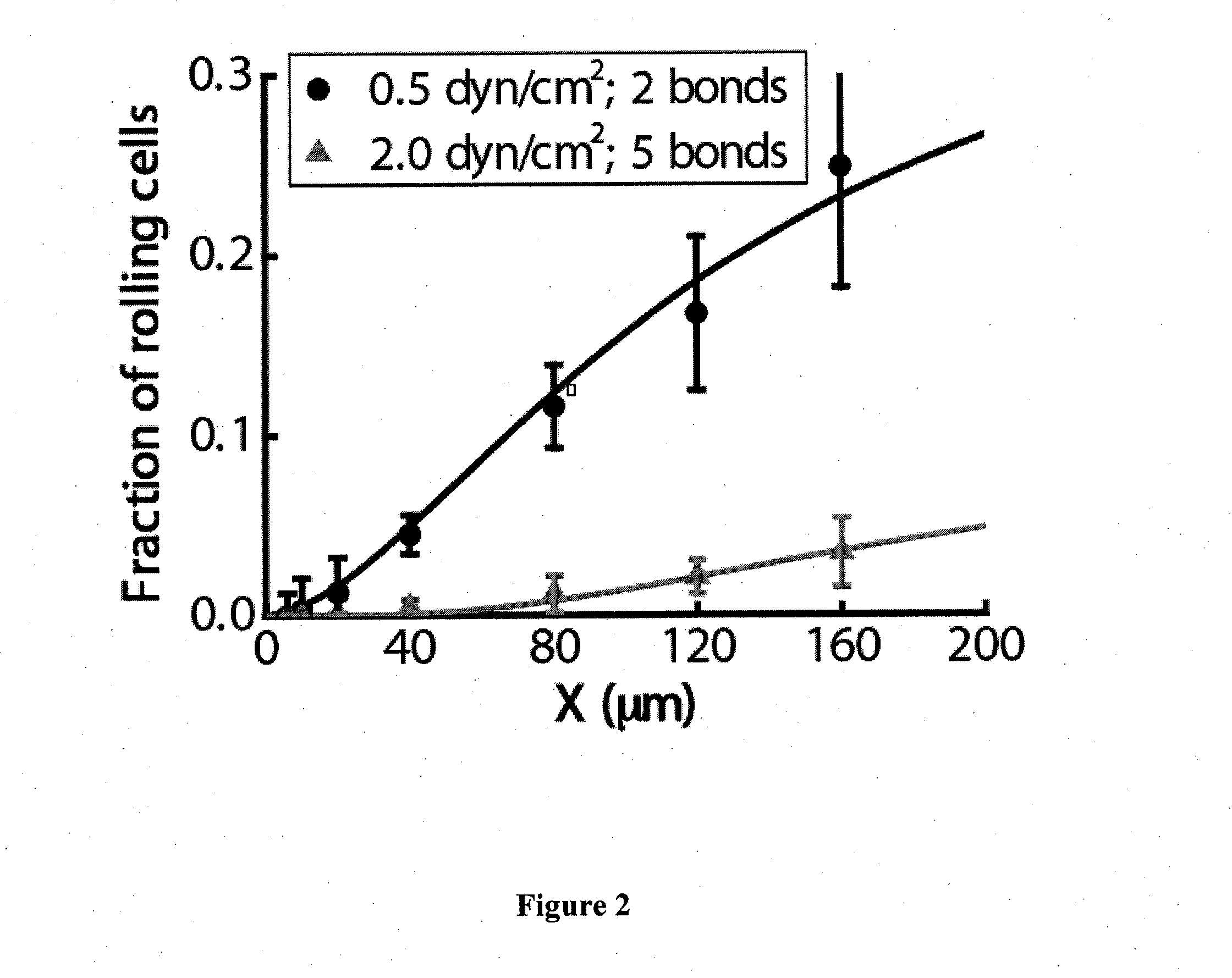
[0056]Development of a microfluidic device for selective capture of free-flowing cells on micro-patterned surfaces. Stripes of fixed width (10 μm) but varying lengths (6, 10, 20, 40, 80, 120 and 160 μm) separated by 100 μm in the direction of flow were patterned on a glass slide using a modified photolithography technique (FIG. 1a) (Ghosh et al. Langmuir 24(15):8134-42, 2008). In brief, a positive photoresist was used to coat a pre-cleaned glass slide surface, which was subsequently exposed to UV light through a chrome mask with the appropriate design. The irradiated regions of photoresist were then dissolved upon incubation with MF-CD-26 Developer. The photoresist-patterned slide was then immersed in 0.1% (v / v) solution of octadecyltrichlorosilane in hexane to render the slide surface hydrophobic. FITC labeled goat anti-human IgG-Fc specific antibody was then added to the slide prior to the immobilization of P- or L-selectin-Ig chimeras at prescribed concentrations (Ghosh et al. La...

example 2

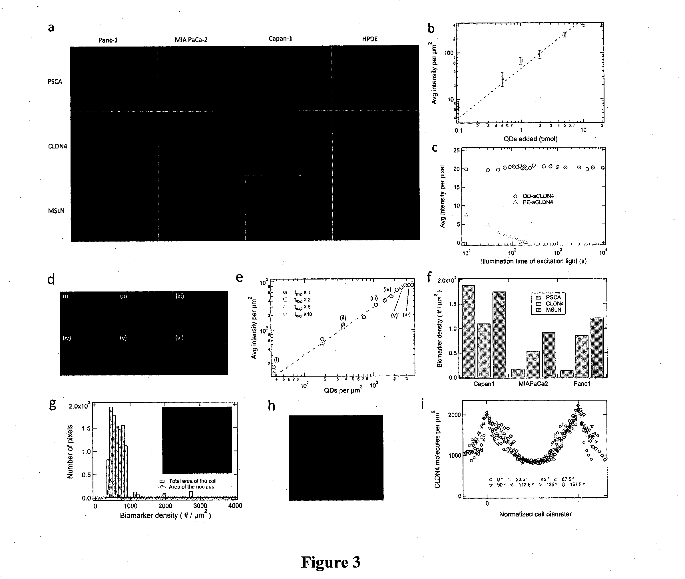
[0060]Quantitative profiling of molecular biomarkers for pancreatic cancer. In preliminary studies we have demonstrated proof-of-principle of quantitative profiling of mesothelin, claudin-4, and PSCA in three pancreatic cancer cells lines. The histologic progression from non-invasive precursor lesions (called Pancreatic Intraepithelial Neoplasia or PanINs) to invasive and metastatic pancreatic cancer is associated with the sequential accumulation of molecular markers (Maitra et al., Adv Anat Pathol 12(2):81-91, 2005; Maitra et al. Mod Pathol 16(9):902-912, 2003; Maitra et al. Annu Rev Pathol 3:157-188, 008; Prasad et al., Cancer Res 65(5):1619-26, 2005). For example, cell surface proteins such as prostate stem cell antigen (PSCA) (Maitra et al. Mod Pathol 16(9):902-912, 2003; Argani et al., Cancer Res 61(11):4320-4324, 2001) are aberrantly overexpressed even at the stage of non-invasive precursor lesions. In contrast, the protein mesothelin is aberrantly overexpressed only on the su...

example 3

[0067]Development of a microfluidic-based device for efficient and selective capture of pancreatic cancer cells: Enumeration of CTCs in peripheral blood of cancer patients has been reported to serve as an indicator of overall survival, disease stage forecasting, and as a promising method for clinical management (Braun et al., N Engl J Med 351(8):824-6, 2004). Nevertheless, detection of CTCs remains challenging due to their extremely low abundance among a high number of circulating blood cells. To this end, most assays employ enrichment steps based on morphometric or immunoseparation methods, which typically provide low recoveries with high purity, or low purity with high recoveries or in other cases, require complex sample processing whose success and reproducibility depend on trained personnel. Microchip technology has recently drawn much attention because of its potential to efficiently and selectively isolate and enumerate CTCs. For instance, a microfluidic approach utilizing an ...


PUM


Abstract

Applications in nanomedicine, such as diagnostics and targeted therapeutics, rely on the detection and targeting of membrane biomarkers. The present invention, in one embodiment, utilizes quantitative profiling, spatial mapping, and multiplexing of cancer biomarkers using functionalized quantum dots. This approach provides highly selective targeting molecular markers for pancreatic cancer with extremely low levels of non-specific binding and provides quantitative spatial information of biomarker distribution on a single cell, which is important since tumors cell populations are inherently heterogeneous. The quantitative measurements (number of molecules per square micron) is validated using flow cytometry and demonstrated using multiplexed quantitative profiling using color-coded quantum dots.

Description

CROSS-REFERENCE TO RELATED APPLICATIONS[0001]This application claims priority to U.S. Provisional Patent Application Ser. No. 61 / 367,188, filed Jul. 23, 2010, the contents of which are hereby incorporated by reference in its entirety.STATEMENT REGARDING FEDERALLY SPONSORED RESEARCH OR DEVELOPMENT[0002]This invention was made with government support under grant numbers US54CA143868 and US54CA151838 awarded by the National Institutes of Health. The government has certain rights in the invention.FIELD OF THE INVENTION[0003]The invention relates to the field of diagnostic testing for cancer. The device and method are useful for detection, stage forecasting and clinical management of cancer. All references cited herein are hereby incorporated in their entirety.BACKGROUND OF THE INVENTION[0004]Profiling Cancer Biomarkers: The detection of cancer biomarkers is important for diagnosis, disease stage forecasting, and clinical management. Since tumor populations are inherently heterogeneous, ...

Claims


Application Information

Patent Timeline
IPC IPC(8): G01N33/574C12M1/34B82Y5/00B82Y15/00
CPCB82Y15/00G01N33/57438G01N2333/4725G01N33/588G01N33/57473
Inventor SEARSON, PETER C.LEE, KWAN HYIKONSTANTOPOULOS, KONSTANTINOSTONG, ZIQIU
Owner THE JOHN HOPKINS UNIV SCHOOL OF MEDICINE
Why Eureka
Try Eureka