Methods of Reducing Myocardial Injury Following Myocardial Infarction

Active Publication Date: 2012-05-17
EAST CAROLINA UNIVERISTY
View PDF0 Cites 0 Cited by
  • Summary
  • Abstract
  • Description
  • Claims
  • Application Information

AI Technical Summary

Benefits of technology

[0007]Embodiments of the present invention provide methods of reducing injury following cardiovascular events or disease in a subject in need thereof, comprising administering to the subject a therapeutically effective amount of an ephrin. Further

Problems solved by technology

The heart generally lacks an endogenous regenerative capacity sufficient for repair after injury.
Consequential left ventricular (LV) remodeling after myocardial infarction (MI) leads to LV dilatation, ultimately leading to heart failure (Pfeffer & Braunwald, 1991; Gaudron et al., 1993; Goldstein et al., 1998; Holmes

Method used


Image

  • Methods of Reducing Myocardial Injury Following Myocardial Infarction
  • Methods of Reducing Myocardial Injury Following Myocardial Infarction
  • Methods of Reducing Myocardial Injury Following Myocardial Infarction

Examples


example 1

Methods

[0078]All procedures were approved by the East Carolina University Institutional Animal Care and Use Committee and the investigation conforms to the Guide for the Care and Use of Laboratory Animals published by the US National Institutes of Health (NIH Publication No. 85-23, revised 1996).

Animals

[0079]Six week old B6 / 1295 breeder pairs were obtained from Jackson Laboratories (Bar Harbor, Maine) to establish an in-house colony (strain #101045). Animals were housed in 12-12 light / dark cycle conditions and received food and water ad libitum.

Surgical Procedure

[0080]Male 8-10 week old mice (22-28g) were anesthetized (20 μl / g Avertin i.p.), intubated, and mechanically ventilated. The left anterior descending (LAD) coronary artery was permanently ligated using 8-0 suture. Sham controls in which the suture was pulled through the heart but not ligated, and either IgG-Fc or ephrinA1-Fc was injected, were done to ensure that there was no injury caused by the injection (data not shown). ...

example 2

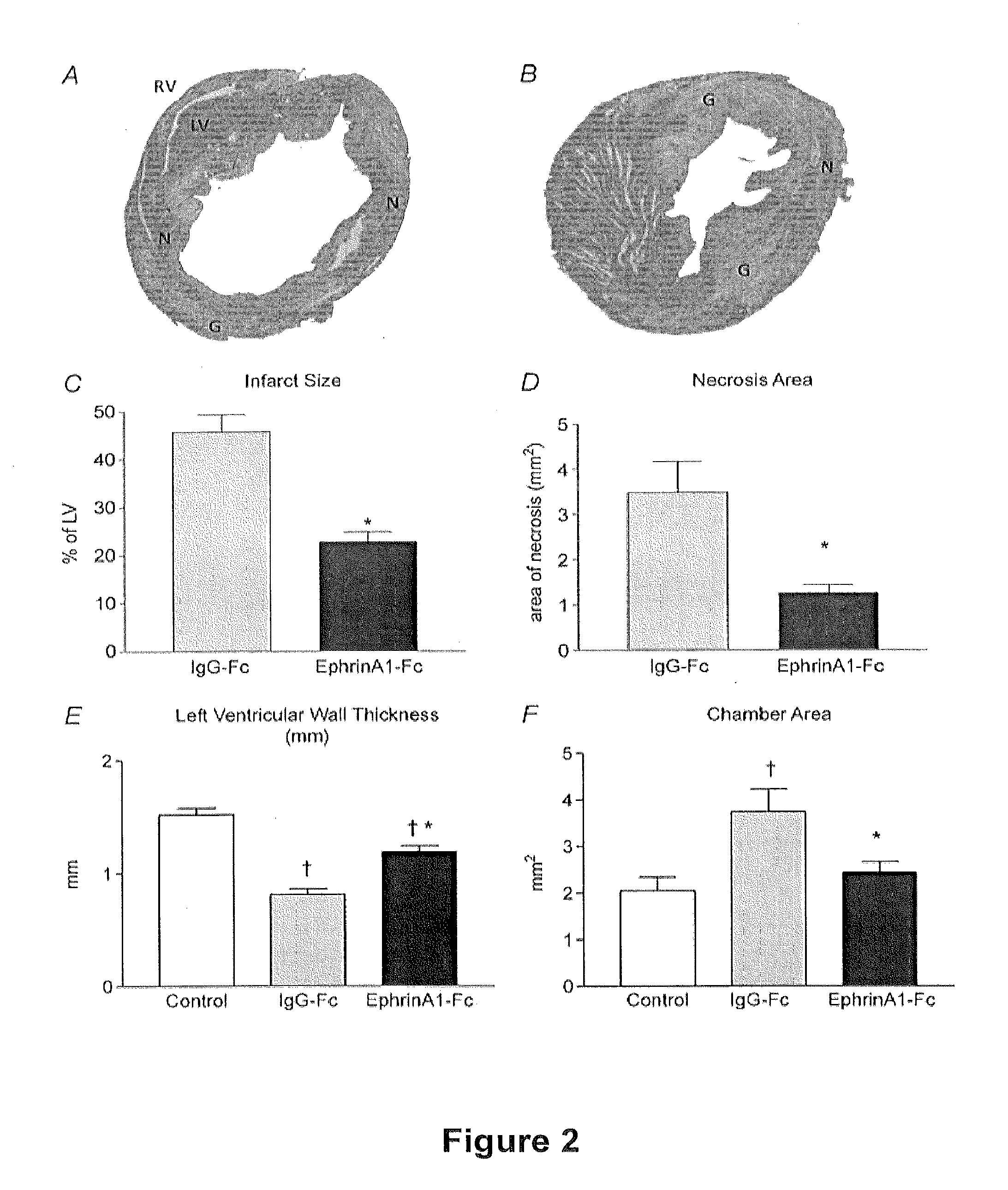
[0091]EphrinA1-Fc Reduces Infarct Size, Necrosis, Chamber Dilation, and Left Ventricular Free Wall Thinning

[0092]EphrinA1-Fc or IgG-Fc was injected into the border zone of the infarct immediately after coronary ligation. Four days after surgery, tissue was collected and either fixed for histology and immunohistochemistry, or frozen for RNA and protein isolation. Overall survival for this study was 70%, and there was no difference in survival between experimental groups. Histological staining and morphometric analyses (FIG. 2) show a 50% reduction in the size of the infarct (expressed as a percent of the left ventricle), 64% less necrotic area, a 35% reduction in chamber dilation, and 32% less thinning of the infarcted left ventricular free wall. There was no significant difference in chamber area between uninjured control hearts and those treated with ephrinA1-Fc at day 4 post-MI.

example 3

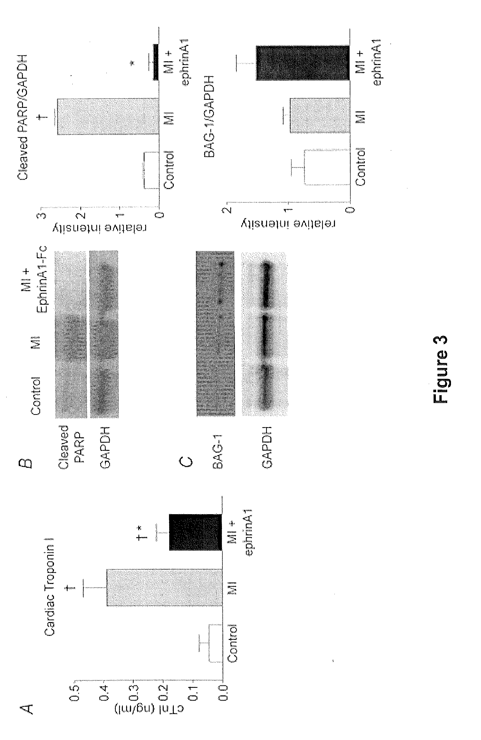
[0093]Cardiac Troponin I Levels Reduced with ephrinA1-Fc Administration

[0094]Serum cTnI levels were measured prior to surgery and at the time of euthanasia (four days post-MI) in the same animals. There was an 89% increase in cTnI levels following MI in vehicle treated hearts. However, cTnI levels in ephrinA1-Fc treated hearts were 54% lower than those from vehicle treated animals (FIG. 3A). There was no significant difference between pre-surgery levels and those of ephrinA1-Fc treated animals four days post-surgery.


PUM

PropertyMeasurementUnit
Timeaaaaaaaaaa
Timeaaaaaaaaaa
Compositionaaaaaaaaaa

Abstract

The present invention discloses methods of reducing injury resulting from cardiovascular disease, such as myocardial infarction, and/or promoting myocardial repair. The methods include administering an ephrin and pharmaceutical compositions including ephrins to a subject. Kits useful for accomplishing the same are also provided.

Description

CROSS-REFERENCE TO RELATED AAPPLICATION[0001]This application claims the benefit of, and incorporates herein by reference in its entirety, U.S. Provisional Application No. 61 / 414,741, filed Nov. 17, 2010.FIELD OF THE INVENTION[0002]The present invention concerns methods of reducing injury resulting from cardiovascular disease or events, such as myocardial infarction, and / or promoting myocardial repair. The present invention further concerns pharmaceutical compositions and kits useful for accomplishing the same.BACKGROUND OF THE INVENTION[0003]The heart generally lacks an endogenous regenerative capacity sufficient for repair after injury. Consequential left ventricular (LV) remodeling after myocardial infarction (MI) leads to LV dilatation, ultimately leading to heart failure (Pfeffer & Braunwald, 1991; Gaudron et al., 1993; Goldstein et al., 1998; Holmes et al., 2005). To reduce this epidemiologic and fiscal burden, it is imperative that strategies be developed to preserve cardiomy...

Claims


Application Information

Patent Timeline
IPC IPC(8): A61K38/17B65D71/00A61P9/00
CPCA61K38/17A61K38/19A61K38/177A61P9/00A61P9/10A61K9/0019A61K45/06C07K14/705C07K2319/30
Inventor VIRAG, JITKA A. I.DRIES, JESSICA L.
Owner EAST CAROLINA UNIVERISTY
Why Eureka
Try Eureka