
Sustained drug delivery system

a delivery system and drug technology, applied in the field of sustained drug delivery system, can solve the problems of high risk of infection, heavy physical/economic burden on patients, and the most often difficult removal of dds carrier thus administered, and achieve the effects of safe and simple implanting into the eye, easy removal, and high safety and economic burden on patients

Active Publication Date: 2012-07-12
TOHOKU UNIV
View PDF1 Cites 9 Cited by
  • Summary
  • Abstract
  • Description
  • Claims
  • Application Information

AI Technical Summary

Benefits of technology

[0041]For a DDS for treating eye disease, a method is disclosed which involves surgically administrating / implanting a DDS carrier and a drug through the vitreous body to the subretina; however, this method places a heavy physical / economic burden on patients, also poses a high risk of infection, and sometimes requires an operator to have an advanced technique. The DDS carrier thus administered is most often difficult to remove when it is made redundant or when it produces side effects. As a method for solving this problem, some transscleral DDSs are disclosed; however, they have had a problem that they are complex systems, have fluidity, cause a feeling of a foreign body because of their large size, or have a short period of slow release.
[0042]An object of the present invention is to provide a DDS for solving the above problems, which can be safely and simply implanted into the eye and can deliver a drug into the eye over a long period of time without the initial burst, particularly in application to the treatment of eye disease. Another object thereof is to provide a highly versatile long-term slow-release DDS also capable of being applied to organs / tissues other than the eye.
[0043]As described above, the DDSs previously disclosed have had a problem that it is inappropriate in the biocompatibility and shape, cannot achieve stable drug release due to the production of the initial burst, or has a short period of slow release. Particularly, the DDSs for intraocular therapy have had a problem that they are complex systems, have fluidity, cause a feeling of a foreign body because of their large size, or have a short period of slow release.
[0044]To solve these problems, the present inventors have made extensive studies, aiming at development of a compact-size stepwise slow-releasing transscleral DDS capable of unidirectionally delivering a therapeutic level of a drug to the choroid and the retina for a long period of time without damage to tissue.
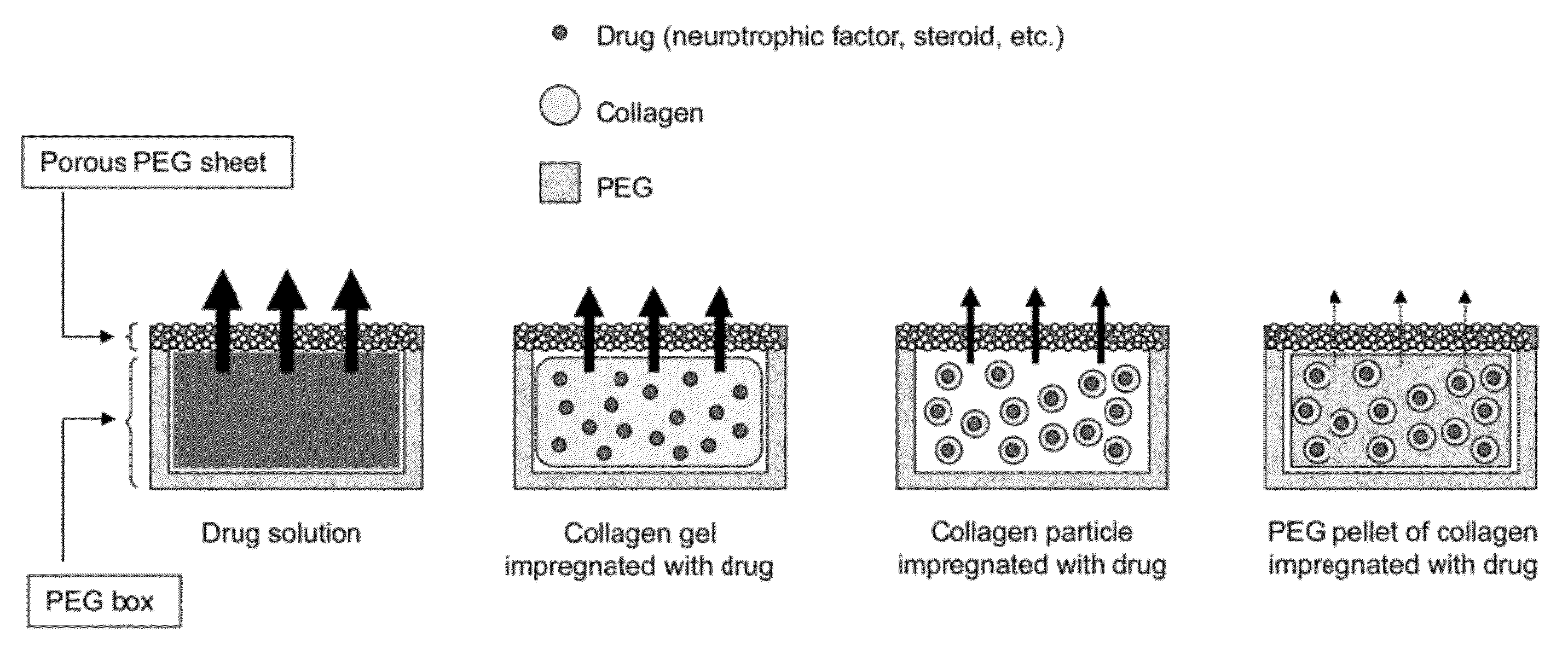
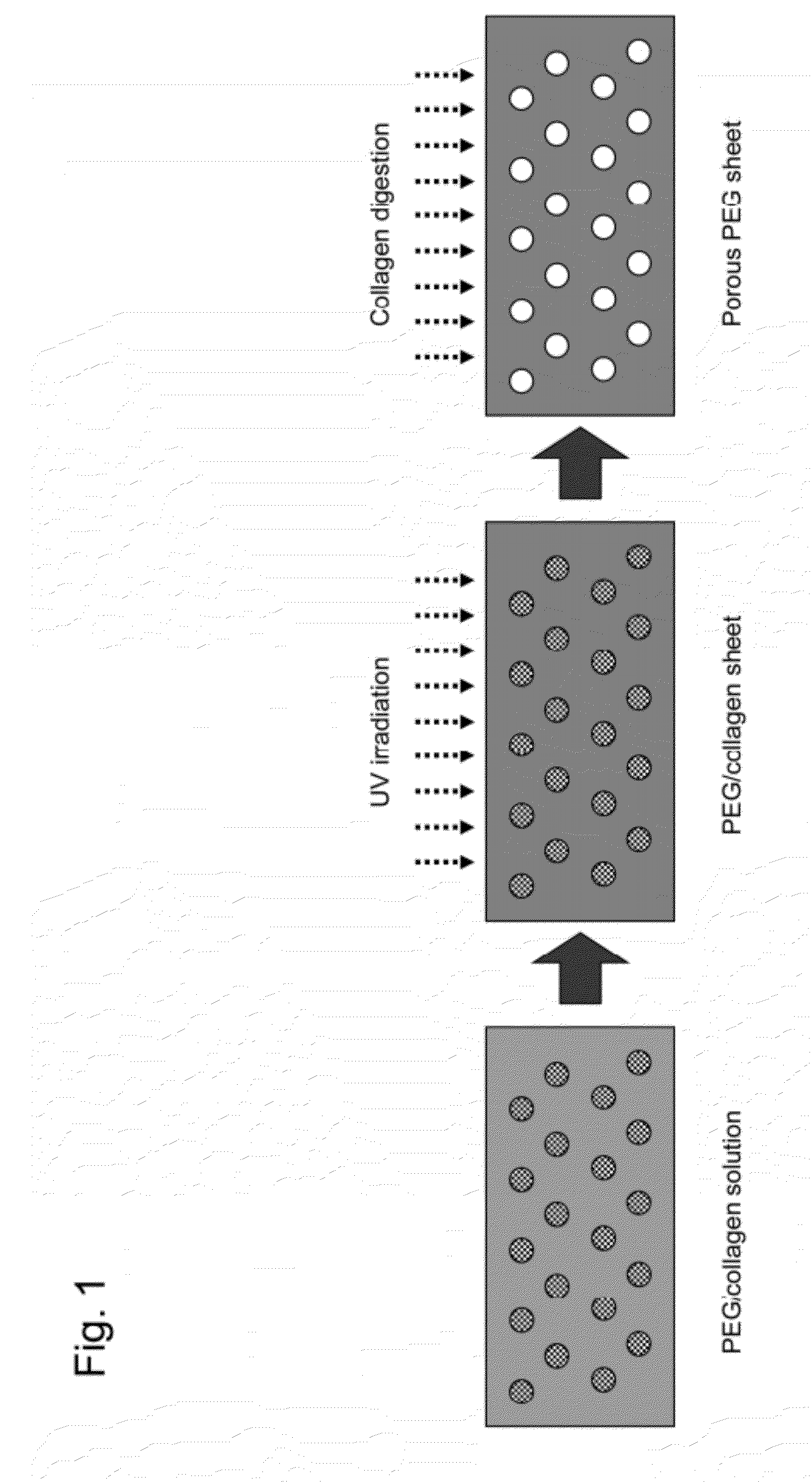
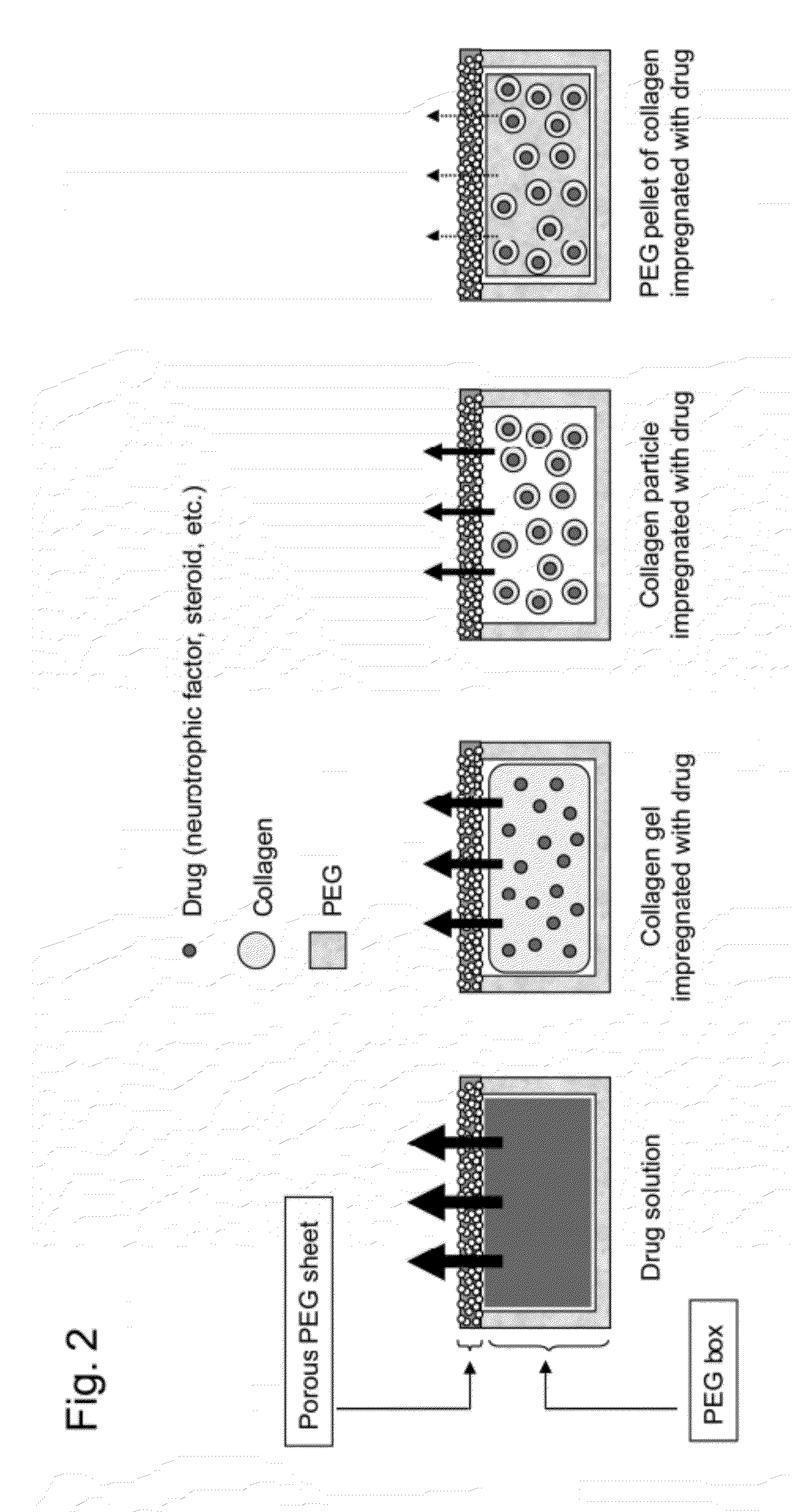
[0045]The present inventors have used collagen and PEG as base materials for DDS. The present inventors have developed a drug reservoir capsule 1 mm or less in thickness and 1 cm2 or less in area to facilitate insertion into the sclera (the sclera as used herein refers to the range from above the choroid to under the conjunctiva, that is, the subsclera, the inside of the sclera, the episclera, the subconjunctiva, and the suprachoroid). The capsule has been produced by capping a box-shaped reservoir prepared from PEG with a porous PEG sheet. The porous PEG sheet has been produced by collagenase digestion using collagen particles as a porogen (a porous mold). The change of the mixing amount of collagen particles can change the pore density. As a result, it has been found that the permeability / slow-release capability of a drug in the capsule can be easily controlled. In addition, it has been found that the drug can be packed in the capsule not in the form of a common solution but in the form of the drug impregnated in collagen (the form of collagen may be a gel or particles) or the resultant collagen wrapped in PEG and pelletized to promote slow release. In other words, this is a new DDS concept of 2 step slow release according to which the slow release of the drug from the collagen occurs in the capsule and the drug is further slowly released through the porous sheet. The combination of the method for packing the drug in the capsule and the porosity of the porous PEG sheet has enabled the period of slow release of 5 years or more to be achieved. In addition, it has been found that the inside of a capsule can be compartmented using a fine processing technology to pack several drugs and the change of the packing form enables the slow release of several drugs at different speeds. Animal experiments have showed that a drug is slowly released in a sustained manner from the device implanted on the sclera.
[0046]Thus, a scleral DDS has been developed which can slowly release a drug for a longer period of time than that for a conventional technique, is easily inserted into the sclera, and can slowly release several drugs at different speeds, thereby accomplishing the present invention.

Problems solved by technology

Although DDS is intended for “feeding a minimum necessary drug to where the demand is when needed”, the site of the implantation thereof has been limited by the inadequate shape / size of DDS, posing a problem that the drug reaches a non-target site to cause side effects.
In addition, some conventional DDSs have produced the initial burst to initially release an excessive amount of a drug or have had an insufficient period of slow release.
For a DDS for treating eye disease, a method is disclosed which involves surgically administrating / implanting a DDS carrier and a drug through the vitreous body to the subretina; however, this method places a heavy physical / economic burden on patients, also poses a high risk of infection, and sometimes requires an operator to have an advanced technique.
The DDS carrier thus administered is most often difficult to remove when it is made redundant or when it produces side effects.
As a method for solving this problem, some transscleral DDSs are disclosed; however, they have had a problem that they are complex systems, have fluidity, cause a feeling of a foreign body because of their large size, or have a short period of slow release.
As described above, the DDSs previously disclosed have had a problem that it is inappropriate in the biocompatibility and shape, cannot achieve stable drug release due to the production of the initial burst, or has a short period of slow release.
Particularly, the DDSs for intraocular therapy have had a problem that they are complex systems, have fluidity, cause a feeling of a foreign body because of their large size, or have a short period of slow release.

Method used


Image

  • Sustained drug delivery system
  • Sustained drug delivery system
  • Sustained drug delivery system

Examples


Embodiment Construction

[0081]The present invention will be described below in detail.

[0082]The therapeutic drug reservoir capsule used in the sustained DDS of the present invention consists of a PEG-made box-shaped reservoir and a porous PEG sheet, and has a capsule structure in which the box-shaped reservoir is closed by a cap made of a sheet-like or box-shaped porous PEG sheet.

[0083]The box-shaped reservoir contains a collagen impregnated with a therapeutic drug or the collagen impregnated with a drug included in PEG and pelletized. As used herein, the form of collagen may be a gel or particles. The drug may be placed in the form of a solution or powder or in the form of a mixture thereof, and the drug may be packed in various forms for applications. The collage impregnated with a drug is also referred to as a drug-embedded collagen or a collagen containing a drug.

[0084]The therapeutic drug contained in the capsule is slowly released to the outside through the portion consisting of a porous PEG sheet of...


PUM

PropertyMeasurementUnit
Therapeuticaaaaaaaaaa

Abstract

Disclosed is a drug delivery system for delivering a drug at a sustained constant rate for a long period, which can be transplanted into an affected part safely and in a simple manner and can deliver a drug to the affected part for a long period. Specifically disclosed is a sustained drug delivery system in which an implant is implanted into a body, wherein the implant is a PEG capsule comprising a box-shaped PEG and a porous PEG sheet.

Description

CROSS-REFERENCE TO RELATED APPLICATION[0001]This is a Continuation of PCT International Application PCT / JP2010 / 063793 filed Aug. 10, 2010, which in turn claims benefit to Japanese Patent Application No. JP 2009-189462 filed Aug. 18, 2009, the entire disclosure of each of which is incorporated herein by reference.TECHNICAL FIELD[0002]The present invention relates to a drug delivery system (DDS) for delivering a drug at a sustained constant rate for a long period. Specifically, it relates to a DDS and a method for treating the eye of a mammal having a disorder or disease of the eye. More specifically, it relates to an implant and a method for sustained administration of a drug into the eyeball. The use of the implant and method described herein enables the sustained administration of a drug to a desired treatment site, specifically into the eyeball.BACKGROUND ART[0003]A drug delivery system (DDS) of the concept of “feeding a minimum necessary drug to where the demand is when needed” h...

Claims


Application Information

Patent Timeline
IPC IPC(8): A61F9/00A61P27/06A61K31/5377A61K31/573A61K31/5575A61P27/02A61K38/04
CPCA61K9/0051A61K9/0002A61K38/185A61K9/4866A61K9/4808A61P27/02A61P27/06A61P9/10
Inventor ABE, TOSHIAKINAGAI, NOBUHIROKAJI, HIROKAZUKAWASHIMA, TAKEAKINISHIZAWA, MATSUHIKONISHIDA, KOJI
Owner TOHOKU UNIV
Learn More