
Multiphoton luminescence imaging of protein crystals

a protein crystal and luminescence imaging technology, applied in the field of protein crystal detection methods, can solve the problems of high false-positive/false-negative rate, limited general applicability, and the inability of uv fluorescence to easily distinguish between disordered aggregates and microcrystalline conglomerates

Active Publication Date: 2012-09-27
PURDUE RES FOUND INC
View PDF1 Cites 2 Cited by
  • Summary
  • Abstract
  • Description
  • Claims
  • Application Information

AI Technical Summary

Problems solved by technology

Consequently, identification of the conditions amenable to the formation of diffraction-quality protein crystals remains a major bottleneck in the sequence to structure pipeline.
The simplest experimental approaches rely on visual inspection or algorithmic analysis of bright-field images, which can be error-prone (high false-positive / false negative rates) and particularly challenging for small (<5 μm) crystals.
However, the deep uv excitation light used for intrinsic UV fluorescence exhibits poor transparency in conventional optical elements and most polymeric materials used for crystallization screenings, posing practical limitations on its general applicability.
Furthermore, UV fluorescence cannot easily discriminate between disordered aggregates and microcrystalline conglomerates.
Moreover, the high-energy photons used for UV-imaging (<280 nm) can induce photochemical damage to proteins during long or repeated exposures through the breakage of disulfide bonds and polymerization of neighboring residues within the crystalline lattice.
Although this technique has the ability to selectively image protein crystals through the protein-specific amide bond at 1550 cm−1, it is difficult to implement on traditional crystallization screening platforms, such as 96 well plates, thus making it a limited and specialized technique.
Other techniques for discriminating between protein and precipitant protein crystals are the “crush test” where the crystals are deemed protein if they disintegrate when touched with a needle and the staining of crystals with Coommasie Brilliant Blue dye, both of which are destructive techniques.
A major disadvantage of fluorophore doping is that protein crystals can routinely exhibit low or moderate doping efficiencies, with the fluorescence from solvated dye increasing the background and reducing image contrast.

Method used


Image

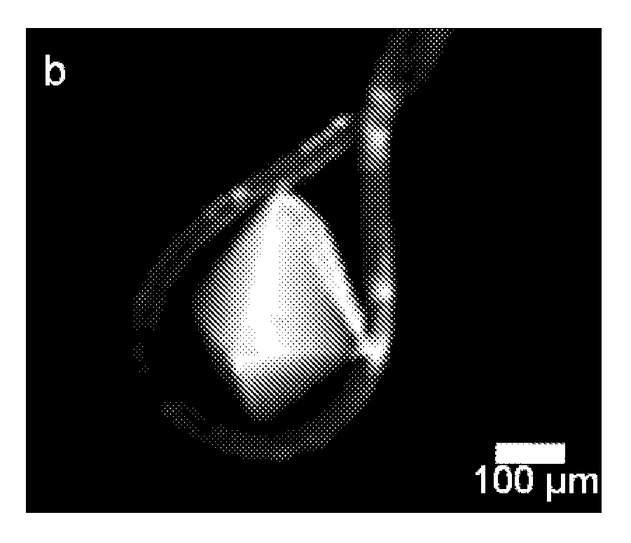
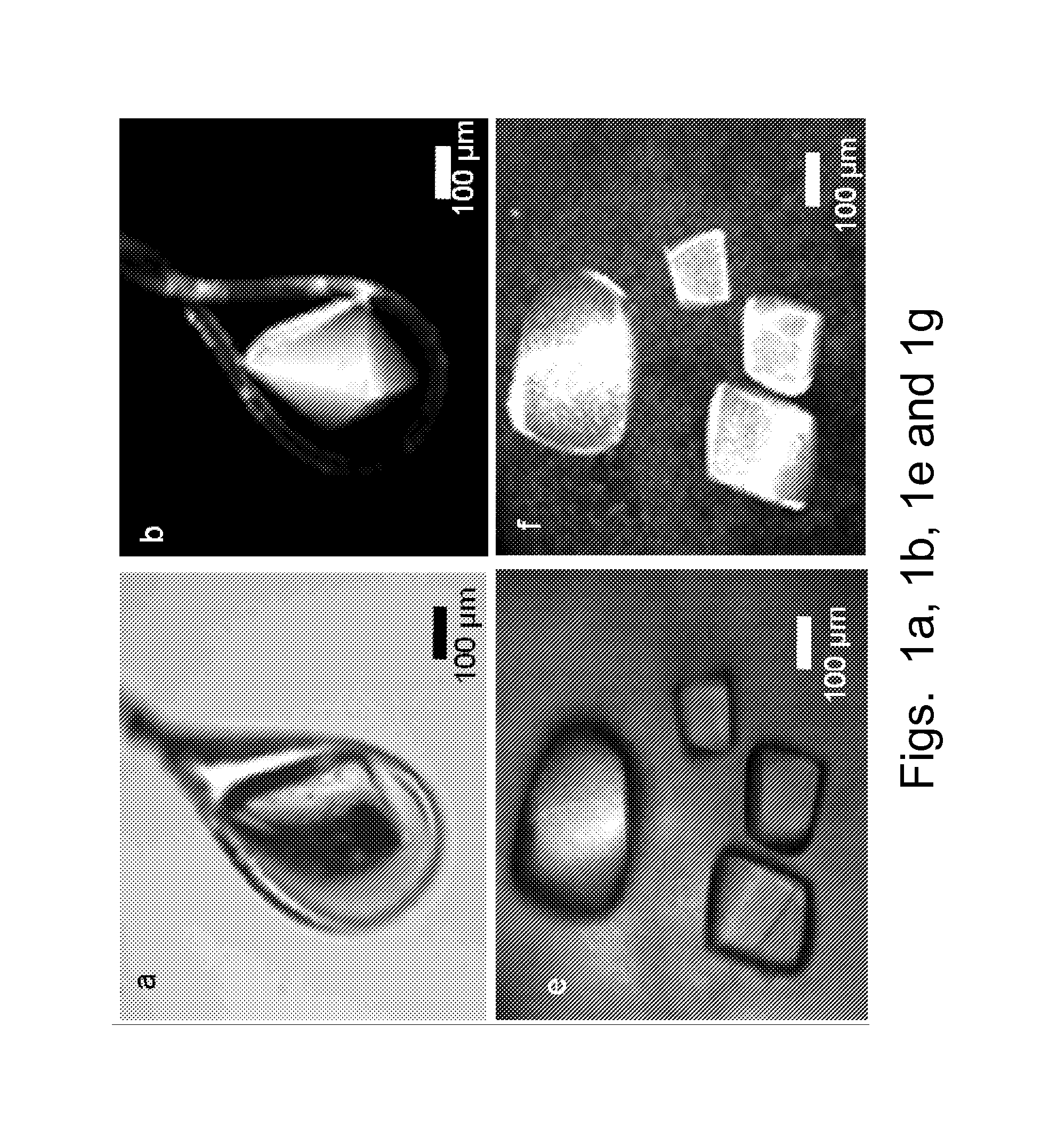
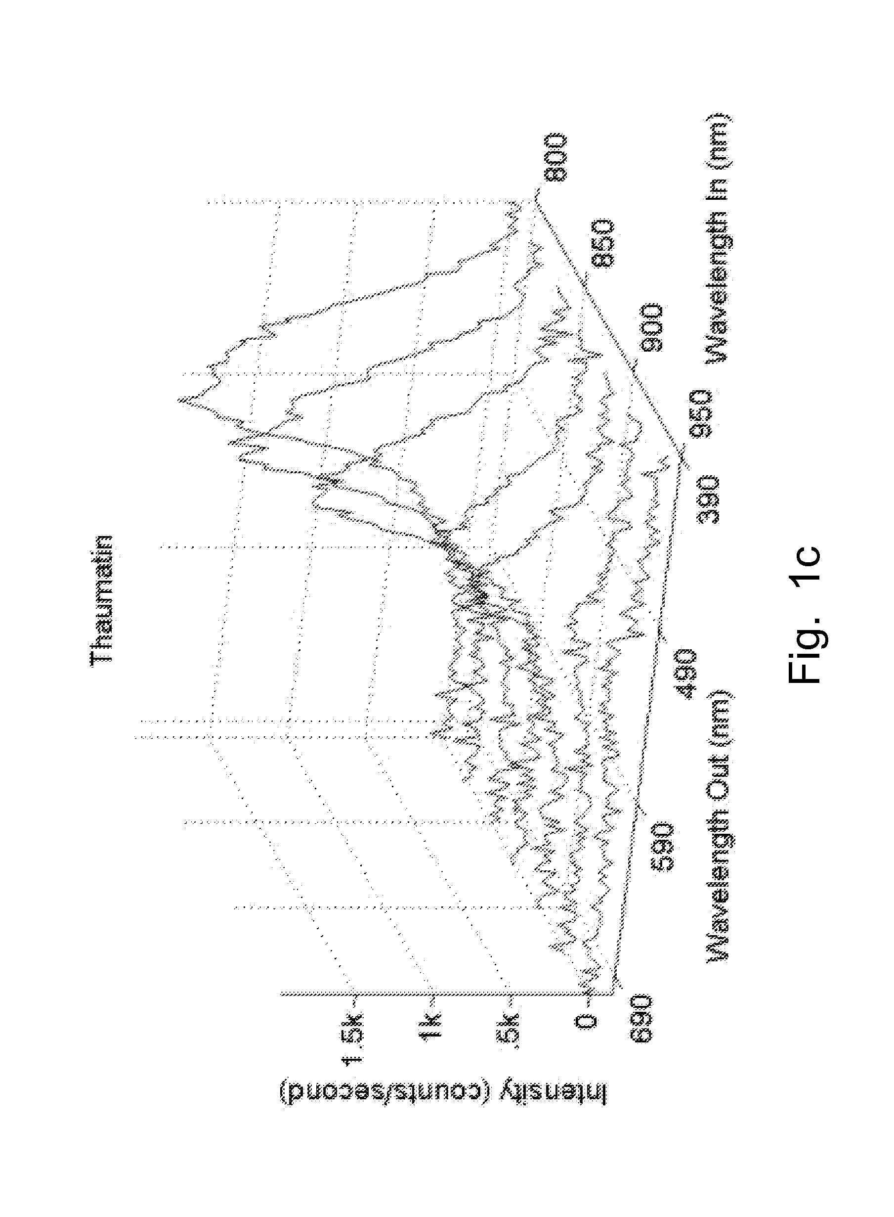
  • Multiphoton luminescence imaging of protein crystals
  • Multiphoton luminescence imaging of protein crystals
  • Multiphoton luminescence imaging of protein crystals

Examples


Embodiment Construction

[0029]The present disclosure provides a method of detection and imaging protein crystals. In particular, multiphoton excited luminescence is employed for selective detection and imaging of protein crystals. The method includes illuminating a sample with a laser; collecting an emission spectrum, evaluating the spectrum, and determining whether the sample contains protein crystals. Preferably, the sample is excited by simultaneous absorption of two or more photons. More preferably, the sample is excited by simultaneous absorption of two or three photons. Preferably, the known spectra of protein crystals are used to evaluate the observed spectrum, and therefore identify the presence of protein crystals.

[0030]The present method can detect any suitable protein crystals, such as super-oxide dismutases (SODs), lysozyme, insulin, myoglobin, deubiquitinating enzymes, and combinations thereof. The method also can be used to image polymers, such as polyamides, amino-dendrimers, etc.

[0031]The i...


PUM


Abstract

A method for detecting protein crystals comprises: illuminating a sample with a laser to produce multiphoton excitation; collecting an emission spectrum; and determining whether the sample comprises protein crystals.

Description

CROSS-REFERENCE TO RELATED APPLICATION AND INCORPORATION BY REFERENCE[0001]The present application claims priority to U.S. Provisional Patent Application No. 61 / 246,334, filed Sep. 28, 2009, the entirety of which is hereby incorporated by reference.[0002]All listed references are hereby incorporated by reference.FIELD OF THE INVENTION[0003]The present disclosure relates to a protein crystal detection method, more specifically to a method of multiphoton excited luminescence imaging of protein crystals.BACKGROUND OF THE INVENTION[0004]Protein structure determination is a key step in developing molecular-level understandings of the role of proteins in cell signaling pathways, which in turn can guide understanding of diseases and the rational design of potential drugs for treatment. High-resolution structures of relatively large proteins are generally obtained by X-ray diffraction from protein crystals. Consequently, identification of the conditions amenable to the formation of diffract...

Claims


Application Information

Patent Timeline
IPC IPC(8): G01N21/64
CPCG01N21/6402G01N21/6486G01N21/6458
Inventor SIMPSON, GARTH J.GUALTIERI, ELLEN J.KISSICK, DAVID J.MADDEN, JEREMY
Owner PURDUE RES FOUND INC
Learn More