Methods of using small RNA from bodily fluids for diagnosis and monitoring of neurodegenerative diseases

a neurodegenerative disease and bodily fluid technology, applied in the field of neurodegenerative diseases diagnosis and monitoring by using small rna from bodily fluids, can solve the problems of late clinical manifestation of disease symptoms, complicated drug development and successful treatment of ad and other neurodegenerative diseases, and complicated development of effective diagnostic methods, so as to improve the effectiveness of currently available treatments, increase clinical trial efficiency, and reduce costs

Active Publication Date: 2012-10-04
DIAMIR
View PDF4 Cites 18 Cited by
  • Summary
  • Abstract
  • Description
  • Claims
  • Application Information

AI Technical Summary

Benefits of technology

[0014]The present invention addresses these and other needs by providing a novel highly sensitive and noninvasive or minimally invasive diagnostic and monitoring methods based on quantification in bodily fluids of synapse and / or neurite small RNAs. The methods of the present invention allow diagnosis and monitoring of neurodegenerative diseases and other neurological disorders prior to occurrence of major morphological changes and massive neuronal cell death and thus have numerous clinical implications. For example, the use of the methods of the present invention can lead to enhanced effectiveness of currently available treatments for neurodegenerative diseases and other neurological disorders as such treatments could be administered at a significantly earlier stage of the disease. The use of the methods of the present invention can also allow development of new effective therapeutic and / or preventive treatments and can decrease costs and increase efficiency of clinical trials associated with such development.
[0038]Such method can further comprise (c) (i) determining that the development of the neuronal pathology in the subject is accelerated if the level of the small RNA is increased in the later obtained bodily fluid sample(s) as compared to the earlier obtained sample(s); (ii) determining that the neuronal pathology in the subject continues to develop at the same rate if the level of the small RNA is not changed in the later obtained bodily fluid sample(s) as compared to the earlier obtained sample(s), and (iii) determining that the development of the neuronal pathology in the subject is slowed down if the level of the small RNA is decreased in the later obtained bodily fluid sample(s) as compared to the earlier obtained sample(s).
[0052]Any of the above methods can further comprise the step of reducing or eliminating degradation of the small RNA.

Problems solved by technology

Drug development and successful treatment of AD and other neurodegenerative diseases are significantly complicated by the absence of effective methods for their early diagnosis and monitoring.
Development of effective diagnostic methods is further complicated by the strong brain potential to compensate for the dysfunction and loss of neurons over a long period of time.
This results in late clinical manifestation of disease symptoms when treatment cannot be very successful due to serious morphologic changes in the brain including the massive loss of neurons.
However, the neuronal loss is a relatively late event, typically following synaptic dysfunction, synaptic loss, neurite retraction, and the appearance of other abnormalities such as axonal transport defects.
New imaging techniques, which are becoming increasingly popular (e.g., positron emission tomography (PET), computed tomography (CT), magnetic resonance imaging (MRI), multiphoton imaging, magnetoencephalography (MEG), electroencephalography (EEG) etc.), are helpful, however, they are currently not sufficiently sensitive and specific for detecting early stages of a disease before major morphological changes occur (Mucke, Nature, 2009, 461:895-897; Mistur et al., J. Clin. Neurol., 2009, 5:153-166; Miller, Science, 2009, 326:386-389; Perrin et al., Nature, 2009, 461: 916-922).
However, these tests are not sufficiently sensitive and specific.
The high invasiveness of the CST collection procedure makes such tests impractical and challenging for everyday clinical use.
Cognitive function testing and brain imaging, which are currently used as main methods for diagnosis of neurodegenerative diseases such as AD, allow only detection of later stages of disease and are not sufficiently specific.

Method used


Image

  • Methods of using small RNA from bodily fluids for diagnosis and monitoring of neurodegenerative diseases
  • Methods of using small RNA from bodily fluids for diagnosis and monitoring of neurodegenerative diseases
  • Methods of using small RNA from bodily fluids for diagnosis and monitoring of neurodegenerative diseases

Examples


example 1

Comparison of Different Methods Used for miRNA Purification from Serum or Plasma

[0102]Since most of the commercially available kits for miRNA isolation have been developed for miRNA purification from cells and tissues various kits are compared with in-house modifications to adjust them for miRNA isolation from serum or plasma. Commercial kits include the miRNeasy kit (Qiagen), the MirVana RNA isolation kit (Ambion / ABI), miRACLE (Agilent), High Pure miRNA isolation kit (Roche), and miRNA Purification kit (Norgen Biotek Corp.). Besides, the in-house techniques based on the use of Trizol (Invitrogen) are developed. In some experiments, miRNA is pre-adsorbed on anion-exchangers, such as Q-Sepharose, or on magnetic beads covered with a RNA-binding material (Q-Sepharose (GE Healthcare), PEI-polyethyleneimine, or other). After Trizol deproteinization, RNA is precipitated with isopropyl alcohol or additionally purified on silica columns. In some experiments, purified RNA is treated with RNA...

example 2

Selection of miRNA for Testing

[0104]Tested miRNAs were initially selected based on literature data on their enrichment in brain compartments and presence in neurites (i.e., axons and / or dendrites and / or spines) and / or synapses (Hua et al., BMC Genomics 2009, 10:214; Liang et al., BMC Genomics. 2007, 8:166; Landgraf et al., Cell. 2007, 129:1401-1414; Lee et al., RNA. 2008, 14:35-42; Schratt et al., Nature. 439:283-289, 2006; Lugli et al., J. Neurochem. 106:650-661, 2008; Bicker and Schratt, J Cell Mol Med., 12:1466-1476, 2008; Smalheiser and Lugli, Neuromolecular Med. 11:133-140, 2009; Rajasethupathy, Neuron. 63:714-716, 2009; Kye, RNA 13:1224-1234, 2007; Yu et al., Exp Cell Res. 314:2618-2633, 2008; Cougot, et al., J. Neurosci. 28:13793-13804, 2008; Kawahara, Brain Nerve. 60:1437-1444, 2008; Schratt G. Rev Neurosci. 2009; 10:842-849) as well as on their involvement in neurite- and synapse-associated processes (The miR-Ontology Data Base: available at the world wide web at ferrolab.d...

example 3

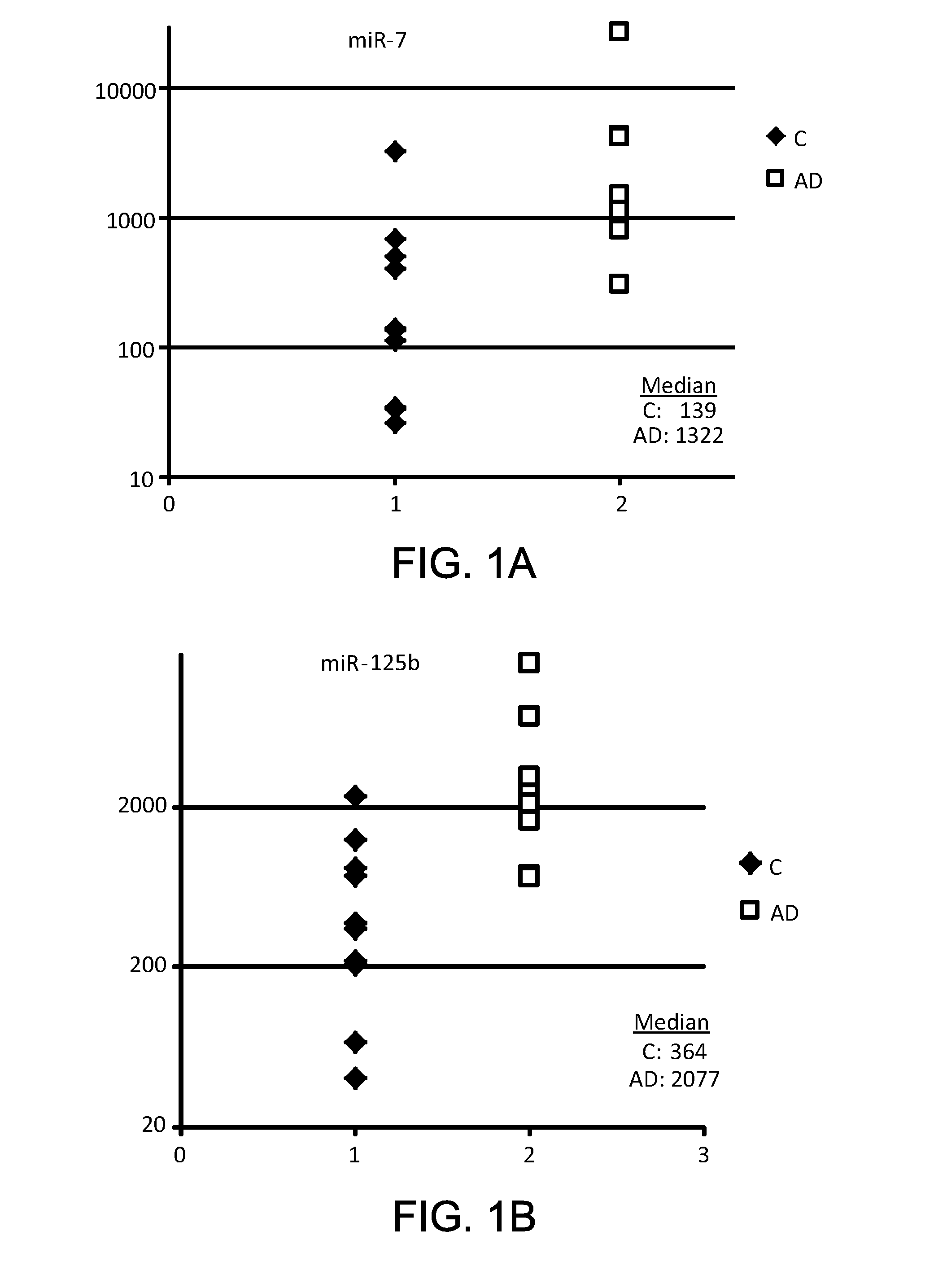
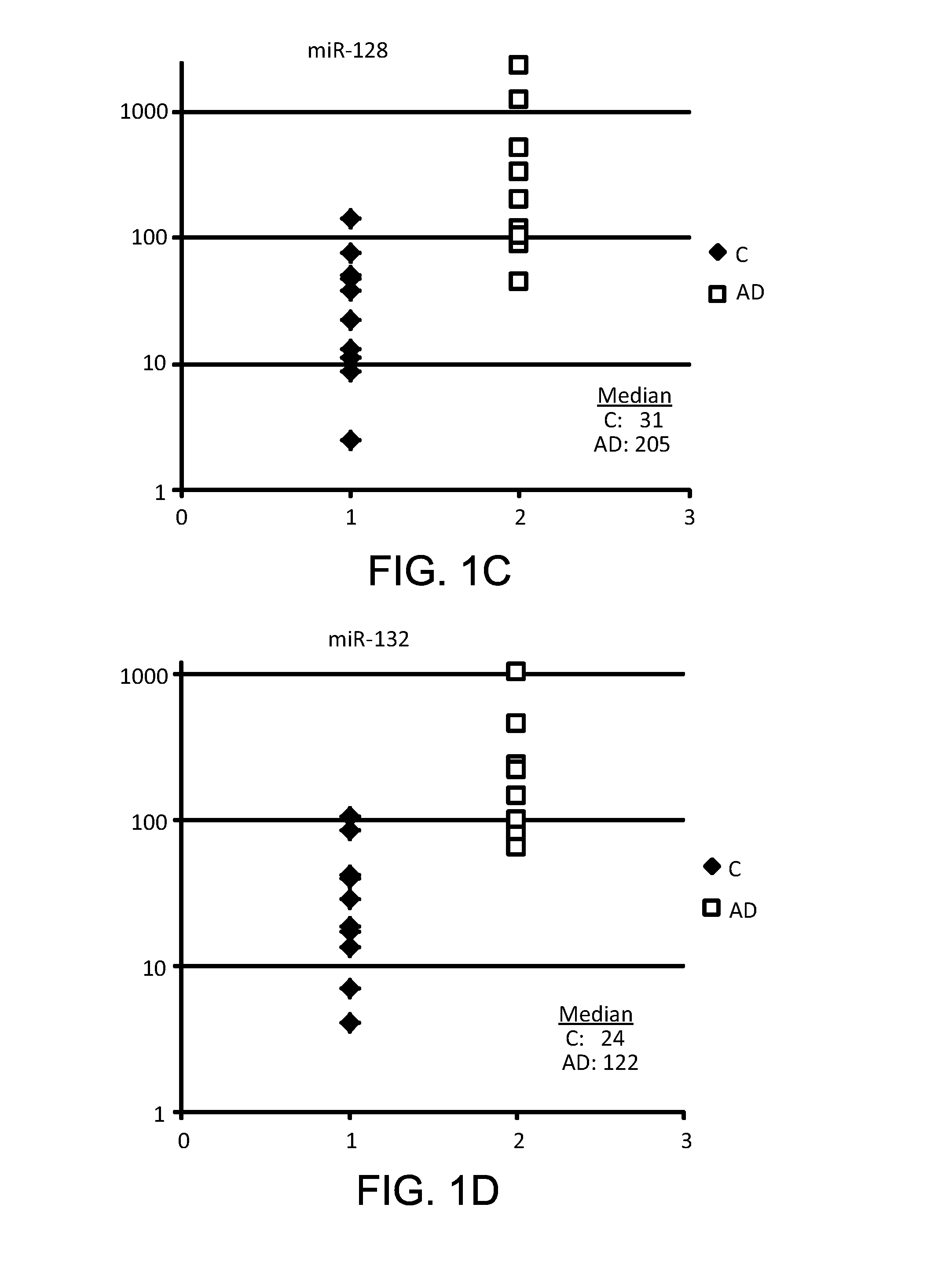
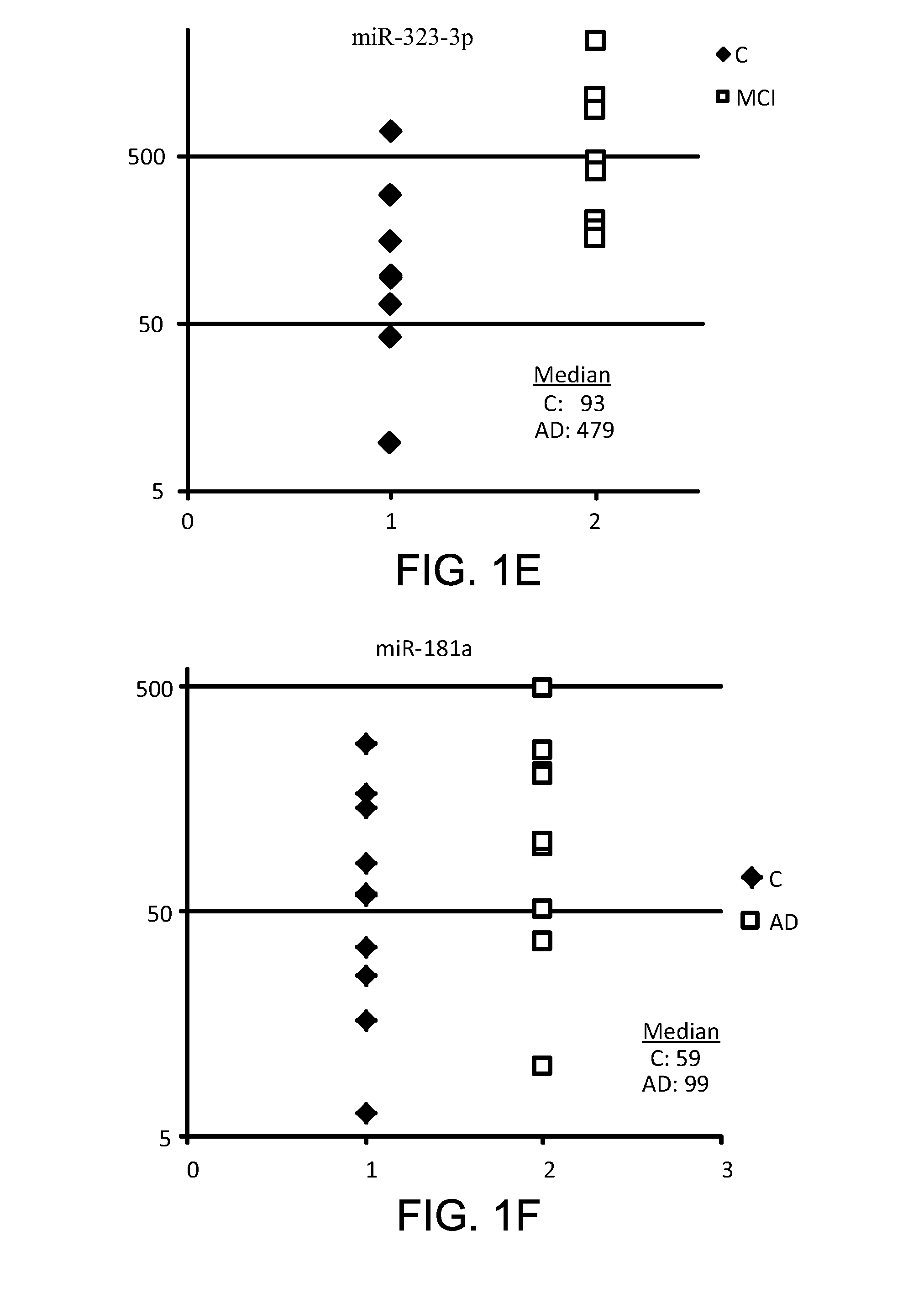
Detection of an Increase in Levels of Synapse and / or Neurite miRNA in Plasma of AD Patients

[0105]Plasma samples were obtained from patients diagnosed with developed AD by cognitive test and brain imaging. Profiles of several neuron-enriched miRNAs from plasma of these patients were analyzed using RT-PCR with primers and probes for each individual miRNA (ABI). The amount of RNA equivalent to 30 μL plasma were taken in each PCR reaction, and 1 / 15 of RT product was taken into final PCR. Thus, the amount of miRNA equivalent to 2 μL plasma was detected. The results were normalized per various miRNA, usually per ubiquitous miR-16, converted into Relative Quantity (RQ) of miRNA according the ABI protocol (2−Δct), and compared with miRNA profiles from age-matched controls. In addition, data obtained with neurite and / or synapse miRNA were compared with data obtained with neuronal body miRNA.

[0106]As shown in FIGS. 1A-G, the data obtained clearly demonstrate that concentrations of many neuron...


PUM

PropertyMeasurementUnit
timeaaaaaaaaaa
timeaaaaaaaaaa
distanceaaaaaaaaaa

Abstract

Described are methods for detection of neuronal pathologies using quantitative analysis in bodily fluids of synapse and / or neurite small RNAs and application of these methods to early diagnosis and monitoring of neurodegenerative diseases and other neurological disorders.

Description

TECHNICAL FIELD OF THE INVENTION[0001]The present invention is directed to methods for noninvasive or minimally invasive detection of pathological changes in brain or other neurons by quantifying neurite and / or synapse small RNA, particularly miRNA, in bodily fluids and application of these methods to early diagnosis and monitoring of neurodegenerative diseases and other neurological disorders.BACKGROUND OF THE INVENTION[0002]Neurodegenerative diseases comprise a large group of pathologies caused by metabolic changes in brain cells, loss of synapses and other compartments of neurons, and finally neuronal death. For review see Neurodegenerative diseases: From Molecular Concepts to Therapeutic Targets. Editors: R. von Bernhardi, N.C. Inestrosa, Nova Publishers, 2008. This group of diseases includes Mild Cognitive Impairment (MCI), Alzheimer's disease (AD), Lewy Body dementia, Parkinson's disease (PD), Huntington's disease (HD), frontotemporal dementia (FTD), vascular dementia, HIV Ass...

Claims


Application Information

Patent Timeline
Patent Type & Authority Applications(United States)
IPC IPC(8): C12Q1/68C40B40/06C40B30/04
CPCC12Q1/6883C12Q1/68C12Q2600/178C12Q2600/112
Inventor UMANSKY, SAMUIL R.SHEINERMAN, KIRA S.
Owner DIAMIR
Why Eureka
Try Eureka