

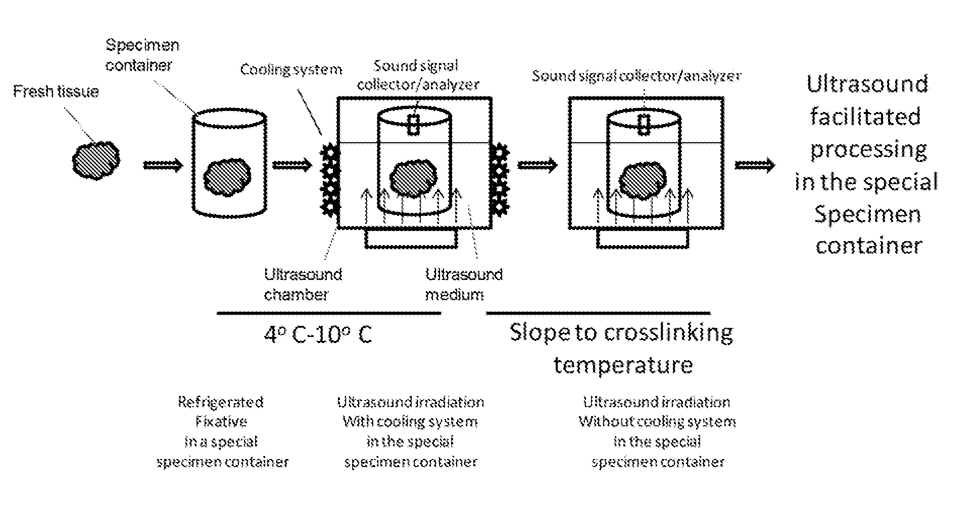
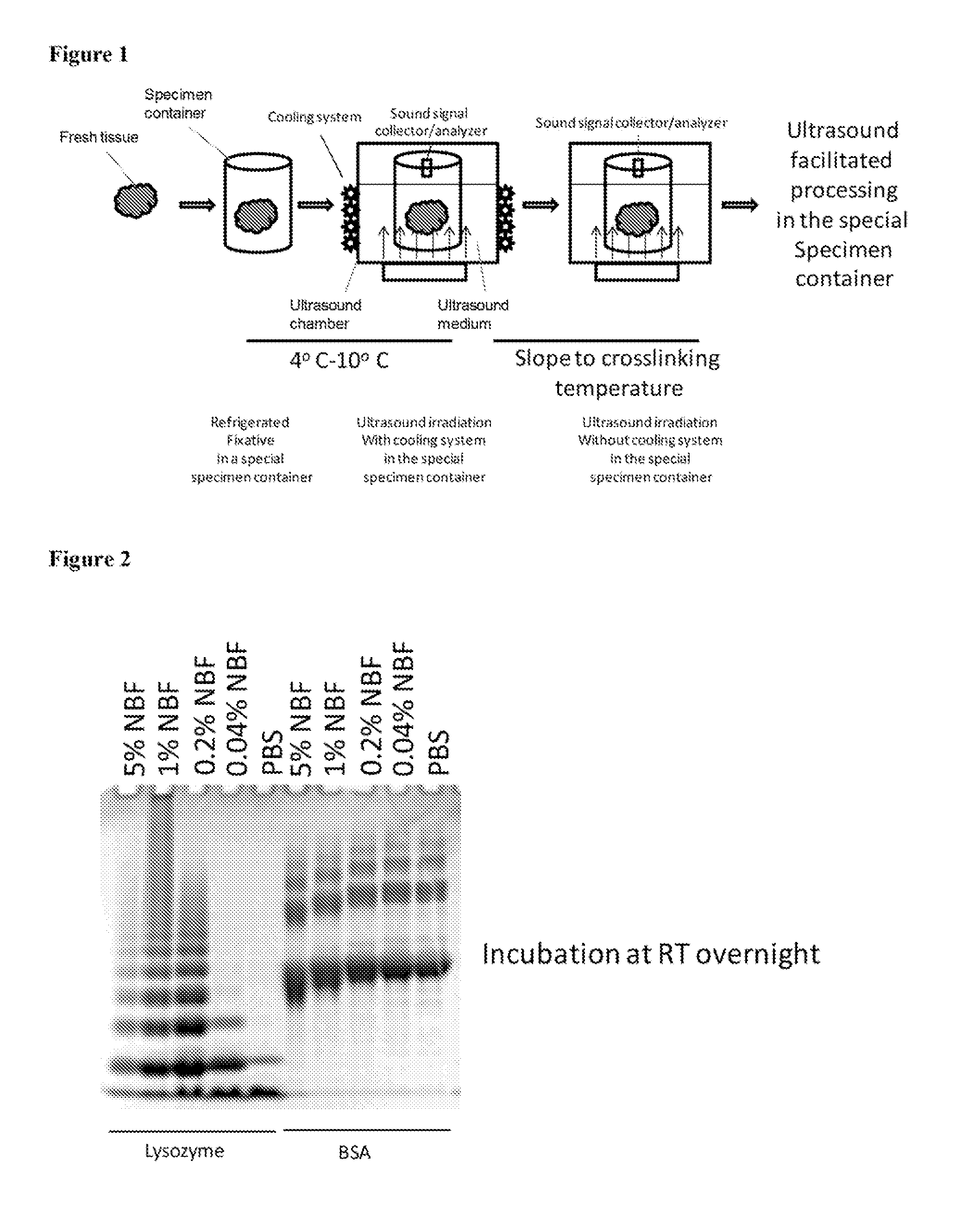
Standardization of tissue specimen preservation by ultrasound and temperature control
a tissue specimen and temperature control technology, applied in the field of tissue specimen preservation standardization and temperature control, can solve the problems of lack of protocol standardization, lack of on-site quality control in tissue fixation, and large specimen size, and achieve the effect of keeping the temperature of tissue specimen and fixative low
- Summary
- Abstract
- Description
- Claims
- Application Information
AI Technical Summary
Benefits of technology
Problems solved by technology
Method used
Image

Examples
example 1
[0064]We treated lysozyme and BSA with NBF for 60 min at various temperatures and immediately loaded the cross-linked products onto SDS gels for separation. For both lysozyme and BSA, cross-linking level gradually increased in the temperature range between 0 degree C. and 40 degree C. As revealed by the SDS PAGE, there is a significant increase in size of oligomers (lysozyme) or in gel mobility (BSA) at 50 degree C., which peaks at around 60 degree C. The inventors noticed that large amount of aggregates began to form when incubating lysozyme with NBF at a temperature greater than 60 degree C. These aggregates were highly intermolecularly cross-linked lysozyme molecules, and could not be separated on SDS PAGE. No aggregates were observed with BSA in NBF at high temperatures, possibly due to the fact that cross-linking in BSA was mostly intramolecular.
[0065]In the case of lysozyme, there was a gradual acceleration of cross-linking speed at temperature of 0-20 degree C. and leveled at...
example 2
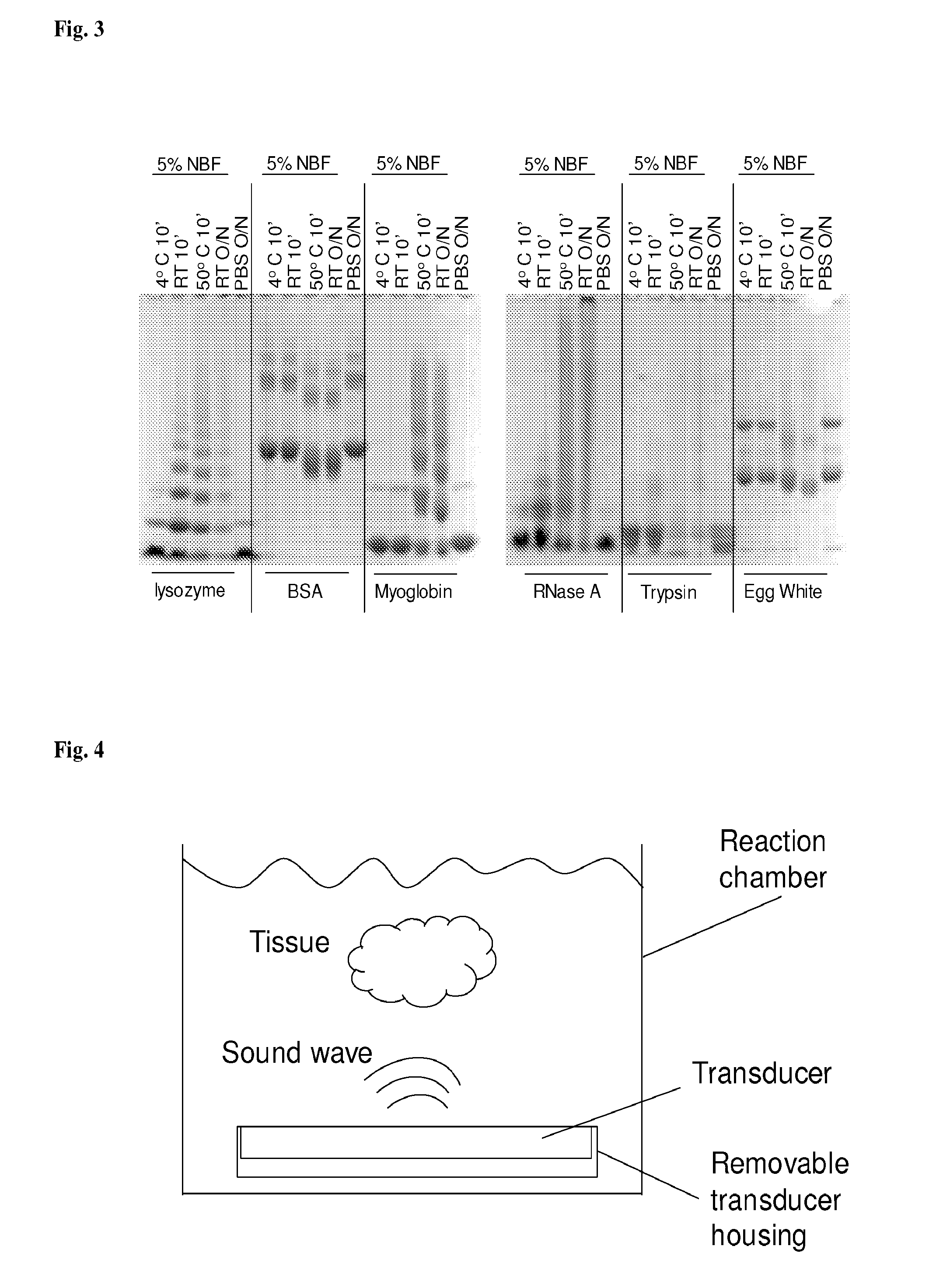
[0066]Significant cross-linking occurs after 10-minute incubation at 50 degree C. In another experiment, we tested cross-linking formation for various proteins when incubated in formalin at 4 degree C., RT, and 50 degree C. for 10 min, in comparison with incubation with formalin at RT overnight. As shown in FIG. 5, cross-linking at 4 degree C. was the slowest for all the proteins tested. And there is a big increase in levels of cross-linking after incubation at 50 degree C. for 10 min, almost comparable to the cross-linking levels after overnight incubation at room temperature. For all proteins except BSA, protein precipitations formed when incubated with formalin at 50 degree C. for 10 min and at RT overnight. This experiment indicated that for many proteins incubation with formalin at 50° C. for 10 minutes lead to significant cross-linking formation. Similar results were also obtained for 5-minute incubations (data not shown).
example 3
[0067]Tissue specimens of 1-4 mm thick were held in tissue cassettes and submerged in a cross-linking fixative (e.g. formalin, 4-25 degree C.) until ultrasound irradiation. Ultrasound is applied to bring the fixative temperature up to 50-80 degree C. alone or in combination with other heating means. The temperature was maintained for 5 to 20 min.
[0068]The tissue specimens were then submerged in a dehydration agent, e.g., 100% ethanol. Temperature of the dehydration agent was brought up to 50-75 degree C. by ultrasound irradiation alone or in combination with another heating means. The temperature was maintained by ultrasound irradiation alone or in combination of another heating means for 5-30 min. This step was repeated at least once.
[0069]The tissue specimens were then submerged in a clearing agent, e.g., xylene or xylene substitute. Temperature of the clearing agent was brought up to 50-75 degree C. by ultrasound irradiation alone or in combination with another heating means. The...
PUM
 Abstract
Description
Claims
Application Information