
Assay for Determining the Type and/or Status of a Cell Based on the Epigenetic Pattern and the Chromatin Structure

a cell type and/or cell type technology, applied in combinational chemistry, biochemistry apparatus and processes, chemical libraries, etc., can solve the problems of fragmentation of the t-lymphocyte subset of regulatory t cells within the tumour microenvironment, lack of reliable technical solutions, and limitations of all three technologies

Active Publication Date: 2013-01-03
EPIONTIS GMBH
View PDF1 Cites 51 Cited by
  • Summary
  • Abstract
  • Description
  • Claims
  • Application Information

AI Technical Summary

Benefits of technology

The present invention provides a method for identifying and quantifying specific types and states of mammalian cells in a sample obtained from a mammal, by analyzing the relative amount of accessible chromatin in regions specific to the cell type. This method is reliable and accurate for identifying and quantifying different types of cells in solid tissue, and can also be used for diagnostic purposes. The invention also provides markers and kits for identifying and quantifying specific cell types. Overall, the invention provides a better understanding of the complex process of gene regulation and cellular differentiation in mammals.

Problems solved by technology

However, understanding the role of T-lymphocytes as major cellular component of the adaptive effector immune response and their counterpart, the T-lymphocyte subset regulatory T cells within the tumour microenvironment remains fragmentary.
In part this deficiency is due to the lack of reliable technical solutions for specific cell identification and quantification in solid tissue.
However, all three technologies are afflicted with limitations when applying them in a truly quantitative manner.
FACS analysis is additionally problematic for solid tissues, since an experimental prerequisite is its dissociation into a single cell suspension.
Despite attempts to automate cell counting by IHC, it is not a reliable method for quantification (Taylor, C. R. and R. M. Levenson, Quantification of immunohistochemistry—issues concerning methods, utility and semiquantitative assessment II.
It is widely acknowledged that those technological difficulties associated with measurements of immune cells in the tumour microenvironment constrain precise studies and the actual immunological status and the prognostic meaning of immune cell infiltration in the tumor environment remain largely unclear.
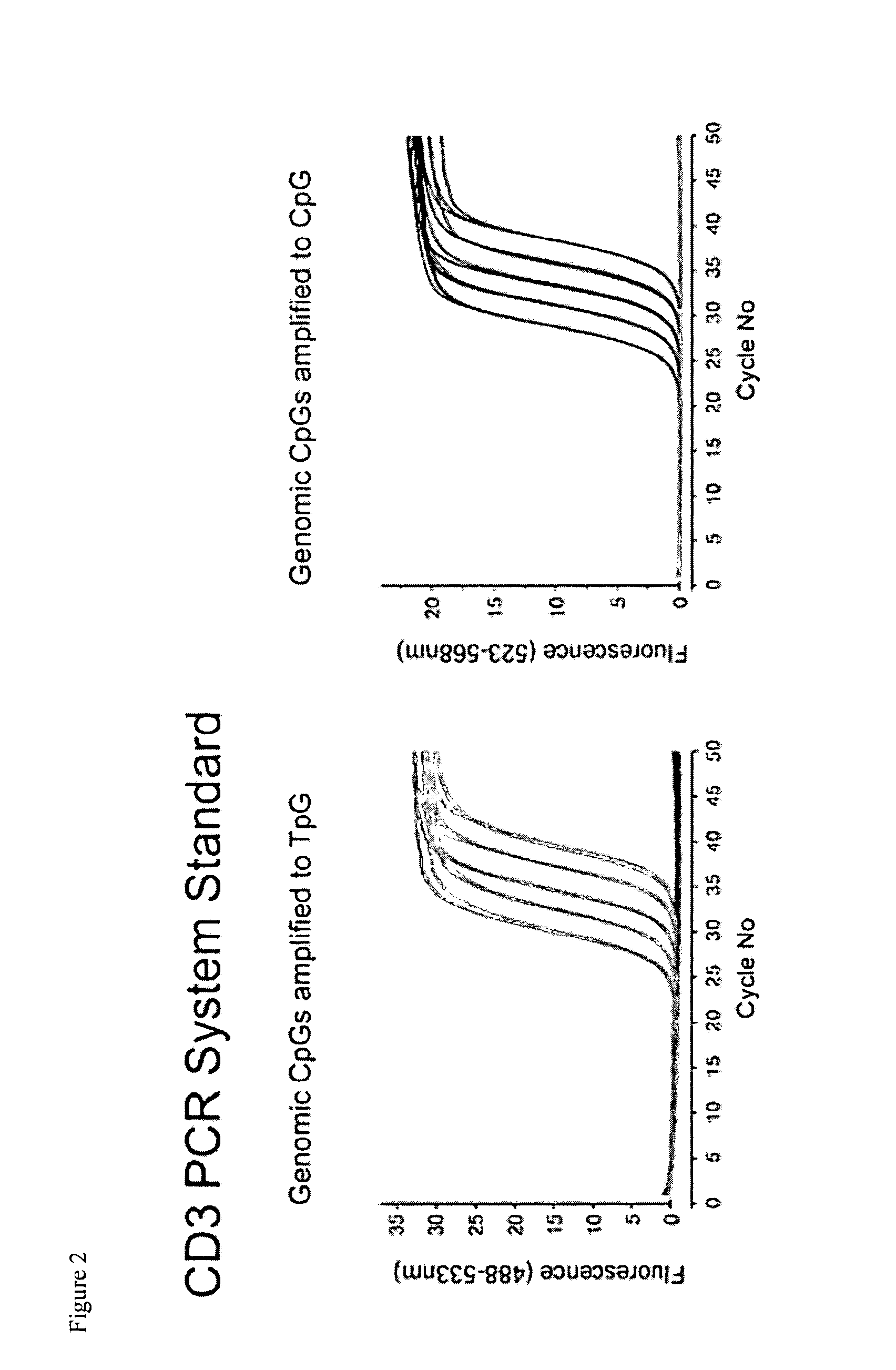
This approach is not possible in other systems, as these are analyzing only one type of gene (e.g. foxp3 tpg is detected and compared with foxp3 cpg).
Any living cell can usually not afford to switch those cycles off, unless it (the cell) is dying or goes into lethal resting.
Specifically, one typically limits the concentration of the probe to very low levels.
Remaining result variability between FACS and epigenetic measurements may be attributed in part to systematic variations, including lacking precision in defining the general leukocyte count by flow cytometry.
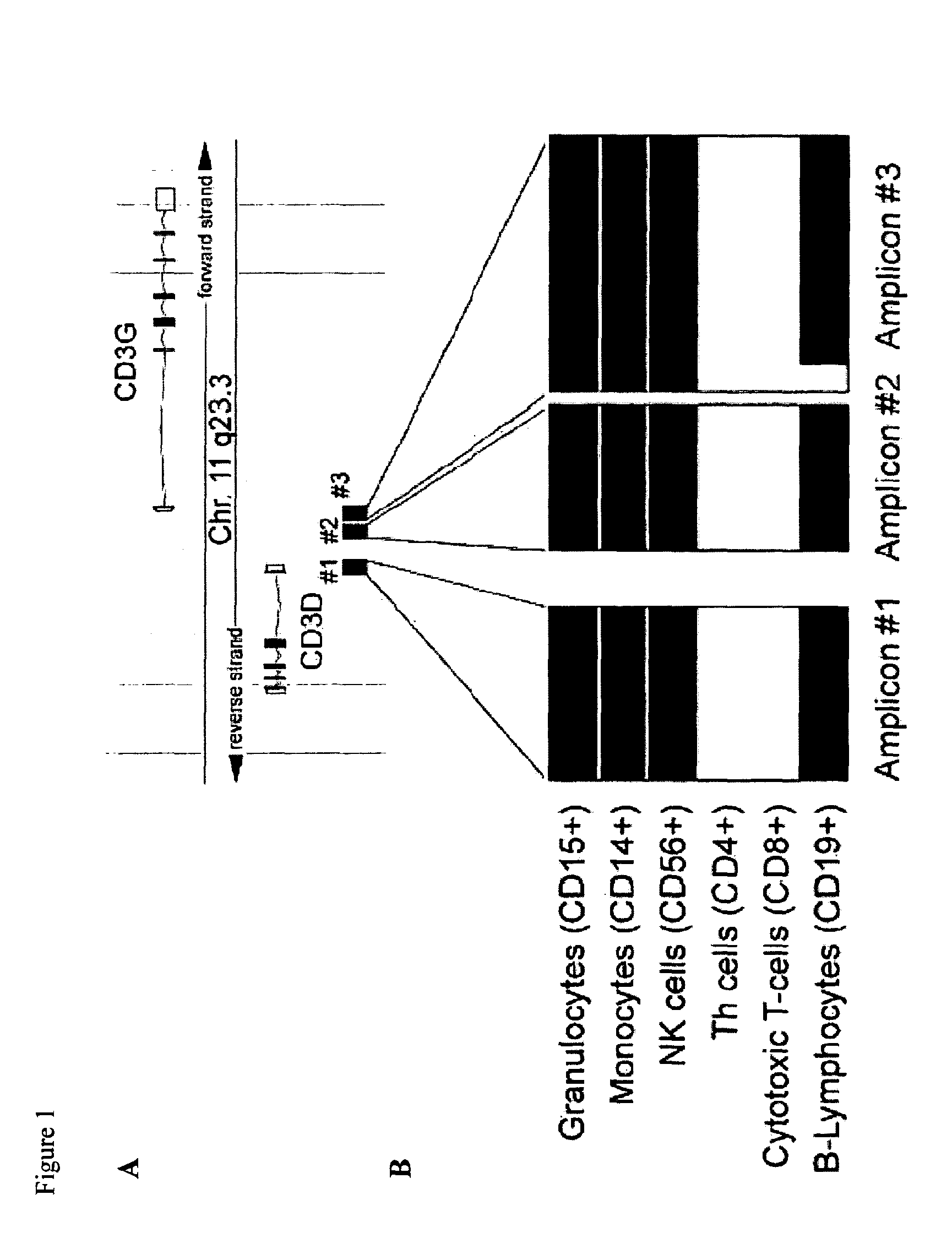
Combining the different datasets uncovers an intrinsic difficulty for the previously reported correlation: the inventors observe a direct linear correlation between Treg number and the overall T-lymphocytes count in all healthy and tumorous tissues.
Therefore, it is not plausible to assume that an increasing CD3 level and, at the same time, decreasing Tregs leads to an improved outcome—at least not if tested univariately as had been done in previous studies.
However, differing results between the present and previous data also require also functional and technical explanations.
Also, higher density of Tregs may occur in particular areas of the tumour, a phenomenon that epigenetic studies cannot pick up.
However, the inventors regard the association of Tregs to CD3 cell counts as a severe inhibition to the efficacy of T cell based drugs, since an unselected homing of CD3 cells without an inhibition of Treg homing threatens to always co-attract activator and its suppressor alike.

Method used


Image

  • Assay for Determining the Type and/or Status of a Cell Based on the Epigenetic Pattern and the Chromatin Structure
  • Assay for Determining the Type and/or Status of a Cell Based on the Epigenetic Pattern and the Chromatin Structure
  • Assay for Determining the Type and/or Status of a Cell Based on the Epigenetic Pattern and the Chromatin Structure

Examples


Embodiment Construction

Materials and Methods

Abbreviations:

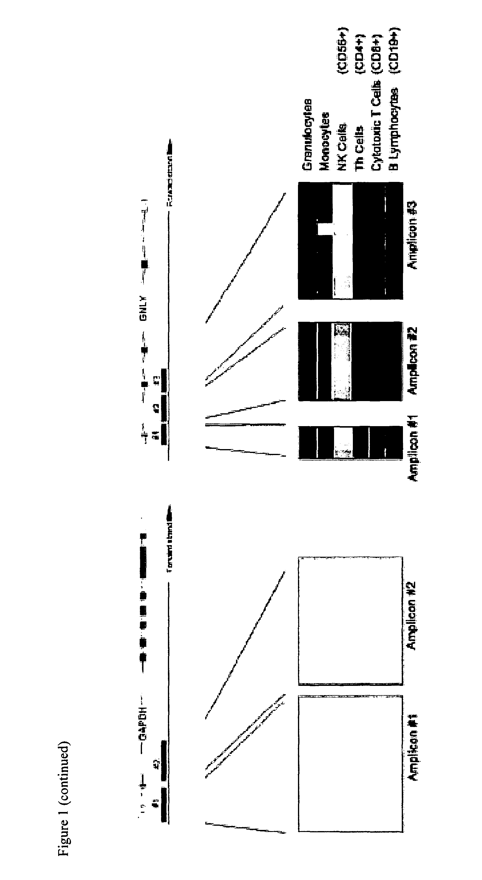
[0100]Amp, amplicon; CD3D, T-cell surface glycoprotein CD3 delta chain; CD3G, T-cell surface glycoprotein CD3 gamma chain; FOXP3, Forkhead box protein P3; GAPDH, Glyceraldehyde-3-phosphate dehydrogenase; GNLY, Granulysin.

Cells and Tissue Samples

[0101]Formalin-fixed and paraffin-embedded tissue samples were retrieved from the archives of the Institute of Pathology, Charité-Universitätsmedizin Berlin, Campus Benjamin Franklin. Representative paraffin blocks of tumour and normal tissue were selected and tissue microarray (TMA) of the colorectal or bronchial carcinoma specimens with the corresponding normal parenchyma were constructed using cores of 1 mm in diameter. Fresh frozen ovarian tissue samples and blood were retrieved from the tumour bank ovarian cancer, Charite-Universitätsmedizin Berlin, Campus Virchow.

Isolation of Genomic DNA

[0102]For purification of genomic DNA from human blood, tissues, the DNeasy Blood and Tissue Kit (Qiagen, Hilden, Ger...


PUM

PropertyMeasurementUnit
Fractionaaaaaaaaaa
Fractionaaaaaaaaaa
Fractionaaaaaaaaaa

Abstract

The present invention relates to a method for identifying a specific type and / or state of a mammalian cell in a sample obtained from a mammal, comprising a) analyzing the relative amount of accessible chromatin in regions that are specific for a cell-type and / or cellular state in the genome of said cell, b) comparing said relative amount of accessible chromatin said in regions with the relative amount of accessible chromatin in regions in the genome of said cell that are unspecific for a cell-type and / or cellular state, and c) deducing the specific type and / or state of said mammalian cell in said sample based on said comparison. Preferably, said identifying further comprises a relative quantification of said specific cell type and / or state based on said comparison. The method can further comprise a diagnosis of a predisposition to a disease or a disease based on said identification. Kits and certain markers in regions of accessible chromatin in the genome are described, too.

Description

[0001]The present invention relates to a method for identifying and / or quantitating the total amount, and / or ratio of a specific type and / or state of a mammalian cell within all cells as present in a sample obtained from a mammal, comprising a) analyzing the relative amount of accessible chromatin in regions that are specific for a cell-type and / or cellular state in the genome of said cell, b) comparing said relative amount of accessible chromatin said in regions with the relative amount of accessible chromatin in regions in the genome of said cell that are unspecific for a cell-type and / or cellular state, and c) deducing the specific type and / or state of said mammalian cell in said sample based on said comparison. Preferably, said identifying further comprises a relative quantification of said specific cell type and / or state based on said comparison. The method can further comprise a diagnosis of a predisposition to a disease or a disease based on said identification. Kits and cert...

Claims


Application Information

Patent Timeline
IPC IPC(8): C12Q1/68C40B30/04
CPCC12Q1/6881G01N33/6875G01N33/56966C12Q2600/154C12Q1/6858C12Q2545/113C12Q2523/125
Inventor OLEK, SVEN
Owner EPIONTIS GMBH
Learn More