System and method for mapping gastro-intestinal electrical activity
a technology of electrical activity and system, applied in the field of system and method for mapping gastrointestinal electrical activity, can solve the problems of inability to reliably achieve analysis of signals obtained, and inability to provide accurate information regarding the normal or abnormal propagation of individual slow wave cycles
- Summary
- Abstract
- Description
- Claims
- Application Information
AI Technical Summary
Benefits of technology
Problems solved by technology
Method used
Image
Examples
example 1
Trial
[0168]Method
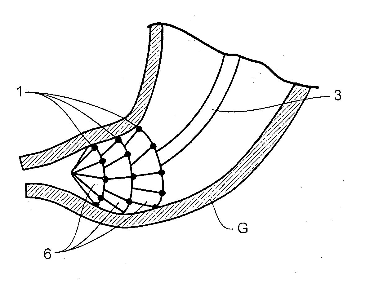
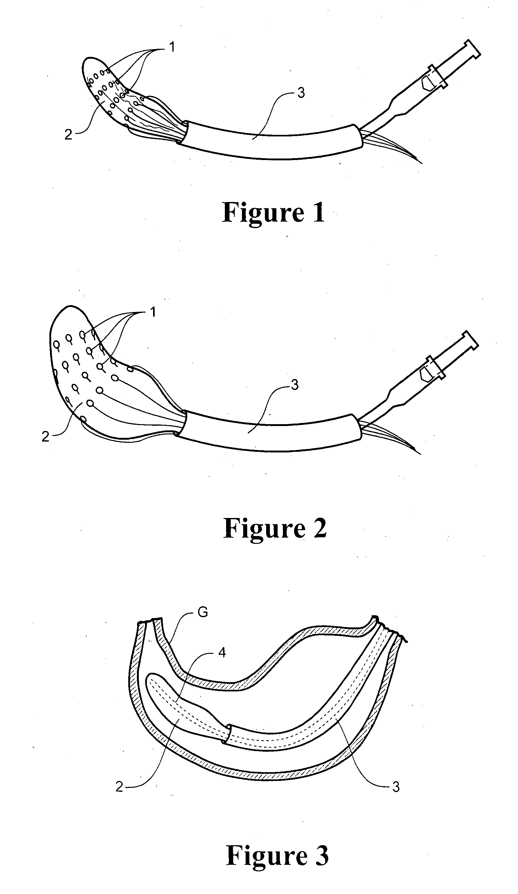
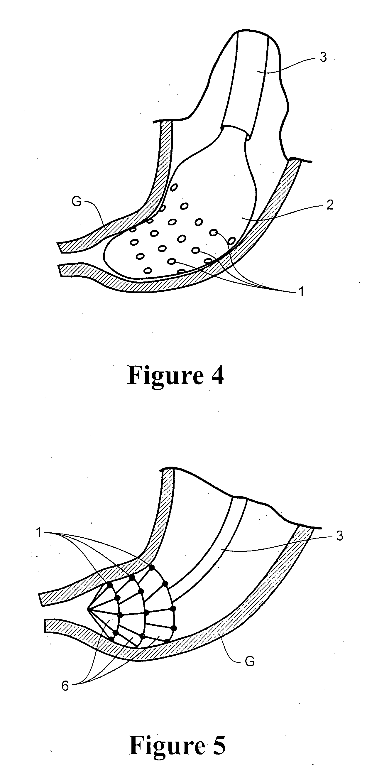
[0169]A mapping catheter was constructed from: a 24 Fr two-way urinary catheter (outer catheter), a 12 Fr nasogastric tube (inner catheter), a latex balloon (standard condom), 32 ECG-dot central pins (stainless steel contact surfaces), 32 copper wire leads (connected to a 68-way SCSI ribbon cable; soldered at each end), and a three-way tap and inflation syringe. The catheters were joined with heat-shrink tubing, and the ECG dots were stuck to the balloon with glue.
[0170]The configuration of the balloon electrode array was circumferential and was numbered as follows (proximal to distal):
7923242930172631151232103527281311146842516122018222119
[0171]The SCSI cable connector pins were connected to port A of the ActiveTwo System (BioSemi, Netherlands). A flexible printed circuit board mounting a number of electrodes was connected to port B, to allow validation against a serosal reference electrode.
[0172]A female weaner cross-breed pig of 39 kg was fasted overnight and ana...
example 2
Trial
[0187]Method
[0188]Flexible PCB multi-electrode recording arrays consisting of copper wires and gold contacts on a polyimide ribbon base were employed. The recording head of each array had 32 electrodes in a 4×8 array, at an interelectrode distance of 7.6 mm.
[0189]Mapping was undertaken in human subjects undergoing upper abdominal surgery, immediately after laparotomy and prior to additional surgical dissection. Up to 6 PCBs (192 electrodes; ˜93 cm2) were used in each experiment, and were held together in ideal parallel alignment. The recording surface of the PCBs were positioned flush with the anterior serosal surface of the stomach. The posterior gastric surface was not mapped. The recording period was 10-15 minutes, usually allowing two adjacent areas of gastric tissue to be mapped.
[0190]Unipolar recordings of 10-15 min duration were acquired using the ActiveTwo System (Biosemi, Amsterdam), at a recording frequency of 512 Hz. The common sense (CMS) and right leg drive (DRL) e...
example 3
FEVT Activation Time Marking
[0200]Slow wave recordings of GI electrical activity were undertaken during surgery in pigs. Recordings were taken with both a high SNR 48 electrode array (resin-embedded, shielded, silver electrodes) and from a lower SNR electrode array (flexible PCBs; unshielded), from the anterior porcine gastric corpus. One 180 second representative data segment was selected from each of five animals: two segments from the high SNR array and three from the low SNR array. Unipolar recordings were acquired from the electrodes via the ActiveTwo System, at a recording frequency of 512 Hz. The common mode sense electrode was placed on the lower abdomen, and the right leg drive electrode on the hind leg. The electrodes array were connected to the ActiveTwo which was in turn connected to a notebook computer. The acquired signals were pre-processed by applying a second-order Butterworth digital band pass filter. The low frequency cutoff was set for 1 cpm ( 1 / 60 Hz); the high ...
PUM
 Abstract
Description
Claims
Application Information
 - R&D
- Intellectual Property
- Life Sciences
- Materials
- Tech Scout
- Unparalleled Data Quality
- Higher Quality Content
- 60% Fewer Hallucinations
Browse by: Latest US Patents, China's latest patents, Technical Efficacy Thesaurus, Application Domain, Technology Topic, Popular Technical Reports.
© 2025 PatSnap. All rights reserved.Legal|Privacy policy|Modern Slavery Act Transparency Statement|Sitemap|About US| Contact US: help@patsnap.com


